Gastric ulcer is the most common chronic disease of the gastrointestinal tract. The number of cases is increasing every year; men aged 25–45 years are most often affected by the disease; in women, the disease is less common. The most dangerous complication of an ulcer is gastric perforation or otherwise perforation.
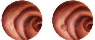
A gastric perforation is a small hole in the wall of the stomach. The edges of the hole are round, even, smooth, the diameter can reach 1 cm. A perforated ulcer is usually localized in the lower wall of the stomach, less often in the side wall.
Food, stomach acid, bile and enzymes can easily enter the abdominal cavity through the perforation and cause a chemical burn. Food particles in the abdominal cavity begin to gradually rot, which provokes the proliferation of bacteria and the development of an acute inflammatory process - peritonitis. Peritonitis develops quickly, within 12–18 hours after the formation of a perforation. Without medical assistance, on the 4th day after the development of peritonitis, the entire body becomes infected, which leads to death.
In the esophagus and small intestine, ulcers and perforations are rare; most often such phenomena are observed in the stomach and duodenum. The exacerbation of the ulcer occurs in the spring and autumn periods, so perforations can often appear at this time. In 20% of cases, the stomach walls are perforated asymptomatically and without the presence of a gastric ulcer. This is where the deadly danger of this disease lies.
Total information
Please note
Gastric perforation occurs in one person per 5 thousand adults - this figure is considered high.
This condition can complicate many stomach diseases, but most often develops with gastric ulcers - in every tenth patient of all who have been diagnosed with this. And among all the conditions that complicate gastric ulcer, it is observed in 15% of all clinical cases.
Important
Most often, young men of working age are affected - from 20 to 40 years old (although in the last 10 years the trend is that patients aged 50 years and older began to suffer more often from this pathology). In general, in men, gastric perforation for various reasons occurs 15-20 times more often than in women.
The peak of gastric perforation occurs in the autumn and spring periods. This is explained by seasonal exacerbation of peptic ulcers of the stomach - it is with this diagnosis that gastric perforation most often occurs.
Violation of the integrity of the stomach for other, non-ulcer reasons accounts for about 10% of all clinical cases - mainly affecting school-age children and young people aged 20 to 30 years.
The occurrence of this pathology not only creates problems in itself, but also signals the fact that the disease is neglected - poor diagnosis or its complete absence
Causes
Gastric perforation most often occurs in diseases and pathological conditions such as:
- long-term and periodically exacerbating gastric ulcer. When finding out in patients how long the ulcer has been observed, it should be remembered that in the elderly there may be no history of ulcers, since this age (from 60 to 70 years, sometimes older) is characterized by a blurred clinical picture of the disease;
- states of shock;
- alcohol abuse;
- non-targeted and uncontrolled use of drugs of certain pharmacological groups;
- binge eating;
- prolonged physical and psycho-emotional stress (although cases of gastric perforation have also been described against the background of an acute, lightning-fast ulcer due to a severe stressful situation);
- infectious agent - in particular, the presence of Helicobacter pylori in the gastric cavity. It should be remembered that this pathogen is found in 95% of patients with peptic ulcers, but acute ulcers in the vast majority of cases are not associated with it in any way.
Factors that directly contribute to perforation of the stomach at the site of ulceration are:
- slow but persistent destruction of all layers of the stomach by hydrochloric acid produced by its own cells;
- increased pressure in the gastric cavity;
- presence of an infectious agent.
Diseases have been identified that most often cause the formation of a hole in the wall of the stomach. This:
- medicinal ulcers (while taking drugs that act aggressively on the stomach wall);
- some endocrine disorders. The most indicative of them is Zollinger-Ellison syndrome - the formation in the pancreas or duodenum of a specific tumor that produces gastrin - a hormone that stimulates the production of pepsin in the stomach, due to which the acidity of the gastric environment increases, and therefore is ulcerogenic (ulcer-forming). ) effect on the gastric mucosa;
- hepatogenic ulcers (occur against the background of changes in the liver);
- pancreatogenic ulcers (formed due to pathology of the pancreas);
- acute ulcer due to Crohn's disease - granulomatous lesions of the entire gastrointestinal tract, including the stomach.
In addition, an acute perforated gastric ulcer can become a complication of diseases of other organs and systems - most often it is:
- acute myocardial infarction (mostly extensive);
- burn disease - changes in many organs and systems due to a burn in a certain location;
- stroke is a violation of cerebral circulation of the ischemic (oxygen starvation) or hemorrhagic (bleeding into the brain tissue) type.
Alternatives. Benefits of stenting for stomach cancer
In case of tumor obstruction of the distal stomach, a stoma can be placed below the narrowing site - an opening that will connect the digestive tract to the surface of the skin. A catheter can be inserted through this hole and the patient can be fed. However, having an ostomy has some disadvantages:
- For many patients, this operation is contraindicated due to advanced tumor process and poor general condition.
- Sometimes, after an ostomy, the patient’s quality of life, on the contrary, decreases.
- Above the stoma and the site of obstruction, digestive juices and saliva continue to accumulate.
- The very presence of a stoma often causes discomfort for patients.
According to some data, only 40% of patients with tumor obstruction of the stomach and duodenum can benefit from an ostomy.
Currently, there is a more modern and less invasive technique for restoring the patency of the gastrointestinal tract - installing a stent (stenting). Most patients tolerate this procedure well.
The only disadvantage of stenting before a stoma is that the effect is less long lasting. The doctor must take this point into account when choosing a palliative treatment method.
Before choosing a treatment strategy, the doctor must carefully assess the patient’s condition, his functional status (using special questionnaires), and take into account all concomitant diseases. If the patient’s condition is satisfactory and the life expectancy is high, the choice often falls on an ostomy.
Course of the disease
Gastric perforation can occur in several forms:
- typical form - with it, gastric contents (partially digested food, gastric mucus, gastric secretions) through a pathological hole that forms in the wall of the stomach, penetrates into the abdominal cavity, reaches the surface of the parietal (lining the stomach from the inside) and visceral (covering the organs of the abdominal cavity) layers of the peritoneum and triggers the development of peritonitis;
atypical form - a perforation forms in the gastric wall, but it is covered by nearby organs and tissues. They can be: the greater omentum (a fatty “apron” hanging freely in the abdominal cavity and covering its organs), the pancreas, the left lobe of the liver, a loop of the small (more often) or large (less often) intestine, folds of the gastric mucosa, a fragment of food;- form with the development of penetration - in this case, the bottom of the perforation does not open into the abdominal cavity, but “rests” on neighboring organs, the ulcer seems to “crawl” onto them - the process of tissue destruction also begins in them.
Most often, gastric perforation occurs in such locations of this organ as:
- front wall;
- back wall;
- greater curvature
- small curvature.
Types of stents
There are different models of gastric stents. It is very important to make the right choice, since the result of treatment, the patient’s quality of life, and the likelihood of developing complications depend on it:
- , self-expandable metallic stents (SEMS) are used for gastric and duodenal cancer Their wall is a metal mesh. Initially, such a stent is in a compressed state. Once installed, it expands and expands the lumen of the organ. The disadvantage of metal stents is that tumors often grow through them, and after some time cause obstruction again.
- There are self-expanding metal stents covered with a plastic membrane that prevents tumor growth. But they often shift.
- Plastic stents are used much less frequently. They are self-expanding and non-expanding. The former are often displaced, and the latter can cause perforation - piercing the wall of the organ.
Symptoms
The clinical picture of perforation of the gastric wall is quite varied and depends on the stage of this pathological condition.
During gastric perforation, three successive stages are distinguished:
- shock;
- false (or imaginary) well-being;
- peritonitis.
The shock stage begins from the moment the stomach wall breaks through and its contents fall on the surface of the peritoneum. The main clinical manifestations of this stage are as follows:
- stomach ache;
- deterioration of general condition;
- change in the patient's behavior.
Characteristics of pain:
it comes suddenly - it is an unbearable pain, which patients characterize as follows: “As if someone had hit you in the stomach with a knife” - that’s why it is also called dagger pain;- by localization - at first it appears in the epigastrium (the upper floor of the abdominal cavity), then quickly spreads throughout the abdomen;
- in terms of irradiation - basically it does not radiate anywhere;
- in strength - the strongest painful;
- in terms of manifestation, it is constant, increasing with the progression of pathological changes in the abdomen due to irritation of the peritoneal layers by acidic gastric contents.
The following changes in the patient's behavior are observed:
- excitation;
- the patients rush about, unable to find a place for themselves, screaming.
The stage of false well-being occurs 6-7 hours from the onset of the disease. The patient feels better, the pain subsides, and sometimes may disappear completely. This stage can last up to twelve hours, but generally lasts for several hours, then the patient gets worse again.
After the stage of imaginary well-being, an inevitable deterioration occurs due to the development of the stage of peritonitis. The following clinical signs are observed:
- increased abdominal pain;
- twilight state;
- a decrease in the amount of urine excreted per day - up to anuria (complete absence).
In the atypical form, when the hole formed in the stomach wall is covered with fragments of neighboring tissues or food, the symptoms are the same, but less pronounced, since the acidic contents of the stomach can enter the abdominal cavity in limited quantities. Also, irritation of the peritoneum may be limited if there are many adhesions in the abdominal cavity - they do not allow acidic gastric contents to spread throughout the abdomen. An atypical course with covered perforation can be:
short-term, when the object covering the hole in the stomach is displaced, and the atypical form is transformed into the usual form of perforation;- long-term - gastric contents enter the abdominal cavity at once, then do not flow out of the stomach, since the hole is closed by nearby organs and tissues (this effect is facilitated by the rapid development of local peritonitis, when a large amount of fibrin is formed, “gluing” neighboring organs to the stomach at the site of perforation) .
note
The covered form of gastric perforation can proceed much easier; in some cases, self-healing has even been observed.
Symptoms of the disease
Perforation of the stomach wall has 3 main stages:
- State of shock.
- False prosperity.
- Peritonitis.
The immediate stage of shock occurs during perforation and entry of gastric contents onto the layers of the peritoneum. A person feels an unbearable “dagger” pain that appears in the upper layers of the abdominal cavity, which quickly spreads to the entire abdominal area. Most patients at this point begin to thrash and scream. The overall severity of the condition increases rapidly:
- pressure decreases;
- pulse slows down;
- the skin becomes moist, cold and pale;
- the patient takes a forced position - on his side, with his knees brought up to his stomach.
After a certain period of time (about 7 hours), the pain subsides, and in some cases disappears completely. Abdominal bloating gradually increases, it becomes less tense, and bowel sounds disappear on auscultation. Arterial hypotension persists, arrhythmia begins to appear and tachycardia increases. The stage of false well-being can last up to 12 hours.
Diagnostics
Important
The symptom of dagger pain, diffuse pain throughout the abdomen, rapid deterioration of the patient’s condition and a history of peptic ulcer disease will allow a diagnosis of gastric perforation to be made with a high degree of certainty.
The diagnosis will be confirmed by physical, instrumental and laboratory examination data. You should be wary of covered perforation - in this case, the symptoms may be blurred, and the diagnosis will be incorrect , but at any moment the covering organs or tissues can be displaced, and the contents of the stomach will re-enter the abdominal cavity (despite the fact that the peritoneum is already compromised).
At the shock stage, physical data are as follows:
upon examination, even by the appearance of the patient, one can ascertain a rapid deterioration in the general condition. In an attempt to alleviate the condition, the patient takes a forced position - lies on his side, pressing his knees to his stomach (the so-called fetal position, which reduces stretching and irritation of the peritoneum). The skin and visible mucous membranes are pale and moist, the tongue remains moist, but is slightly coated with a white coating, the amount of which will increase as the pathology progresses;- when palpating (feeling) the abdomen, there is pronounced tension in the abdominal wall, severe pain throughout the entire abdomen, but symptoms of peritoneal irritation are either absent or mild;
- when percussing (tapping) the abdomen, pain is also noted throughout the abdomen;
- during auscultation (listening with a phonendoscope) of the abdomen - the data at this stage are not very informative, although a weakening of peristaltic noise may already appear.
- when monitoring hemodynamics, the pulse becomes rarer (sometimes the lower limit of normal, which has never been observed in a particular patient), blood pressure decreases.
At the stage of imaginary well-being, physical data will be as follows:
- upon examination, it is visually recorded that the patient is getting better, he is more active, is no longer in a forced position, can change his position, but still covers his stomach with his hands. The skin and mucous membranes are moderately pale, the tongue is moist, coated with a moderate amount of white coating;
- when palpating the abdomen, its tension subsides slightly, but bloating appears and gradually increases, pain is somewhat less than in the shock stage;
- upon percussion - pain throughout the abdomen;
- upon auscultation, peristaltic noises sharply weaken and disappear, a state of “silent abdomen” develops;
- when monitoring hemodynamics, the pulse rate increases, arrhythmias appear, blood pressure remains reduced (almost at the same level as in the previous stage).
At the stage of peritonitis, the following physical examination data are determined:
- upon examination, it is very noticeable that the patient’s general condition is deteriorating, the deterioration is progressing. The patient is inhibited, the reaction to what is happening around is slow. The stomach is swollen. The skin acquires a characteristic earthy-gray color, is covered with sweat that is sticky to the touch, and there is a pronounced pallor of the visible mucous membranes. The tongue is dry, covered with white, and as the disease progresses, with a dirty coating;
- when palpating the abdomen, pronounced tension in the anterior abdominal wall is revealed (surgeons say: “The stomach is like a board”), severe pain throughout the abdomen, symptoms of peritoneal irritation are observed;
- On auscultation of the abdomen, there are no peristaltic sounds. In rare cases, isolated bowel sounds may be observed.
The main methods for confirming gastric perforation are instrumental diagnostic methods, namely:
- survey fluoroscopy and graphy of the abdominal organs - thanks to it, it is possible to identify gas and effusion in the abdominal cavity, which appeared here due to their penetration from the stomach;
- Ultrasound examination of the abdominal organs (ultrasound) - it can be used to detect effusion in the abdominal cavity;
esophagogastroduodenoscopy (EFGDS) - using an endoscope, a pathological hole in the wall of the stomach is visually identified. EGD should be carried out with caution so that if the endoscope accidentally enters the lumen of the hole, it does not expand it and does not aggravate the release of gastric contents into the abdominal cavity;- diagnostic laparoscopy - performed if doubts arise when making a diagnosis, as well as if an atypical (covered) gastric perforation is suspected;
- diagnostic laparotomy - if the symptoms are unclear or vague, surgeons may open the anterior abdominal cavity for diagnostic purposes. When perforation is confirmed, diagnostic laparotomy automatically becomes a routine medical operation, during which the hole in the stomach wall and the consequences of the perforation are eliminated.
Laboratory diagnostic methods are not decisive in the diagnosis of gastric perforation, but they are used in complex diagnostics to obtain an overall picture - the following are used:
- general blood test - an increase in the number of leukocytes and ESR confirms the progression of the inflammatory process during peritonitis;
- general urinalysis - with severe peritonitis, the amount of urine decreases, the specific gravity increases, erythrocytes and leukocytes are detected in the urine.
Typology of perforated ulcers
According to clinical forms there are:
- typical, in which gastric juices and food debris enter directly into the abdominal cavity;
- atypical, in which the hole is covered by other organs.
Depending on the location of the damage, perforation can be combined, pyloroduodenal, or in different parts of the stomach (anterior or posterior wall).
Perforation can occur with bleeding, including from mucosal defects. With this combination, the symptoms are blurred, the picture is atypical. Painful sensations are blurred or absent. These patients are often diagnosed late, increasing the risk of fatal consequences.
With a covered form of perforation, the symptoms are less pronounced and the patient may refuse hospitalization and surgical intervention, and the doctor will not be able to make the correct diagnosis. Rare cases of scarring of the defect have been reported. Basically, an abscess of the omentum occurs, which leads to damage to the walls of the stomach, a fistula is formed and fulminant peritonitis develops. The patient may be in a state of acute toxic shock.
Differential diagnosis
Many of the symptoms that occur with gastric perforation are also characteristic of other diseases and conditions that are included in the group of pathologies of the acute abdomen. Differential (distinctive) diagnosis of gastric perforation should be carried out with such pathologies as:
- acute appendicitis;
- acute cholecystitis;
- acute pancreatitis;
- pancreatic necrosis;
- hepatic colic;
- renal colic;
- destruction (decay) of a malignant tumor;
- abdominal trauma;
- blockage of the mesenteric vessels of the intestine.
Also, perforation of the stomach in some cases should be distinguished from diseases not only of the digestive system, but also of other systems - most often these are:
- rupture of an aneurysm (protrusion) of the abdominal aorta;
- myocardial infarction;
- lower lobe pneumonia or pneumonia with another localization, complicated by pneumothorax (breakthrough of air into the pleural cavity) or pleurisy (inflammation of the pleural layers).
How to recognize?
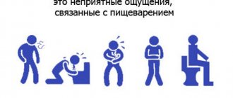
Due to impaired digestion of food, its stagnation and rotting, various unpleasant symptoms are observed:
- lack of appetite;
- heartburn;
- frequent nausea;
- vomiting with impurities of undigested food;
- belching rotten;
- bad breath.
A feeling of heaviness, bloating and stretching in the stomach after a small amount of food and water are also signs of a problem. This condition does not go away during the day. The person notes general weakness, malaise, dizziness, palpitations, rapid pulse, tachycardia, restlessness, anxiety, pale skin and cold sweat.
If this condition continues for a long time, then disruption of the processes of digestion and absorption of nutrients leads to a decrease in body weight and a deficiency of proteins, vitamins and minerals.
Get over it. The most common questions about the functioning of the stomach Read more
Treatment
In case of gastric perforation, the patient is urgently hospitalized in the surgical department, where he is prepared for surgery . Conservative therapy is also used - its purpose is:
- preparing the patient for surgery;
- correction of disorders that have occurred in the body due to peritonitis.
Conservative therapy is based on the following:
- hunger;
- placement of a nasogastric tube to remove stomach contents;
- massive infusion therapy - saline solutions, electrolytes, glucose, protein preparations, blood components (fresh frozen plasma) are injected intravenously;
- antibiotic therapy - to prevent the consequences of infection of the peritoneum, and if it has already developed - to eliminate them;
- hormonal drugs - they are used for hemodynamic disturbances, which can develop as a result of the patient’s state of shock.
The purpose of the operation is to stop the flow of gastric contents into the abdominal cavity. In case of perforation, the following surgical methods are used:
- suturing the resulting gastric opening. It is performed for acute ulcers, when there has been no prolonged protracted course of peptic ulcer disease, which means there are no pronounced changes in the wall of the stomach. Suturing is also performed in elderly patients when a more extensive operation is dangerous (due to surgical and anesthetic risks);
- excision of the edges of the ulcer and suturing the hole;
- if there is a history of ulcers, then, in addition to suturing the perforation or suturing with excision of the edges of the ulcer, the vagus nerve is transected, which will reduce acid formation in the stomach and the risk of further development of a peptic ulcer;
- if perforation is caused by significant changes due to long-term peptic ulcer disease, then partial removal of the stomach is performed;
- If there is a perforation of the ulcer in the final, pyloric part of the stomach, then its plastic surgery is performed.
note
Recently, endoscopic treatment of this pathology has become increasingly popular, when an endoscope is inserted into the abdominal cavity through a small hole in the abdominal wall and tamponade (covering) of the hole formed in the gastric wall is performed.
If gastric perforation is caused by a peptic ulcer, then after surgery the patient should be examined more carefully and conservative treatment of the peptic ulcer should be prescribed. It is based on:
- dietary nutrition;
- antibiotic therapy aimed at eliminating Helicobacter pylori;
- discontinuation of non-steroidal anti-inflammatory drugs that provoke the formation of stomach ulcers;
- enveloping drugs that protect the gastric mucosa from aggressive factors.
Such treatment is necessary to prevent re-perforation of the stomach wall.
Classification
According to the clinical course:
- The typical form is the leakage of contents into the free abdominal cavity.
- Atypical form - the defect is covered by the omentum or adjacent organ.
According to pathological and anatomical characteristics:
- Perforation of acute ulcers. An acute ulcer can occur as a complication of other diseases, for example, myocardial infarction, stroke, with extensive burns, or taking certain medications.
- Perforation of chronic ulcers. Is a complication of peptic ulcer disease
According to the location of the perforated ulcer:
- Gastric ulcer (anterior wall, posterior wall, lesser or greater curvature).
- Pyloroduodenal ulcer
- Combined form (ulcer in both the stomach and duodenum)
- Perforation of peptic ulcers anastomosis
Prevention
The basis for preventing gastric perforation is the so-called secondary prevention - that is, the prevention of all diseases and conditions that may be complicated by perforation of the gastric wall. First of all, you should beware of:
- stomach ulcer;
- alcoholic damage to the gastric wall;
- improper use of certain medications (primarily non-steroidal anti-inflammatory drugs);
- prolonged psycho-emotional stress.
You should also quit smoking, as nicotine robs the stomach tissues - it impairs their blood supply, and therefore their ability to regenerate (recovery).
Patients with gastric ulcers should be registered at a dispensary and periodically examined by a gastroenterologist.
What causes perforation
Typically, acute ulcers are not associated in any way with H. pylori infection. Confirmation of a peptic ulcer in 95% indicates its Helicobacter pylori etiology. An acute ulcer can be considered a stage of gastric ulcer. In addition to H. pylori, gastric perforation can be provoked by:
- Solinger-Elison syndrome;
- medicinal ulcers and other endocrine diseases;
- pancreatogenic, hepatogenic and other types of acute ulcers (for example, Crohn's syndrome).
For the appearance of a hole in the gastric wall, its destruction in all layers by hydrochloric acid and an increase in pressure in the abdominal area are of great importance. Performative chronic ulcer is considered a stage in the progression of gastric ulcer disease. This pathology occurs in the absence of necessary therapy.
Forecast
The prognosis for perforation of the stomach wall is always serious, since changes in the peritoneum due to the action of the acidic contents of the stomach occur quickly, and the deterioration of the general condition of the body is aggravated due to painful shock. Delay in making a correct diagnosis worsens the patient's chances.
At the age of 20-45 years, mortality due to gastric perforation ranges from 1 to 5%. In elderly patients, this figure can increase several times.
Kovtonyuk Oksana Vladimirovna, medical observer, surgeon, consultant doctor
9, total, today
( 181 votes, average: 4.54 out of 5)
Inguinal hernia: symptoms, causes, removal surgery
Wound infection: what is it, types and methods of treatment