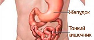
Everything in the human body is interconnected. In order for any of its functions to be realized, the combined work of several organs or even systems is necessary. An example is the digestive tract. Food enters the stomach and is digested in its cavity. While this process is happening, she cannot go any further. Once the stomach has completed its function, food must descend into the cavity of the duodenum. In order for this to happen, a special hole must open to allow the partially digested lump to pass through. All other organs work on this principle, this applies to the urinary, reproductive and other systems. The sphincter plays an important role in this. What is this? Thanks to this organ, the transition of substances (digestive bolus, bile, pancreatic juices, feces, urine, sperm) from one cavity to another is carried out.
Sphincter - what is it?
From Greek, the name of this organ is translated as “I compress.” Already from the name it becomes clear that sphincters are able to change their shape, that is, relax or, conversely, tense. With their participation, many processes occur in our body. To understand what their significance is, it is necessary to answer the question: “Sphincter – what is it?” This organ consists of muscle tissue, and therefore has a pronounced ability to contract. Thanks to sphincters, we can control some of the natural processes of our body, eat, speak. In the cardiovascular system, their role is played by valves that discharge blood, deposit it, etc.
Sphincter inflammation
Sphincteritis - inflammation of the mucous membrane of the sphincter - occurs quite often in patients with chronic infectious invasions of the gastrointestinal tract. In women, rectal sphincteritis is most often diagnosed, since this area is located near the genitals, which increases the risk of ascending tissue infection. Other causes of pathology in women of any age include:
- chronic inflammatory processes of the anus (hemorrhoids, proctitis, paraproctitis);
- imbalance of intestinal microflora;
- anal fissures;
- autoimmune disorders.
Rectal sphincteritis
The risk group for the development of rectal sphincteritis includes elderly patients, women who practice anal sex, or suffer from diseases in which neuromuscular transmission is disrupted. If a woman has previously undergone surgery on the intestines or bile ducts, the risk of inflammation of the anal sphincter is more than 40%.
Manifestations of sphincteritis in women can be different, but most often these are frequent and painful “false” urges to have a bowel movement, a change in the color of the stool (it becomes dark brown), the appearance of foam or a large amount of mucus on the surface of the feces. The urine also changes: it becomes cloudy and darkens. A sediment may form at the bottom of the container in which the urinary fluid is collected.
At the doctor
It is necessary to treat any sphincter diseases, regardless of their location, in a timely manner. If a woman delays seeing a doctor, necrosis (death) of certain areas of the muscle valve may begin. If inflammation spreads to neighboring cells and tissues, acute intoxication and blood poisoning may develop. For confirmed necrotic processes, purulent abscesses, and sepsis, surgical treatment followed by supportive care is indicated.
Classification of sphincters
Each sphincter is a circular muscle, however, they have differences. These organs can be classified according to various criteria. There are anatomical and functional sphincters. The first group represents formed organ structures, the presence of which is obvious. The latter are found in the body in smaller quantities. A striking example is the lower esophageal sphincter, which does not have a clear anatomical structure and, as a result, cannot be visualized. Nevertheless, it performs the same function: if necessary, it narrows or expands the organ cavity. The following classification is based on muscle type. There are striated and smooth muscle sphincters. The first are arbitrary, that is, controlled by the human consciousness. The second are involuntary sphincters, they are represented by muscle sphincters that close and open the lumen of the digestive tract. Depending on belonging to a particular system, another classification is distinguished. It includes digestive, excretory and visual sphincters.
Functions
The sphincter has 3 main functions:
- Regulation of the flow of bile and pancreatic juice into the duodenum. While a person is hungry, the sphincter tone is increased. As soon as food from the stomach begins to enter the duodenum, the sphincter resembles a pump, as it releases bile in a continuous stream, this lasts from a few seconds to 1 minute - depending on the volume and nature of the food.
- Prevents backflow (reflux) of intestinal contents into the ductal system of the glands. During the resting phase, very little bile is secreted into the duodenum, approximately 18 drops per minute. The tone of the sphincter is such that intestinal contents cannot flow back.
- Promotes the accumulation of bile in the bladder. The sphincter constantly maintains a pressure difference between the gallbladder and the duodenum. During periods of rest, the work of the sphincter looks like a slow pump: as soon as a sufficient amount of bile has collected, the sphincter squeezes a few drops out of the bladder, allowing a new portion of bile to enter the bladder. When the gallbladder muscles contract, the sphincter relaxes, and vice versa.
Be sure to read: Stomach and duodenal ulcers: signs and treatment methods
What is a sphincter? Characteristics of individual species
Based on the classification, which is based on belonging to a functional system, it can be understood that each of the groups has several subtypes. A sphincter is a muscle capable of moving a substance not only from the cavity of one organ to another, but also between its parts. Most of all, this fact concerns the digestive system, which is why it includes many similar organs. These include: the upper and lower esophageal, duodenal, small and large intestinal sphincters, as well as the pylorus. A separate group included in the digestive tract are the constrictor muscles of the biliary and pancreatic systems. The genitourinary organs contain the internal and external urethral sphincter. Only one such muscle belongs to the visual system. Thanks to this sphincter, the pupil dilates and contracts.
Human sphincters [edit | edit code ]
In human anatomy, the best known sphincters are the following ( due to the fact that sphincters often separate two organs, in the list below they may be included in both of these organs and thus duplicate
):
Sphincters of the digestive system [edit | edit code ]
There are about 35 different sphincters in the digestive system [2].
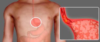
Esophageal sphincters [edit | edit code ]
- Upper esophageal sphincter
- Lower esophageal sphincter ( synonym
Cardiac sphincter)
Sphincters of the stomach [edit | edit code ]
- The pylorus is a muscle in the pylorus that controls the evacuation of gastric contents into the duodenum.
Sphincters of the duodenum [edit | edit code ]
- Pyloric sphincter
- Bulboduodenal sphincter
- Suprapapillary sphincter
- Prepapillary sphincter
- Infrapapillary sphincter
- Ochsner's sphincter
- duodenojejunal sphincter
Sphincters of the biliary and pancreatic systems [edit | edit code ]
- Sphincter of Oddi (lat. sphincter Oddi) is a smooth muscle sphincter located in the large duodenal (Vaterian) papilla, regulating the flow of bile and pancreatic juice into the duodenum, and also protecting the ducts from reflux of intestinal contents. The sphincter of Oddi includes:
- Sphincter of Westphal (sphincter of the major duodenal papilla), ensuring separation of the ducts from the duodenum
- Sphincter of the common bile duct
- Pancreatic duct sphincter
- Sphincter of Mirizzi - located at the confluence of the cystic and common bile ducts
- Lütkens sphincter - located at the junction of the cystic duct into the neck of the gallbladder
- Helly's sphincter is a circular muscle located in the minor duodenal papilla and plays the role of a valve for the accessory (Santorini) pancreatic duct
Sphincters of the colon [edit | edit code ]
- Ileocecal sphincter (lat. sphincter ileocaecalis) - sphincter between the small and large intestines.
- Busi's sphincter (synonyms: colocecal Busi's sphincter, cecum-ascending sphincter) is a sphincter located on the border between the cecum and ascending colon
- Hirsch's sphincter is a thickening of the muscular membrane of the ascending colon at the border of its middle and upper third.
- The Cannon-Boehm sphincter is a sphincter that separates the proximal (initial) third of the transverse colon from the central one;
- Cannon's sphincter is a sphincter that separates the central third of the transverse colon from the distal (terminal) one;
- Sphincter Bally (synonym: distal sphincter of the descending colon) is a sphincter located on the border of the descending colon and sigmoid colon in humans
- The sigmoid rectal sphincter (O'Burn-Pirogov-Moutier sphincter) separates the sigmoid colon from the rectum.
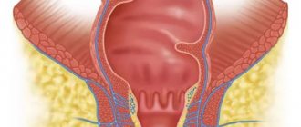
- Sphincters of the anus (lat. sphincter ani):
- External sphincter of the anus (lat. sphincter ani externus) - a sphincter formed by striated muscles, contracted voluntarily (that is, controlled by consciousness);
- The internal sphincter of the anus (lat. sphincter ani internus) is a smooth muscle, involuntarily contracted sphincter.
Sphincters of the excretory system [edit | edit code ]
- Internal urethral sphincter (lat. musculus sphincter urethrae internus).
- External urethral sphincter (lat. musculus sphincter urethrae externus).
Sphincters of the visual system [edit | edit code ]
- Pupil sphincter (lat. musculus sphincter pupillae).
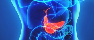
Sphincters of the biliary and pancreatic systems
This group is of great importance in medical practice. Since pancreatitis is a very dangerous disease, it is necessary first to determine the functional activity of these muscles. The largest of them is the sphincter of Oddi. It is located in the papilla of Vater and is necessary for the flow of bile and pancreatic juice into the lumen of the duodenum. With weak functional activity of this sphincter, reverse reflux of these substances may occur. In this case, corrosion and gradual death of the pancreas occurs. The sphincter has 3 components. Each of them represents a small muscle and departs from a specific organ, then they merge in the area of the papilla of Vater. These parts are the sphincters of Westphal (separates the cavity of the duodenum), the common bile and pancreatic ducts.
User's diary Elena Vladimirovna Astafieva
SPHINTERS OF THE DIGESTIVE SYSTEM [gastrointestinal sphincters]
(Greek: σφνγγω - compress, tighten). A sphincter is a structure that controls the flow of a substance (liquid or gas) through a certain area of a hollow organ. Flows controlled by sphincters can be: the flow of a mixture of gases through the respiratory tract, the flow of blood through the blood vessels (bloodstream), the flow of exocrine gland secretions (saliva, pancreatic juice, bile), the flow of nutrients, chyme and feces through the gastrointestinal tract, flow of urine through the urinary tract, etc. The main characteristics of a flow can be flow density (the amount of substance passing per unit time per unit cross section of the flow) and flow speed (the amount of substance passing per unit time). The flow of matter through the sphincters is controlled by certain nerve centers. Based on control signals, sphincters can partially or completely change the characteristics of the flow of substances through the sphincters and, thus, ensure the normal function of the organs of which they are part. The sphincters have more or less the same base and very diverse supporting structures. All sphincters have a muscular basis. It is a local accumulation of muscle fibers that are arranged circularly, spirally and/or longitudinally. These can be smooth muscle fibers as well as striated muscle fibers. Contraction or relaxation of the muscle base partially or completely closes or opens the sphincter and thus ensures a unidirectional flow of substance through the sphincter. In addition, contraction of the sphincter muscle can actively increase/decrease fluence and flow velocity. The flow of matter through the sphincters is controlled by controlling the contraction and relaxation of the muscle base of the sphincters. For this purpose, myogenic, neurogenic and humoral mechanisms are used together or separately. It is obvious that any sphincter contains neuronal structures for the perception of afferent information, its transmission to the regulator, and the transmission of efferent information (control) from the regulators to the actuator. Contraction of the muscle base of different sphincters can be controlled without the participation of consciousness, that is, involuntarily, with the participation of consciousness, that is, voluntarily, as well as in a combined way. Depending on the location of the sphincters, they can be designated as terminal sphincters and intermediate sphincters . The terminal sphincters are located at the entrance and exit of the flow. For example, the final sphincters include the sphincters of the oral cavity - the entrance to the gastrointestinal tract and the sphincters of the rectum with the anus - the exit from the gastrointestinal tract. All other sphincters located throughout the gastrointestinal tract are classified as intermediate sphincters. The muscular base of the sphincter may be part of the wall of a hollow organ. This is the organ's own (internal, intrinsic) sphincter . The muscular basis of the sphincter can be an independent structure that interacts with the organ and is located near the surface of its walls. This is an improper (external, extrinsic) sphincter organ . The combined sphincter has its own muscle base and additional muscles that interact with its own muscle base. In the early days of studying sphincters, it was believed that they contained nothing but a muscle base. Later it was found that sphincters can have a variety of auxiliary structures. In addition to the muscular base, sphincters located in the wall of the organ may, as an auxiliary structure, have (in the submucosa) a local accumulation of blood vessels (arteries and veins). Controlling the degree of filling of these vessels with blood can be direct or indirect by an additional mechanism for controlling the flow of substances through the sphincter. An additional direct physical control mechanism is expressed in the promotion of density of flow blocking by the sphincter. An additional indirect biochemical control mechanism is to provide the sphincter muscle base with nutrients, oxygen and to remove the end products of sphincter muscle metabolism. As an auxiliary structure, sphincters may have folds of mucous membrane, designed for more successful functioning of the sphincters. These folds, to one degree or another, act as valves. They are often called that, although they do not work independently, but in interaction with the sphincter, they are a structure subordinate to the sphincter.
Scheme. Sphincters of the gastrointestinal tract . Modification: 1. NIDDK Image Library. URL: https://www.catalog.niddk.nih.gov/ImageLibrary/, 2. Zaprudnov A.M. Motor-evacuation disorders of the digestive tract and the use of prokinetics in childhood, Pediatrics. Supplement to the journal Consilium Medicum. 2006, 8, 2.
Note. The sphincters, shown with yellow dotted lines, are numbered in red numbers.
See table in a separate window: Sphincters of the gastrointestinal tract. Diagram 1 shows the main sphincters of the gastrointestinal tract : 1 - upper esophageal sphincter, 2 - middle esophageal sphincter, Hacker sphincter 1 , Shatsky ring 2 3 - lower (cardial) sphincter of the esophagus, 4 - valve of the pyloric sphincter of the stomach, 5 - bulboduodenal sphincter , 6 - suprapapillary sphincter, Kapandzhi sphincter 3 , 7 - prepapillary sphincter, Ochsner sphincter 4 , 8 - duodenojejunal fold of Treitz 5 , 9 - ileocecal sphincter of Varolius 6 , ileocecal sphincter, Varoliev sphincter, ileocecal valve, 10 - proxy minor cecal ascending sphincter blind intestines, Busy's sphincter 7 , 11 - sphincter of the base of the celiac process, Gerlach's valve 8 ), 12 - distal cecal-ascending sphincter, Hirsch's sphincter 9 , 13 - right (proximal) sphincter of the transverse colon, Cannon's sphincter 10 -Bam 11 , 14 - middle sphincter of the transverse colon, Horst's sphincter 12 , 15 - left (distal) sphincter of the transverse colon, Cannon's sphincter 10 , 16 - proximal sphincter of the descending colon, Payr's sphincter 13 -Strauss 14 , 17 - distal sphincter of the descending colon, Bally's sphincter 15 , 18 - median sphincter of the sigmoid colon, sphincter of Rossi 16 - Mutier 17 , 19 - sphincter of the transition of the sigmoid colon to the rectum, sigmarectal sphincter, O'Bairn sphincter 18 - Pirogov 19 - Mutier 17 , 20 - proximal (third) sphincter of the rectum , Nelaton's sphincter 20 , 21 - internal involuntary sphincter of the rectum, 22 - external voluntary sphincter of the rectum.
Notes 1 Victor Hacker, von Hacker, Viktor (Victor) Ritter, 1852-1933, Austrian surgeon 2 Richard Schatzki, Richard Schatzki 1901-1992, radiologist, USA. 3 Kapanji, Kapanji, Kapanci, Y., Italian scientist. 4 Ochsner, Ochsner Albert John - surgeon, USA, 1858-1925. Textbook edited by him: Surgical diagnosis and treatment (vv 1-4) - Ochsner, Albert John, URL: https://www.archive.org/details/surgicaldiagnosi01ochsuoft 5 Treitz, Wenzel, pathologist, Bohemia, 1819-1872 . 6 Varolius, Constanzo Varolius (Varolius), Italian anatomist, 1543-1575. 7 Busse, Otto, German pathologist, 1867-1922. 8 Gerlach, Gerlach Joseph von, German anatomist, 1820-1896. 9 Hirsch, JS, German doctor. 10 Cannon Walter B., radiologist, USA, 1871-1945. 11 Böhme Arthur, German physician, 1878-?. 12 Horst, Hörst?, German doctor?. 13 Payr, E. Payr, German surgeon, 1871-1946. 14 Strauss, Strauss N., German physician, 1868-1945. 15 Balli R., Italian radiologist. Balli R.: The sphincters of the colon. Radiology, 1929, 12, 6, 484-495. 16 Rossi, K., Italian radiologist. 17 Moutier, Moutier Francois, French gastroenterologist. 18 O'Beirne J., ?. 19 Pirogov Nikolai Ivanovich, Russian surgeon, naturalist, teacher and public figure, 1810-1881. 20 Nélaton, Nélaton Auguste, French surgeon, 1807-1873. Diagram 2 shows the sphincters of the bile ducts and pancreatic ducts. In more than two-thirds of people, the terminal portions of the common bile duct and the main pancreatic duct merge into a common channel, the pancreaticobiliary duct . It forms an extension called the hepatic-pancreatic ampulla ( 21 tubercles of Vater, Vater's tubercle, ampulla of Vater). In less than one third of people, in addition to the main pancreatic duct, which opens into the cavity of the hepatopancreatic ampulla, there may be an accessory pancreatic duct (accessory duct of Santorini 22 , or 23 ). It is observed as the minor duodenal papilla (papilla or Baldwin's papilla 24 , minor duodenal papilla). The sphincter of the main duodenal papilla is located in the distal part of the papilla . At the exit of the accessory duct, in the lesser duodenal papilla, is the sphincter of the accessory pancreatic duct (Helly sphincter 25 ). The hepatopancreatic ampulla is a complex and variable morphological structure formed by the fusion of the main pancreatic duct and the common bile duct. This is a single channel for the flow of bile and pancreatic juice into the cavity of the duodenum. It is called the pancreaticobiliary duct . The hepatopancreatic ampulla contains three sphincters, which are collectively called the sphincter (complex) of Oddi 26 (although Glisson's sphincter was the first to describe it ) .
Scheme. Hepatic-pancreatic ampulla . Modification: Feldman M., LaRusso N. F., eds. Feldman's GastroAtlas Online. URL: https://www.gastroatlas.com/index.aspx
The terminal sphincter of the common bile duct (Boyden's sphincter 28 , sphincter choledochus of Boyden) is a structure anatomically isolated from the other sphincters of the sphincter of Oddi complex 26 . The sphincter of the common bile duct is divided into two distinct parts. The supraduodenal (pancreatic) part is located before entering the wall of the duodenum. The intramural part is located in the wall of the duodenum. The length of the pancreatic part is ~4 ÷ 7 mm, the length of the intramural part is ~3 ÷ 5 mm. The thickness of these parts of the sphincter is ~144.5 µm. Elements of the sphincter can continue into the base of the 21st papilla of Vater. The length of the continuation section can be ~15 ÷ 19 mm. The sphincter of the common bile duct consists of three layers of smooth muscle fibers. The fibers of the inner layer are arranged circularly, the fibers of the middle and outer layers are arranged longitudinally (in a spiral) relative to the main axis of the duct. Circular muscle fibers begin in the pancreatic part of the duct, then surround its intramural part and pass to the base of the ampulla. The longitudinal (spiral) muscle fibers of the sphincter of the common bile duct have a descending and ascending direction. Descending muscle fibers are independent. They cover the inner layer of circular muscle fibers on all sides and are the middle muscle layer of the sphincter. The descending muscle fibers enter the intramural part of the sphincter of the common bile duct, spirally weave into the sphincter of the hepatopancreatic ampulla and end at the same level with the circular fibers of the sphincter of the hepatopancreatic ampulla at the opening of the papilla. The thickness of the descending longitudinal fibers covering the ampulla of the papilla is ~20.4 µm. The ascending muscle fibers are a continuation of the longitudinal muscle layer of the duodenum. They cover the middle layer of descending intrinsic longitudinal muscle fibers. Thus, the ascending muscle fibers form the outer, third muscle layer of the sphincter. The outer layer of ascending muscle fibers is an auxiliary structure of the sphincter. These fibers end at a distance of ~4 ÷ 6 mm proximal to the beginning of the circular fibers of the supraduodenal part of the sphincter of the common bile duct. The sphincter of the main pancreatic duct , or Westphalian sphincter 29 , is a structure anatomically isolated from the other sphincters of the sphincter of Oddi complex 26 . The distal part of the main pancreatic duct, located in the hepatopancreatic ampulla, is called the main pancreatic duct (duct of Wirsung 30 ). At the junction of the pancreatic duct with the common bile duct in the area of the ampulla of Vater, the pancreatic duct has an independent sphincter - the sphincter of the main pancreatic duct. It contains circular, semicircular and longitudinal smooth muscle fibers. Independent circular and semicircular fibers make up the inner layer of the sphincter. The outer longitudinal layer is formed by muscle fibers extending from the muscular layer of the duodenum. The length of the sphincter of the main pancreatic duct is £5 mm, thickness ~34.4 µm. All three sphincters located in the hepatopancreatic ampulla: the common sphincter of the hepatopancreatic ampulla, the terminal sphincter of the common bile duct and the sphincter of the pancreatic duct are collectively called the sphincter (complex) of Oddi 26 . In addition to the terminal sphincter located in the hepatopancreatic ampulla, the bile ducts also have sphincters. The sphincter of the cystic bile duct (Lutkens sphincter 31 , Lutkens' sphincter) is a circular bundle of smooth muscle fibers in the neck of the gallbladder at the junction of the neck with the cystic bile duct. Sphincter of the common bile duct (Mirizzi's sphincter 32 , Mirizzi's sphincter in common hepatic duct) is located in the area of the common bile duct, located immediately after the confluence of the hepatic bile duct and the cystic bile duct. The sphincter of the hepatopancreatic ampulla (common sphincter of the papillary ampulla, Westphalian pylorus 29 ) is a structure anatomically isolated from the other sphincters of the sphincter of Oddi complex 26 . The sphincter of the hepatopancreatic ampulla consists of circular and spiral muscle fibers that surround both the common bile duct and the pancreatic duct. The distribution of muscle fibers along the ampulla of the papilla is uneven. Muscle fibers are concentrated in two places: at the base of the 21st of Vater and at the mouth of the papilla. Therefore, the sphincter of the hepatopancreatic ampulla is divided into two sphincters: the sphincter of the base of the papilla of Vater and the sphincter of the orifice of the papilla . The sphincter of the base of the papilla is ~73.4 µm thick, the sphincter of the orifice of the papilla is ~55 µm thick, and the thickness of the layer of circular fibers between them ( papillary erector , accessory structure) is ~39 µm.
- 2572
Sphincters of the colon
As you know, the digestive tract consists of several sections, in each of which a certain process of food processing occurs. In the cavity of the small intestine, the nutrients needed by the body are absorbed. In the future, the food is considered to be completely digested. Therefore, in the large intestine, only water is absorbed, as well as the formation and movement of feces. In each of the departments there are sphincters that retain (and subsequently let through) processed products of the body. The anal orbicularis muscles are of greatest importance. There are 2 types of them: involuntary and voluntary and sphincter. The first consists of smooth muscle tissue and passes feces into the ampulla of the rectum. The external anal sphincter goes directly into the environment and is controlled by the human consciousness.
Disruption of the digestive sphincters
If the circular muscles work poorly, problems arise throughout the body. With insufficient activity or complete atrophy of any sphincter, the entire system to which it belongs suffers. If this concerns the upper parts of the digestive tube, then reverse reflux of the contents is observed. This is manifested by the appearance of frequent vomiting of eaten food, bile, and acidic substances. Its character depends on the section in which the reflux occurred (esophagus, stomach, duodenum). Disruption of the sphincters of the lower part of the digestive tract is manifested by prolonged stagnation of feces and abdominal pain. If this condition is not diagnosed in time, intestinal obstruction may develop.
Diseases of the sphincter of Oddi
Dysfunction
Main article: Sphincter of Oddi dysfunction
The most common disorder occurs in 2 forms:
- stenosis (anatomical narrowing) of the sphincter - inflammation, fibrosis, proliferation of the mucous membrane, injury from small gallstones, pancreatitis;
- primary functional dyskinesia associated with impaired general motor function of the intestines.
It is impossible to distinguish these forms clinically; sometimes this is revealed only at section (autopsy). The international criteria for sphincter of Oddi dysfunction (SOD) are:
- painful episodes last 30 minutes or more against the background of normal health;
- there must be at least one painful attack during the year;
- the pain is persistent, the intensity forces you to consult a doctor, your ability to work is impaired;
- There are no other diseases of the digestive system that could cause such pain.
The international classifier identifies 3 types of biliary (bile) dysfunction and one type of pancreatic dysfunction. These types of dysfunctions are delimited based on the results of instrumental and laboratory examinations.
Spasm
This is a variant of dysfunction that occurs according to the hyperkinetic type.
It manifests itself as recurring pain in the abdomen (abdominal), which is accompanied by dyspeptic disorders - nausea, vomiting, lack of appetite, alternating constipation and diarrhea. The pain radiates to the back or shoulder blades and does not go away with changing body position and taking antacids. The pain occurs several times a week, at night, is provoked by fatty foods, 2-3 hours after eating, body temperature does not rise. The patient usually knows which food triggers the attack.
Spasm of the sphincter of Oddi is more common in women, the basis is increased tone of longitudinal and transverse muscle fibers. Due to the spasm, the pressure in the gland ducts increases, which causes clinical symptoms.
Doctors noticed that a typical patient is a woman from 30 to 50 years old, of asthenic build, underweight, and highly emotional.
Postcholecystectomy syndrome
The most common type of DSO, found in 45% of all patients who have had their gallbladder removed.
The symptoms are almost the same as for biliary colic: severe pain in the right hypochondrium, which can radiate to the lower back, right shoulder blade or forearm, nausea, repeated vomiting mixed with bile. Compared to colic, the intensity of pain and dyspeptic symptoms is somewhat less. The reason is a narrowing of the bile ducts, small stones of the common bile duct, accompanying inflammation of the gastrointestinal tract. Be sure to read:
Duodenal ulcer: how is the pathology manifested and treated?
After removal of the gallbladder, sphincter insufficiency occurs when bile continuously flows into the lumen of the duodenum. If the sphincter tone increases even slightly, the pressure in the entire bile tract also increases. This leads to the development of pain syndrome. Typically, painful attacks recur (due to poor diet and other harmful conditions) 3 or 5 years after cholecystectomy.
Fecal and urinary incontinence
If the patient complains of uncontrolled defecation or urination, then a malfunction of the sphincters should be suspected. Involuntary release of fluid can occur with poor contractility of the orbicularis muscle. If the bladder is not able to retain urine in its cavity, then they speak of sphincter insufficiency, which can be true or false. The same applies to the act of defecation. If the external anal sphincter malfunctions, a person cannot control the process, and fecal incontinence occurs. Each of these pathologies causes not only physical, but also moral harm, so it is necessary to seek surgical help in time.
Artificial sphincters
In some situations, it is impossible to bring the orbicularis muscle into action. Most often this occurs with advanced diseases or with congenital absence or removal of this organ. In such cases, an artificial sphincter is used. In most cases, such operations end favorably and make life much easier for patients.
So, sphincter - what is it? This muscle is of great importance for the body, contributes to its functions and prevents many pathological conditions. Now you know what a sphincter is. Photos of this organ are presented in the article.