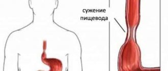
Esophageal obstruction is a pathological condition in which the movement of food through the esophagus is impaired due to its narrowing. The narrowing can be caused by scar changes (stenosis), external pressure due to pathological changes in neighboring organs (external compression) or blockage of the organ lumen by a tumor or foreign body (obturation).
There are many reasons for this pathology. It is known that esophageal cancer, which sooner or later leads to obstruction of the organ, is in third place among all malignant neoplasms of the gastrointestinal tract and in sixth place among all malignant tumors. Regarding benign causes of esophageal obstruction, cicatricial narrowing caused by burns accounts for approximately 69%, while cicatricial changes due to chronic esophagitis account for approximately 22%.
Classification of esophageal obstruction
By origin, obstruction can be congenital or acquired. Congenital obstruction (esophageal atresia) can be either a consequence of genetic pathology or the result of a violation of intrauterine development. Acquired develops as a consequence of various pathological processes.
Depending on the pathological process that caused the obstruction, it can be benign or malignant. Malignant is an obstruction caused by cancer of the esophagus or mediastinal organs; benign - arising for reasons not related to oncological pathology.
In addition, obstruction of the esophagus is distinguished depending on the degree of narrowing of the lumen of the organ:
- the diameter at the narrowest point is 9–11 mm, a medium-caliber endoscope fits;
- diameter 6–8 mm, fiberoptic bronchoscope passes through;
- diameter 3–5 mm, passes through an ultra-thin fiberscope;
- diameter <3 mm, impassable for endoscopic equipment.
Classification of the disease
Dyskinesia is caused by impaired contraction of the thoracic region or improper functioning of the esophageal sphincter. With the disease, contractions of the esophageal muscles increase or decrease.
Hypermotor disorders are classified as follows:
- When individual parts of the esophagus are involved, segmental esophagospasm occurs.
- If there are problems with all parts of the esophagus, diffuse esophagospasm develops.
- Motor disorders of a nonspecific nature.
If the lower esophageal sphincter is not functioning properly, the doctor may diagnose gastroesophageal reflux disease, cardiospasm, or segmental esophageal spasm. It is important to start treating all types of diseases on time.
Causes of esophageal obstruction
All possible causes of obstruction of the esophagus can be reduced to several main groups.
Malignant neoplasms of the esophagus itself or mediastinal organs (trachea, large bronchi, lymph nodes). As the tumor grows, the lumen of the organ is blocked, disrupting the movement of the food bolus.
Post-burn stenoses . These are scar narrowings of the esophagus left after chemical burns. They occur after accidental or intentional ingestion of concentrated acids or alkalis. Alkalies are especially dangerous in this regard: unlike acids, they do not form a scab that prevents the substance from penetrating deep into the wall of the organ, but, on the contrary, loosen it. Because of this, burns are deeper and have more serious consequences.
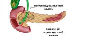
Recurrent esophagitis . These are chronic inflammations of the esophagus caused by one reason or another. Constant inflammation sooner or later leads to scar changes, which, in turn, lead to a narrowing of the lumen of the organ.
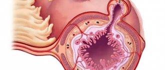
Esophageal rings . These are thin concentric protrusions into the lumen of the esophagus, which include all three layers of the wall: mucous, submucosal and muscular. The reasons for their appearance are not fully known. Some authors suggest that these are inflammatory changes, others believe that esophageal rings are associated with impaired motility of the organ. Most often they are asymptomatic, but if they form in large quantities, they can disrupt the patency of the esophagus.
Congenital pathologies . The so-called esophageal atresia is a developmental defect that is detected in the coming hours after the birth of the child. An operation is required to re-form the esophagus.
Others _ This group includes such relatively rare causes of esophageal obstruction as tuberculosis, scleroderma, tertiary syphilis, trauma and foreign bodies, esophageal diverticula and other pathologies.
Causes of the disease
According to experts, it is impossible to say with certainty the cause of dyskinesia. Determining the factors contributing to the onset of the disease is very difficult, but very important. Without eliminating the cause, all other symptomatic treatment will be useless, except for maintaining the patient’s relatively normal condition. Known causes are divided into primary and secondary.
Primary causes of dyskinesia
Primary dyskinesia occurs independently, without additional influence from other diseases and does not depend on the specific structure and functioning of the organ, except for congenital anomalies in its structure. Provoked by disorders of the nervous system. The main primary causes are:
- stress;
- psychoemotional disorders;
- neuroses;
- nervous breakdowns;
- alcohol abuse;
- heredity.
Secondary causes of dyskinesia
Esophageal dyskinesia can develop against the background of other gastrointestinal diseases.
Secondary dyskinesia occurs as a consequence of another disease, and not independently. Secondary reasons are considered:
- Diseases of the esophagus:
- inflammation of the mucous membrane (esophagitis);
- the presence of a diverticulum;
- hernia;
- the occurrence of neoplasms.
- Diseases of other organs or entire systems:
- scleroderma - damage to connective tissue in the body;
- muscle tissue dystrophy;
- peptic ulcer;
- chronic inflammation of the gallbladder;
- diabetes;
- serious disorders of the nervous system.
- Taking hormonal drugs such as Thyroliberin, Glucagon, Somatostatin, etc.
Symptoms of esophageal obstruction
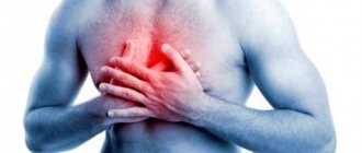
The main symptom of esophageal obstruction is dysphagia, or difficulty swallowing. Depending on the degree of narrowing of the organ lumen and the nature of the food, the severity of this symptom may vary. Thus, liquid or semi-liquid food can freely pass into the stomach, while solid food immediately causes chest pain, belching, and vomiting. As the lumen narrows, the symptoms increase. Since the patient cannot eat properly, he loses weight, and signs of nutritional deficiency appear (anemia, edema, osteoporosis, etc.).
Separately, it should be noted the symptoms of obstruction caused by esophageal rings. This pathology is not characterized by long-term persistent swallowing disorders. Usually the problem occurs suddenly, during a hurried or dry meal. The attack is relatively short-lived and resolves after drinking fluids or vomiting. It often occurs when eating fried meat; in foreign literature it is sometimes called “steak syndrome.” Months or even years may pass between episodes of swallowing problems caused by esophageal rings.
Treatment
At the initial stage, it is necessary to eliminate the factors that provoke the development of the disease. These include stress, physical activity, alcohol, smoking, eating cold, hot and spicy foods (spices, hot sauces, carbonated drinks). The patient is recommended to streamline his daily routine: avoid overwork, eat on time in a calm environment, get good sleep on a high pillow with his head elevated.
Diet
The most common treatment method is a special diet. It involves eating small portions (no more than three hundred grams) 5-7 times a day. The prepared food in this case should avoid the undesirable properties described above. Dishes are suitable liquid or crushed (mashed potatoes, soup), warm, not spicy. Products enriched with vegetable coarse fiber are not recommended in the patient's diet. For bottle-fed infants, antireflux mixtures help eliminate symptoms expressed by vomiting and regurgitation.
Non-drug treatment
Therapy without the use of medications is to prevent surges in intra-abdominal pressure. For this purpose, it is necessary to reduce emotional and physical stress. The patient is advised to avoid overeating, lifting and carrying heavy objects, and physical exercises involving bending the body are strictly prohibited. It is unacceptable to wear clothes that tightly tighten the chest and throat.
Drug treatment
The use of medications is aimed at restoring the motor function of the esophageal tube. There is also a fight against the spastic state of smooth muscles, which often results in paralysis of the esophagus. Metoclopramide and Betanectol increase muscle tone, and to eliminate spasms, doctors recommend a number of drugs, the most popular of which are Papaverine and Drotaverine. In the primary form of the disease, which occurs against the background of nervous breakdowns and stressful conditions, it is recommended to take sedatives.
To increase the stimulation of gastric peristalsis, aceclidine, carbacholine, strychnine and other drugs are used. During this treatment, food and oral medications are prohibited.
A special place is occupied by various medicinal herbs (more about traditional treatment methods in the following paragraphs). Ginseng root and Eleutherococcus extract are excellent for treating atony and increasing the tone of the esophageal tube. Regular chamomile, mint, and popular valerian root are very valuable for easing spasms.
Spa treatment is of great benefit. Visiting resorts that offer guests a range of services, including physiotherapy and procedures involving the use of mineral waters, will have a beneficial effect on the condition of the gastrointestinal tract.
Surgical intervention
This treatment method is rarely used. In cases where diet and medications are powerless to eliminate the disorder.
Traditional methods
Traditional methods are very widespread. Several of the most popular recipes are highlighted.
- Take in equal quantities: anise, celandine, St. John's wort, corn silk, knotweed herb, coriander. The plants are mixed, 600 ml of boiling water is poured. After half an hour of infusion, the mixture is ready for use. Drink three times a day, regardless of the time of meal.
- You need corn silk, bear's ears, smooth herb, beans and knotweed grass - equal parts of everything. Mix and pour one glass of hot water. Use the strained mixture throughout the day. Don't forget to let it sit for one hour first.
- Thoroughly grind and mix: three-leaf cotton wool leaf, celandine, chamomile. Pour a tablespoon of the resulting mixture into a glass of boiled water. The mixture is infused for about an hour. Drink 100 ml morning and evening.
- Necessary ingredients: a couple of parts of buckthorn, three knotweed, chamomile extract, St. John's wort, sandy immortelle leaf. The plants are mixed and filled with one liter of cold water. The mixture is infused for about twelve hours. After boiling for five minutes, strain thoroughly. We drink one glass before breakfast, and drink the rest in small portions after each subsequent meal.
Diagnosis of esophageal obstruction
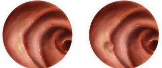
In modern conditions, the main method for diagnosing esophageal obstruction is esophagoscopy. This is an endoscopic examination, during which the doctor can assess the degree of narrowing of the esophagus, its patency, and the condition of the mucous membrane. If necessary, during esophagoscopy you can take biopsy material - tissue for further examination under a microscope.
If endoscopy is not possible due to lack of equipment or extreme narrowing of the esophagus, radiography is used to diagnose obstruction. The patient is given a mixture to drink containing barium salts, which are impenetrable to x-rays. After this, a series of photographs are taken to assess the level and extent of narrowing of the esophagus, as well as the motility of its walls.
If technically possible, CT and MRI may be recommended, which can more accurately determine the duration and degree of narrowing of the esophagus, but due to their high cost they are used relatively rarely.
If the patient has signs of malnutrition, clinical and biochemical blood tests are recommended to assess the degree of trophic disorders in the body. They allow you to identify possible deficiencies of iron, protein, and other micro- and macroelements.
Diagnostics
To make a correct diagnosis and start treatment, contact a gastroenterologist and endoscopist. Specialists conduct the necessary examination:
- fluoroscopy of the esophagus;
- esophageal manometry;
- esophagoscopy;
- examine feces and urine;
- endoscopic examination.
An endoscope is used to examine the inaccuracies of the internal organ. To check the motor function of the esophagus, the Carvalho test is performed.
After the examination results are obtained, the doctor determines the correct diagnosis and prescribes treatment.
Treatment of esophageal obstruction
Treatment of esophageal obstruction is exclusively surgical. The traditional method is bougienage. Special dilators - bougies - are inserted into the lumen of the esophagus, changing their diameter from smaller to larger. Balloon dilatation is also used - a balloon is inserted into the lumen of the esophagus, which, expanding, stretches the walls of the esophagus. It is also possible to dissect the scar during endoscopic surgery. In case of tumor lesions of the esophagus and the impossibility of radical surgery, the part of the tumor that blocks the lumen of the organ is excised, and a stent is installed in this area, which prevents re-narrowing of the esophagus.
As part of complex therapy, drugs for parenteral nutrition (intravenous administration of nutrients), vitamin and mineral complexes, and gastroprotectors are prescribed to help restore the mucous membrane after surgery (Rebagit).
Prognosis and prevention of esophageal obstruction
The prognosis largely depends on the nature of the narrowing of the esophagus and the possibility of parenteral nutrition. Compared with stenoses caused by a malignant tumor, benign ones usually have a better prognosis. There is no specific prevention of pathology.
[1] A.V. Yankin. Esophageal cancer: from statistics to diagnosis. Practical Oncology, 2003.
[2] V.L. Belevich, D.V. Ovchinnikov. Benign stenosis of the esophagus and its anastomoses: state of the problem. Bulletin of the Russian Military Medical Academy, 2012.
Prevention
Preventive measures against such a disease consist of following several simple rules:
- lead a healthy lifestyle;
- follow your doctor's recommendations regarding nutrition;
- exclude overeating and avoid performing heavy physical activity immediately after consuming food;
- limit yourself from stressful situations;
- sleep so that your head and forearms are several degrees higher than the lower half of your body;
- promptly treat those diseases that can cause esophageal dyskinesia. To prevent exacerbations, it is best to undergo therapy in a sanatorium-resort setting;
- undergo preventive examinations with a gastroenterologist several times a year.
The prognosis of such a disorder is individual for each patient and depends on several factors, in particular the type of disease, its duration and the nature of its course. In any case, such a pathology significantly worsens a person’s quality of life.