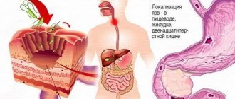
In adolescents and adults, foreign bodies in the esophagus are most often lumps of food and bones. Pathology is almost always associated with compaction of food. The risk is increased in edentulous adults, severely ill patients, and people with predisposing diseases of the esophagus.
Typically, a foreign body of the esophagus gets stuck in one of three places of physiological narrowing:
- in the area of the cricopharyngeal muscle (the place where the pharynx enters the esophagus);
- in the projection of the aortic arch;
- at the junction of the esophagus and the stomach.
Less commonly, foreign bodies are localized in the area of the bifurcation of the trachea and the upper opening of the chest. 90% of foreign bodies pass further into the stomach on their own.
Signs and symptoms of an esophageal foreign body
In case of acute entry of a foreign body into the esophagus (up to 1 day), the following symptoms occur:
- dysphagia – a condition when the process of swallowing becomes difficult or impossible;
- pain when swallowing and drooling;
- suffocation;
- retching and vomiting;
- saliva mixed with blood.
If a foreign body remains in the esophagus for a long time:
- Respiratory symptoms predominate (as a result of swelling of the tissue adjacent to the esophagus, blocking the flow of air through the trachea): cough, stridor and hoarseness.
- Common symptoms: restlessness and irritability;
- weight loss up to anorexia;
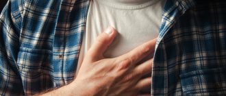
- chest pain and fever;
- the sensation of a foreign body usually corresponds to the level at which the body is lodged in the esophagus.
In case of complications (perforation of the esophagus - loss of integrity), redness, swelling and crepitus appear in the neck area (a characteristic cracking or crunching sensation is felt upon palpation).
In 20% of cases it does not manifest itself at all.
Symptoms of esophageal injury
The varied symptoms of esophageal damage are usually divided into local and general. Local manifestations include pain along the esophagus, aggravated by the act of swallowing, dysphagia, hoarseness, subcutaneous emphysema, infiltration and hyperemia of the soft tissues of the neck, pain on palpation, pneumothorax, muscle tension in the anterior abdominal wall. Common manifestations include pallor and cyanosis of the skin, shortness of breath, cold sweat, tachycardia, chills, and hyperthermia.
Each type of damage corresponds to a certain set of clinical signs, but most of them are observed at a late stage, already with the development of purulent complications.
Damage to the esophagus by foreign bodies in most cases does not have clear clinical symptoms. The esophagus has the ability to pass rather large and sharp objects without much damage. Sharp objects (fish bones) often get stuck when they pierce the wall of the esophagus with one sharp end. There is pain when swallowing, solid food cannot pass through the esophagus (dysphagia). Symptoms are more pronounced when the cervical esophagus is damaged. The most common symptom is pain along the esophagus with irradiation to the back of the head (with perforation of the cervical spine), interscapular (with perforation of the upper and middle thoracic sections) or epigastric region (with perforation of the lower thoracic and abdominal sections of the esophagus).
As a rule, the pain sharply increases with swallowing and palpation of the soft tissues of the neck. However, a similar pain syndrome is characteristic of any foreign body in the lumen of the esophagus (without damage or with non-penetrating damage).
Patients often experience chest pain, dysphagia, “unease” in the esophagus, a scratching sensation when swallowing food, and hoarseness. With massive foreign bodies, clinical signs of airway compression may be observed. In case of perforation of the esophageal wall, the clinical picture of mediastinitis develops. Similar symptoms are observed with damage to the esophagus caused by esophageal cancer, congenital cysts and fistulas of the neck, thyrotoxicosis (Graves' disease).
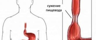
The clinical picture of chemical and thermal damage to the esophagus is always clear. Typically, burns to the esophagus occur as a result of accidental or intentional (for the purpose of suicide) ingestion of alkali, acid and other caustic liquids. Acid and alkali, acting on the mucous membrane of the esophagus, cause its severe destruction. An acid burn causes more superficial damage to the wall of the esophagus. The acid can pass into the stomach, where it also causes burns to the mucous membrane. Alkali causes the deepest damage to the wall of the esophagus. After healing, scar tissue forms in the area of chemical damage, which leads to obstruction of the esophagus or stomach.
In the clinical picture, dysphagia comes first. There are burn marks in the corners of the mouth and on the tongue. It is necessary to ask the patient or people around him about what he drank.
Causes
Foreign objects get stuck due to a discrepancy between the size of the foreign body and the diameter of the esophagus or in response to damage to the wall, the presence of structural disorders of the organ (stenosis, stricture).
Food getting stuck in the esophagus is usually caused by 3 reasons:
- pathology of the muscular apparatus of the esophagus;
- violation of neuromuscular regulation;
- changes in the lumen of the organ.
With eosinophilic esophagitis, jamming occurs periodically.
Foreign body of the esophagus in a child
A foreign body of the esophagus in children in 80% of cases occurs at a young age, especially up to two years. Coins account for 80% of all foreign bodies ingested.
Symptoms in children:
- refusal to eat;
- stridor (wheezing, noisy breathing) and cough;
- inflammation of the upper respiratory tract;
- pain in the neck, throat.
A predisposing factor is narrowing of the esophagus. Rarely in children, the pathology is complicated by esophageal-aortic and esophageal-tracheal fistulas.
Diagnostics
How is esophageal foreign body diagnosed?
If there are no breathing difficulties and the patient’s condition is satisfactory, the doctor prescribes diagnostics in the form of X-ray examination and esophagoscopy. The research results make it possible to assess the integrity of the esophagus, the degree of its damage, and select treatment tactics.
Diagnosis of foreign bodies of the esophagus
Diagnosis of the presence of foreign objects is based on data about the swallowed object. The physical examination should focus on the extent of the lesion:
- Complications to look for include: esophageal obstruction, perforation, and bleeding;
- a thorough examination of the oropharynx - looking for erythema, inflammation of the upper respiratory tract, throat or abrasions on the palate;
- direct and indirect laryngoscopy;
- Auscultation of the lungs will reveal the presence of stridor and wheezing;
- the presence of peritonitis or intestinal obstruction during examination of the abdomen (palpation, clinical picture).
Laboratory tests are not required.
Treatment
There are three main principles for managing patients with foreign bodies stuck and/or causing damage to the esophagus.
1. If there is no spontaneous advancement, the foreign body must be removed. If there is no spontaneous removal, the foreign object can in most cases be removed using a laryngoscope and Magill forceps or a flexible pediatric endoscope and special instruments. Sometimes the object is advanced into the stomach for safer manipulation when removing it (for example, to close a pin). The meat can often be removed using the proteolytic enzyme papain. The need for surgical removal of a foreign body occurs in less than 5% of cases.
In children, it is usually recommended to pass the Fogarty catheter below the foreign body, inflate the balloon, and then remove the catheter while pushing the foreign body with the balloon. The danger of this method is the possibility of an object slipping into the larynx or trachea, which will cause airway obstruction.
2. The esophagus should be carefully examined, either directly (using an endoscope) during removal of the object, or after its removal, if rupture is not suspected; if perforation is expected, then the study is carried out using water-soluble contrast.
3. In all age groups of patients, with the exception of children, the need to remove foreign objects should be carefully considered.
R. E. Barney, J. R. Mackenzie
Published
Special research methods
- X-ray of the chest and neck is necessary for all patients with suspected foreign object intrusion (to localize it, regardless of symptoms). Foreign bodies in the esophagus are located in the frontal plane, and in the trachea - in the sagittal plane.
With perforation of the esophagus, air is found in the retropharyngeal space, in the soft tissues of the neck or in the mediastinum.
- Examination of the esophagus with contrast materials to identify materials transparent to x-rays, which allows:
- detect changes in the contour of the barium column;
- assess the passage of the contrast solution into the stomach, which makes it possible to identify partial or complete obstruction.
Important! Oral contrast agents with significant esophageal obstruction may increase the risk of aspiration. Barium can also coat the mucosal layer and interfere with subsequent endoscopy.
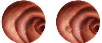
- Endoscopy is the best method for identifying the location and removal of foreign bodies from the esophagus and stomach. In addition, it is possible to check the surrounding mucous membrane of the esophagus for the presence of pathology.
- Computed tomography detects foreign bodies that are not detected by other methods.
- A metal detector may be used to detect swallowed metal objects, especially coins.
Treatment and diagnosis
Diagnosis involves promptly undergoing a series of research procedures that will reveal the location of the object, its shape and the degree of damage to the mucous membrane. The most commonly performed are x-rays and endoscopy. However, most often a basic examination and history is sufficient.
Additionally, the following can be used for examination:
- thorough and specialized examination of the pharynx, larynx, esophageal tube;
- esophagoscopy;
- fistulography;
- CT scan;
- ultrasonography;
- tests to identify inflammatory processes;
- mirror laryngoscopy;
- pharyngoscopy;
- rigid endoscopy;
- fibroendoscopy.
Based on the data obtained, decisions are made regarding the removal of the foreign object.
Treatment directly depends on the degree of damage that was caused by swallowing a foreign object. So, it may include:
- removal by surgery;
- removal of an object during endoscopy;
- use of anesthetics;
- use of antibiotics;
- washing with solutions that promote rapid healing and relieve inflammation;
- sulfa drugs.
The main danger is considered to be the moment when an object digs into the walls of the esophagus, thus provoking heavy bleeding. In such cases, serious surgical intervention is required. During the operation, it is necessary not only to remove the foreign body, but also to return the esophagus to its previous appearance and functionality. In some cases, it is possible to receive disability.
Removal of foreign bodies from the esophagus
- If foreign bodies are found in the upper and middle parts of the esophagus, extraction is required.
Asymptomatic patients with coins or smooth objects in the distal esophagus require observation for 24 hours to ensure that the foreign bodies have passed into the stomach.
- A dense piece of food that completely blocks the esophagus requires emergency removal. Attempting digestion with proteolytic enzymes is not recommended due to the serious risk of injury involved, including esophageal perforation and aspiration.
- Sharp or pointed objects should be removed regardless of their location.
- Button batteries require emergency removal regardless of their position in the esophagus. Potassium hydroxide and mercury often leak from them and can cause burns to the mucous membrane within 4-6 hours.
Prognosis and prevention
The prognosis for a foreign body in the stomach is usually favorable - in most cases it is not life-threatening. Prevention of the appearance of foreign bodies in children consists of closer monitoring by relatives. In adults, prevention of foreign bodies includes careful monitoring of food quality, calm eating without distractions (reading, Internet, TV shows) and timely identification and treatment of mental pathologies.
I. Belov, G. Adzhigitov. Foreign bodies of the gastrointestinal tract. Medical newspaper, 2004.
A. G. Beburishvili, V. V. Mandrikov, A. N. Akinchits. Foreign bodies of the gastrointestinal tract. Educational and methodological manual. Volgograd, 2007.
Methods for removing a foreign body of the esophagus
- Extraction using a Foley catheter under fluoroscopic control:
- an effective and safe method in experienced hands;
- a Foley catheter is inserted through the nose into the esophagus, placing the end of the catheter and the balloon under the foreign body under fluoroscopic control;
- the balloon is filled with barium contrast agent and the catheter is slowly removed;
- The method is contraindicated in case of chronic swallowing of foreign bodies, in non-contact patients, in case of sharp or pointed objects.
- A distal esophageal foreign body (small without sharp edges) should be pushed into the stomach using a Foley catheter.
- Endoscopic removal is a favorite method for removing various foreign bodies:
- if the foreign body remains for more than 2-4 days, use is difficult due to concomitant irritation and swelling of the esophagus;
- in difficult cases (children, psychiatric patients, foreign body difficult to remove), general endotracheal anesthesia is required.
- Administration of intravenous glucagon:
- reduces the tone of the sphincter of the lower third of the esophagus without affecting the contraction of the esophagus;
- often helps food bolus located in the distal part of the esophagus pass into the stomach;
- recommended for cases of esophageal obstruction lasting less than 24 hours.
- Use of gas-forming substances:
- recommended for cases of esophageal blockage lasting less than a day.
- can be combined with the administration of glucagon, after which gas-forming substances are given orally.
- Surgical intervention:
- used in cases where a foreign body cannot be removed by other means;
- approximately 2% of all patients require surgical treatment;
- The most common causes of surgery are toothpicks and bones.
Conservative treatment of pathology
After removing the foreign body of the esophagus (ICD-10 code was indicated earlier), detoxification and anti-inflammatory therapy is prescribed. For this purpose, solutions and tablet medications are used. If the patient's condition is severe, electrolytes and colloidal drugs should be administered. Experts also prescribe antacids and antibacterial medications to reduce the impact of negative factors on the damaged esophagus.
Therapy may include the use of medications necessary for the patient’s recovery.
- “De-Nol” promotes the regeneration of damaged cells, prevents acid production, and normalizes blood circulation.
- "Iberogast", which allows you to normalize the process of food passage, eliminate dysphagia, reduce acidity, and restore muscle tone.
- “Omeprazole”, “Almagel”, which reduce the process of inflammation and suppress the production of acid that impedes healing.
For detoxification the following are used: “Gluconeogenesis”, “Reomacrodex”, “Reopoliglyukin”. Drug therapy should also be supplemented with vitamin complexes. During the recovery period, the patient is advised to use immunostimulants - “Prodigiozan”, “Immunal”, “Tactivin”, “Thymogen”.
Drug treatment of esophageal foreign body
- EZ Gas: 30 ml solution (NaHCO3, citric acid and simethicone) orally. The resulting gas can push food through.
- Glucagon: 1-2 mg intravenous bolus after a small test dose to determine sensitivity. Will allow you to relax the tone of the muscles of the esophagus and lower sphincter.
To reduce the pressure of the lower esophageal sphincter, the following drugs can be used: Verapamil, Nitrates, Buscopan.
If the wall of the esophagus is damaged, antibiotic therapy is carried out with broad-spectrum drugs against flora.
Urgent Care
When a foreign object gets stuck in the esophagus, the airway can become blocked. In this case, the victim requires immediate assistance. Experts recommend using the “5 + 5” technique. What does he mean?
- It is necessary to apply 5 blows to the victim in the area of the shoulder blades on the back.
- Perform the Heimlich maneuver - make 5 pushes to the diaphragm.
- These steps should be repeated until the victim’s breathing is completely restored.
The Heimlich maneuver must be performed in compliance with certain rules.
- It is necessary to take a position behind the victim, clasp his stomach with your hands, and lean forward slightly.
- Form one hand into a fist and place it in the area above the navel.
- With your other hand, grab your fist and press hard.
- It is necessary to repeat the pushes until the foreign body is displaced and breathing is restored.