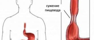
The esophagus is a hollow tube that serves to connect the pharyngeal opening and the stomach. Despite its simple structure, the role of the organism in the physiology of digestion is great. The anatomical structure of the esophagus is such that the diameter of its lumen is not the same everywhere. Natural narrowings do not interfere with patency.
The reasons that cause pathological narrowing of the esophagus can be very diverse, from neurotic spasm during a stressful situation to strictures due to esophageal cancer.
Symptoms of narrowing of the human esophagus are associated with impaired movement of food through the esophagus: dysphagia, belching, pain, esophageal vomiting. Narrowing can occur both with diseases of the organ itself and with pathology of organs located nearby. To diagnose narrowing of the esophageal opening, X-ray examination with contrast and endoscopic methods are used. Depending on the etiology, treatment can be conservative (diet therapy, drug treatment, physiotherapy, treatment with traditional methods) and surgical (bougienage, endoscopic surgery, abdominal surgery).
Anatomical and physiological narrowings of the esophagus
In the upper section, the muscular lining of the esophagus is represented by transversely striated muscle fibers, and the rest is smooth muscle. Muscle fibers are arranged in two layers. The longitudinal muscle layer is composed of external fibers, and the internal muscle fibers form a circular layer. Contraction and slight thickening of the inner layer creates five natural narrowings: three anatomical and two functional.
These narrowings do not affect the function of the esophagus. Their clinical significance and peculiarity is that it is in these places that swallowed foreign bodies are found. In addition, anatomical and physiological narrowing of the esophagus is a favorite localization of many pathological processes.
Anatomical narrowings
Human anatomy is such that in those places where the esophagus passes close to other organs, the internal circular layer of muscles forms small thickenings - these are anatomical narrowings of the esophagus, which are constant, are detected both during a person’s life and during pathological examination:
- pharyngeal narrowing - the place of the initial segment of the organ;
- bronchial constriction - the place of contact of the esophagus with the left bronchial trunk;
- diaphragmatic - the place of passage through the diaphragm.
How many physiological constrictions does a person have?
Physiological narrowing of the esophagus is not constant. The reason for their occurrence is a spasm of the muscle layer. These narrowings can only be detected during life. There are two physiological constrictions in humans:
- aortic – the place of contact with the aortic arch;
- cardiac - transition of the organ to the stomach.
Treatment and rehabilitation
The tactics to combat the disease are determined depending on the causes of its occurrence. For example, with a scar form, bougienage is used - the introduction of special tubes that stretch the tissue. This method of treating esophageal disease has a significant drawback - after some time, the disease will return and there will be a need to repeat the procedure.
- In the presence of cancerous tumors, surgical intervention cannot be avoided, regardless of whether the “tube” itself or nearby systems are affected. The removed tissue is usually replaced with colon tissue. It is difficult for patients to undergo surgery, but if the disease is detected early, the prognosis can be very favorable.
- Whatever the cause of the lesion, treatment always includes rehabilitation and proper care for the patient.
- The doctor prescribes a diet that promotes a speedy recovery.
- The patient needs to monitor his weight, strictly follow a diet, and give up harmful foods, the list of which will be indicated by the doctor.
Physical and anatomical narrowings do not need to be eliminated, being natural.
Classification
Table 1. Classification according to etiological characteristics
| Etiology | Causes | Characteristics of pathological narrowings | |
| Congenital | Embryogenesis disorders | Hypertrophy of the muscle layer; the appearance of fibrous and cartilaginous rings in the structure of the esophagus; the appearance of serous films obliterating the lumen. | |
| Purchased | Functional | Psycho-emotional and neurological disorders (esophagospasm) | There are no organic changes. |
| Organic | Severe infections, trauma, radiation injury, GERD, hiatal hernia, tumor processes | Violation of the integrity of all layers of the wall of the esophagus, leading to scarring and narrowing of the organ. | |
Table 2. By localization of narrowing
| Type of narrowing | Anatomical location | Target organ |
| High | Pharyngeal (cervical) | Esophagus |
| Average | Place of bifurcation of the trachea and the location of the aortic arch | |
| Low | Epiphrenal (cardiac) | |
| Combined | Esophageal-gastric junction extending to both organs. | Esophagus and stomach |
Table 3. According to the degree of narrowing of the esophageal opening:
| Degrees of narrowing of the esophageal opening | Constriction diameter | Passability for food | Cross-country ability for diagnostic equipment |
| Stage of selective esophageal patency | Up to 1.1 cm | Almost any food passes except coarse, large pieces. | It is possible to insert a medium caliber endoscope |
| Compensated stage | 0.4-0.6 cm | Semi-liquid and liquid food passes through. | Through the narrowing it is possible to carry out a fiber bronchoscope |
| Subcompensated stage | Less than 0.4 cm | Only liquid food passes. | Only an ultra-thin fiber endoscope can be inserted |
| Obliteration of the esophagus | 0.1 or less or completely obliterated | Complete disruption of the passage of food through the esophagus - the inability to swallow water and saliva. | Impossible to insert even for the thinnest fiberscope |
Esophageal stenosis: types, causes, symptoms, treatment
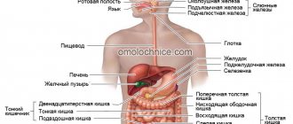
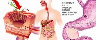
Food, saliva and water from the oral cavity enter the stomach through the esophagus - a hollow muscular-mucosal tube 3-4 mm thick and about 25-30 cm long, which has its own peristalsis, thanks to which food moves from the pharynx to the stomach. The importance of the esophagus in the overall harmonious functioning of the body is difficult to overestimate, since any, even minor, dysfunction entails serious disruptions in the functioning of the entire digestive system and requires timely optimal treatment. Today we will talk about a fairly common pathology of this organ - stenosis.
ANATOMY OF THE ESOPHAGUS AND THE ESSENCE OF THE PROBLEM
Esophageal stenosis is a violation of the patency of the esophagus due to its pathological narrowing due to various reasons, which has an extremely negative effect on the functioning of the human digestive system, and in the case of obliteration (complete closure of the lumen of the esophagus) is fraught with catastrophic consequences, including death.
The esophagus has three sections of different lengths: cervical (5-8 cm), thoracic (15-18 cm) and abdominal (1-3 cm). Throughout its entire length, the esophageal tube has a different diameter. On average, the width of its lumen is 2-3 cm, but it has three natural physiological narrowings: at the junction of the pharynx and the esophagus, the diameter of the esophageal tube is about 1.4 cm; in the area where the left main bronchus and aorta are closely adjacent to the esophagus, the width of its lumen usually does not exceed 1.6 cm; in the place where the esophagus connects to the stomach through an opening in the diaphragm, its diameter varies from 1.6 to 1.9 cm. It is in places of physiological narrowing of the esophagus that foreign objects most often get stuck, pathological narrowing of its lumen (stricture) or protrusion of the wall occurs ( diverticula).
TYPES OF ESOPHAGAL STENOSIS
There are several classifications of esophageal stenosis. Etiologically, congenital esophageal stenosis, caused by pathology of intrauterine development of the fetus, is distinguished, and acquired. The first type of stenosis accounts for 10% of the incidence, the second - 90%.
Depending on the location of the stricture (narrowing), there are high stenosis (in the cervical region), medium (at the site where the aorta adjoins the esophagus), low (in the lower third of the esophagus, above the hole in the diaphragm) and combined (at the site where the esophageal tube enters the stomach ). Based on the length (extent) of the narrowed sections of the esophagus, strictures of less than 5 cm (short), more than 5 cm (long), more than 2/3 of the length of the esophagus (subtotal), and total are distinguished.
Based on the severity of narrowing of the lumen of the esophagus, there are 4 degrees of stenosis:
I. esophagus narrowed to 1.1 -0.9 cm;
II. diameter of the esophageal tube - 0.8-0.6 cm;
III. lumen width - 0.5-0.3 cm;
IV. The diameter of the narrowed esophagus is only 0.2-0.1 cm, in the worst case it is completely closed (obliterated).
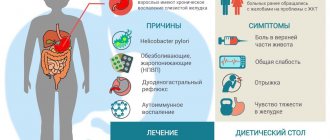
CAUSES
Intrauterine pathologies of the development of the mucous membrane of the esophagus, cartilage, muscle tissue, etc.
- Any diseases of the esophagus: erosions, ulcers, hernias, benign or malignant neoplasms.
- Stomach diseases: chronic gastritis, gastroesophageal reflux disease (GERD), ulcers, etc.
- Inflammatory processes in the esophagus caused by the presence of a foreign body in it, for example, a stuck fish bone or an accidentally swallowed small object.
- Any injuries to the esophagus: external penetrating wounds with damage to the esophageal tube, chemical burns of the esophageal mucosa when aggressive substances enter (for example, household chemicals, acids, poisons, etc.), accidental injuries during any surgical procedures, etc. , after which scars form on the esophageal mucosa.
- Complications of such severe infectious diseases as scarlet fever, diphtheria, tuberculosis, syphilis, etc.
- External pressure on the esophagus of nearby organs, for example, with an aortic aneurysm, enlarged lymph nodes, mediastinal tumors, etc.
SYMPTOMS
The symptoms of esophageal stenosis are pronounced, so its diagnosis, as a rule, does not cause difficulties, although in the process of making a diagnosis, some diseases (for example, stomatitis, pharyngitis), protrusion of the walls of the esophagus (diverticula) and the presence of a foreign body are excluded.
The main symptom of esophageal stenosis is disturbances in the swallowing process (dysphagia). In total, there are 4 degrees of dysphagia:
- I. periodic discomfort when eating solid foods;
- II. the ability to swallow only very well chewed and semi-liquid food;
- III. Only liquid food passes through the esophageal tube;
- IV. The esophagus does not even allow water and saliva to pass through.
Other characteristic symptoms of esophageal stenosis:
- increased salivation,
- regurgitation after eating,
- vomit,
- chest pain,
- attacks of suffocation and coughing.
If it is impossible to eat normally for a long time due to stenosis of the esophagus, a person sharply loses weight, which is accompanied by general weakness, fatigue, low performance, psychological depression, and depression.
TREATMENT AND PREVENTION
Therapy for stenosis begins after confirmation of the preliminary diagnosis using endoscopy and x-ray examination. During the studies, the localization and extent of the stricture, the degree of narrowing of the esophagus are established, and the exact causes of stenosis are confirmed or determined.
During treatment, until the condition of the esophagus normalizes, the patient is recommended to eat semi-liquid and liquid foods, and, if necessary, take antacids and astringents. In case of complete obliteration of the esophagus, nutritional enemas are prescribed, and in the most severe cases, a hole (fistula) is made in the stomach, a rubber tube is inserted into it, the end of which is brought out and attached to the skin with a plaster. Through this tube, using a funnel, nutritional mixtures recommended by the attending physician are directly poured into the stomach.
To treat esophageal stenosis, the bougienage method
— gradual expansion of the lumen of the esophagus using flexible or metal rods (bougies) of different diameters (in ascending order) or balloon dilatation. If it is impossible to expand the esophagus in this way, extended strictures or dense scars on its mucosa are dissected with electrosurgical instruments. It is also possible to perform endoprosthetics by introducing a self-expanding stent into the esophagus. In difficult cases, it is necessary to remove narrowed sections of the esophagus, replacing them with gastric or intestinal grafts.
The effectiveness of treatment depends on the causes of esophageal stenosis, its severity and the individual characteristics of the patient (for example, the presence of concomitant diseases). With benign strictures of the esophagus, as a rule, it is possible to achieve a stable positive result and restoration of the functions of the esophagus. For esophageal cancer, which caused its stenosis, the prognosis is disappointing.
To avoid stenosis of the esophagus and its severe consequences, doctors recommend always chewing food thoroughly, avoiding stomach diseases (gastritis, ulcers, GERD, etc.) and treating them promptly, and avoiding injuries to the esophagus, foreign bodies and aggressive chemicals getting into it.
Medical office magazine No. 198 Author: Zhenya Orlova Photo: Yandex Pictures
Clinical signs and symptoms of esophageal narrowing
The clinical picture of narrowing of the esophagus is mainly determined by a violation of the act of swallowing.
The most characteristic signs of this condition are the symptoms:
- Dysphagia (in Latin dysphagia) is a disorder of swallowing. There are four degrees of dysphagia: periodic difficulties in swallowing solid foods;
- the ability to swallow only semi-liquid food;
- patients can only swallow liquid food;
- it is impossible to swallow even saliva or water.
Dysphagia can be constant or remitting. It is believed that in case of esophageal cancer, dysphagia is constant and increasing. However, if the tumor disintegrates or ulcerates, the degree of dysphagia may decrease briefly. In children with a congenital form of esophageal stenosis, signs of dysphagia may appear already at the first feeding.
Dysphagia is called paradoxical when solid food is swallowed more easily than solid food; this symptom is characteristic of achalasia cardia.
- Thoracalgia. Pain is often localized in the retrosternal region; more often it accompanies the act of swallowing, but may not be associated with it.
- Feeling of fullness and pressure. Such sensations occur when food accumulates above the narrowing site.
- Increased salivation (salivation).
- Esophageal vomiting.
Symptoms
The very first symptom is difficulty swallowing solid food - dysphagia. The severity of the stenosis is judged by the severity of dysphagia.
| Stage | How it manifests itself |
| Stage 1 | Occurs only periodically, is not accompanied by pain or the pain is mild. There is slight discomfort when passing the lump when eating fast, hot, or spicy. |
| Stage 2 | The pain is almost always pronounced; only liquids and semi-liquid foods such as broths, soups, and jelly are painless. |
| Stage 3 | The patient cannot eat any food, only drink liquid: sharp pain and vomiting immediately occur. |
| Stage 4 | The inability not only to eat, but also to drink, including swallowing one’s own saliva. This condition requires emergency hospitalization in a surgical hospital, and in some cases, in intensive care. |
Expert opinion
Sevastyanov Roman
General practitioner, hepatologist, gastroenterologist, highest qualification category. Site Expert
Nausea and vomiting, increased or, conversely, decreased saliva production are also common. Chronic inflammatory diseases of the bronchopulmonary system often develop. With a high degree of stenosis, a prolonged dry cough, spasms in the larynx, and sometimes suffocation occur.
What research methods can be used to see narrowing of the esophagus?
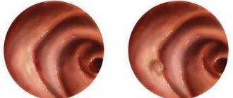
If there are clinical signs of esophageal stenosis, then to confirm the diagnosis, the patient undergoes endoscopic and x-ray examinations; with the help of these examinations, narrowing of the esophagus can be seen:
- Esophagoscopy confirms the fact of esophageal stenosis and its level. The mucous membrane is examined using an endoscope. The cause of the stenosis is determined. If necessary, an endoscopic biopsy is performed.
- X-ray with barium allows you to trace the passage of a contrast suspension, see places of narrowing of the esophagus, examine the contours of the organ, its relief and motility.
Scar narrowing of the esophagus
RSP or cicatricial narrowing of the esophagus are growths of connective tissue in the wall, leading to its deformation. The process can affect different levels of the organ and have different lengths.
RSP arise as a result of acute and chronic esophagitis, when the muscular layer is involved in the inflammation process.
ICD 10: K 22.2 Obstruction of the esophagus; T 28.6 Chemical burn of the esophagus
The main reasons for the development of cicatricial narrowings:
- Acute corrosive esophagitis resulting from damage to the esophagus by caustic chemicals. The lumen of the esophagus in the area of scar tissue is uniformly narrowed. Extension is determined above the area of stenosis.
- Peptic strictures are a result of GERD, which can be the outcome of many pathologies of the lower esophageal sphincter. With a long course of esophagitis, the process can spread to the muscle layer and even to the peri-esophageal tissue. Usually this is a long process, but sometimes it develops very quickly. The radiograph reveals an extended cylindrical stricture (may exceed 5 cm), the folds of the mucous membrane are thickened and tortuous.
- Postoperative strictures do not develop in the esophagus itself, but at the sites of anastomoses performed during gastrectomy, between the esophagus and intestines, or the gastric stump.
Hiatal narrowing
The most common complication of hiatal hernia is erosive-ulcerative esophagitis, as a result of constant exposure of the esophagus mucosa to stomach contents. Damage to the organ wall can be very deep, and the muscle layer is often involved in the process. The outcome of such inflammation is the formation of scars. Hiatal narrowing of the esophagus is a stenosis of the organ against the background of a long-standing hiatal hernia - a hernia of the esophagus.
Causes of the disease
There are other causes of stenosis that are not natural. In this case, the phenomenon needs treatment. They come in two types.
Benign
These include:
- benign neoplasms;
- narrowing of the esophagus due to the presence of scars (cicatricial);
- mechanical damage;
- the disease can be caused by a chemical burn resulting from ingestion of substances such as alkalis, acids, etc.
Cicatricial stenosis can be caused by reflux esophagitis, which causes the release of stomach contents into the “tube”. This leads to tissue erosion and inflammation.
Often, narrowing of the esophagus is provoked by external pressure on the organ, for example, with heart disease, when the left atrium expands, or an aortic aneurysm.
The disease can be caused by an enlarged thyroid gland or cardiospasm, but such cases are quite rare.
Malignant
This category includes a cancerous tumor of the organ itself or nearby systems. This reason is common.
Circular narrowing
CSP is an x-ray symptom characterized by a limited narrowing of the organ with smooth, clear contours; dilated areas of the esophagus are defined above and below the narrowing. If such an x-ray picture is detected, it is necessary to exclude endophytic cancer of the esophagus. This form of tumor leads early to CSP.
The contours of the narrowing are smooth and clear due to the connective tissue in the tumor mass. If “undermining” is detected at the place where the narrowed part transitions into the widened part, the diagnosis becomes very likely.
Operations on the esophagus for narrowing
Treatment is carried out either by a thoracic surgeon or by a specialist in endoscopic surgery. The question of how to treat a particular patient is usually decided collectively.
Surgical treatment, in case of benign stenosis, includes bougienage and balloon dilatation.
If the scars are very dense, they are excised using various endoscopic techniques.
When treating inoperable patients with malignant tumors of the esophagus, according to the latest clinical recommendations, endoscopic esophageal stenting is performed.
In cases of extended and recurrent stenosis, resection of the narrowed section of the esophagus is performed, followed by esophagoplasty.
If the patient's condition is severe and abdominal surgery is not possible, gastrostomy is performed as a necessary measure to provide nutrition to the patient.