general description
Based on etiology, mechanical, thermal, chemical and spontaneous damage to the digestive tract should be recognized. A larger group consists of mechanical damage. They appear as a result of stuck foreign bodies, various instrumental manipulations, wounds with cold steel and firearms, severe closed trauma, and a jet of compressed gas. Thermal and chemical damage (burns) occur as a result of accidental or intentional ingestion of hot and chemical solutions. Another group consists of so-called spontaneous ruptures of the esophagus.
Causes
Esophageal rupture most often occurs due to:
- frequent endoscopic examinations;
- chemical burns;
- ingress of foreign bodies, especially sharp ones;
- trauma and penetrating wounds;
- with careless performance of various operations, and as a result of injury to the esophagus.
In rare cases, frequent vomiting or severe and prolonged coughing can lead to ruptures. Labor, or rather strong pushing, can cause illness. Against the background of an epileptic attack, a rupture may also occur.
Symptoms of esophageal injury
The varied symptoms of esophageal damage are usually divided into local and general. Local manifestations include pain along the esophagus, aggravated by the act of swallowing, dysphagia, hoarseness, subcutaneous emphysema, infiltration and hyperemia of the soft tissues of the neck, pain on palpation, pneumothorax, muscle tension in the anterior abdominal wall. Common manifestations include pallor and cyanosis of the skin, shortness of breath, cold sweat, tachycardia, chills, and hyperthermia.
Each type of damage corresponds to a certain set of clinical signs, but most of them are observed at a late stage, already with the development of purulent complications.
Damage to the esophagus by foreign bodies in most cases does not have clear clinical symptoms. The esophagus has the ability to pass rather large and sharp objects without much damage. Sharp objects (fish bones) often get stuck when they pierce the wall of the esophagus with one sharp end. There is pain when swallowing, solid food cannot pass through the esophagus (dysphagia). Symptoms are more pronounced when the cervical esophagus is damaged. The most common symptom is pain along the esophagus with irradiation to the back of the head (with perforation of the cervical spine), interscapular (with perforation of the upper and middle thoracic sections) or epigastric region (with perforation of the lower thoracic and abdominal sections of the esophagus).
As a rule, the pain sharply increases with swallowing and palpation of the soft tissues of the neck. However, a similar pain syndrome is characteristic of any foreign body in the lumen of the esophagus (without damage or with non-penetrating damage).
Patients often experience chest pain, dysphagia, “unease” in the esophagus, a scratching sensation when swallowing food, and hoarseness. With massive foreign bodies, clinical signs of airway compression may be observed. In case of perforation of the esophageal wall, the clinical picture of mediastinitis develops. Similar symptoms are observed with damage to the esophagus caused by esophageal cancer, congenital cysts and fistulas of the neck, thyrotoxicosis (Graves' disease).
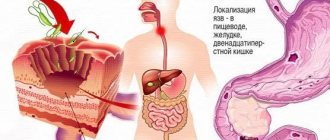
The clinical picture of chemical and thermal damage to the esophagus is always clear. Typically, burns to the esophagus occur as a result of accidental or intentional (for the purpose of suicide) ingestion of alkali, acid and other caustic liquids. Acid and alkali, acting on the mucous membrane of the esophagus, cause its severe destruction. An acid burn causes more superficial damage to the wall of the esophagus. The acid can pass into the stomach, where it also causes burns to the mucous membrane. Alkali causes the deepest damage to the wall of the esophagus. After healing, scar tissue forms in the area of chemical damage, which leads to obstruction of the esophagus or stomach.
In the clinical picture, dysphagia comes first. There are burn marks in the corners of the mouth and on the tongue. It is necessary to ask the patient or people around him about what he drank.
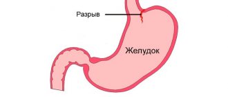
Esophageal rupture: reasons for the formation of a through hole
Traumatic perforation and spontaneous rupture of the esophagus have different causes. This is a rare pathology; 1% of all cases occur in the thoracic surgery department. In males, the gap occurs 3 times more often than in females.
In most cases, pathology occurs as a consequence of chronic diseases of the esophageal tube or incorrectly performed medical examinations. Spontaneous perforation occurs as an independent disease in isolated episodes.
A type of esophageal rupture is also a condition called Boerhaave's syndrome (more about it).
Rupture due to trauma
A traumatic lumen in the esophagus can form as a result of therapeutic and diagnostic manipulations:
- fibrogastroduodenoscopy - a method of examining the esophageal tube, stomach, duodenum using a flexible endoscope to assess the condition, the effectiveness of treatment, take a biopsy, remove foreign bodies;
- tracheotomy is an emergency surgical operation to restore air access to the trachea in case of sudden blockage of the respiratory tract;
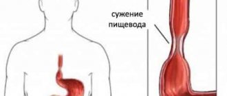
- bougienage - a medical procedure to expand the alimentary tract with a special probe (bougie), done for stenosis of the esophagus;
- cardiodilatation - artificial expansion of the junction of the cardiac part of the stomach and the esophagus during spasm;
- tracheal intubation - insertion of a special tube into the trachea for artificial ventilation of the lungs when air is obstructed in the respiratory tract;
- surgical procedures in the cervical region, chest, and abdominal organs.
Other types of injury:
- internal burn of the esophagus, including chemicals;
- ulcers or swelling on the walls of the tube;
- stuck foreign bodies inside the cavity;
- penetrating injuries of the chest, neck.
Causes of spontaneous rupture
Factors that provoke spontaneous rupture of the esophagus are associated with disruption of the muscular layer of the esophagus. Changes occur with reflux disease, with drug-induced esophagitis, with ulcers as a result of infection in AIDS patients.
- increased vomiting after eating, drinking liquid or alcoholic beverages - intraesophageal pressure rises to 200 mmHg;
- repeated gag reflex with impaired functions of the vomiting center in the brain;
- increased intra-abdominal, then intra-esophageal pressure - a condition that occurs during lifting heavy objects, straining during defecation, straining during childbirth, as well as uncontrollable coughing, epileptic seizures, and blunt trauma to the abdominal area.
Banquet rupture of the esophageal mucosa
The reflux of stomach contents or gases into the esophagus causes an increase in internal esophageal pressure. Perforation occurs in its weakest area, which is located on top of the diaphragmatic ring. This circumstance occurs when restraining the gag reflex at the table while eating. This pathological condition is called banquet esophagus.
Perforated damage in the esophagus can be complete, when all layers of the wall are affected, and incomplete, with damage to one or more membranes.
It is important! Esophageal tube rupture is considered an emergency, often requiring emergency surgical intervention!
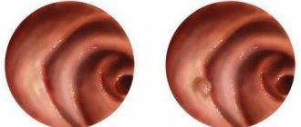
Diagnosis of esophageal injury
Three methods are of greatest importance in diagnosing diseases of the esophagus: x-ray examination, esophagoscopy and motility studies. The main method is x-ray examination, which should precede the others. The study begins with fluoroscopy, necessarily supplementing it with radiography. If necessary, resort to X-ray cinematography and tomography. Esophagoscopy should be undertaken in cases where it is necessary to clarify the diagnosis, for example, with esophageal cancer, it is necessary to take a piece of the tumor for histological examination.
In addition, a smear should be made to study the cytological picture, which in some cases makes it possible to clarify the diagnosis even with a negative result of histological examination. Esophagoscopy is also used for therapeutic purposes to remove foreign bodies and dilate strictures of the esophagus. Esophagoscopy is performed either with a rigid esophagoscope (Brünings, Friedel, etc.) or with a flexible one with fiber optics. For diagnostics, it is more rational to use a flexible tool, while for bougienage a rigid one is needed.
The study of esophageal motility is an important additional method for diagnosing a number of benign diseases - cardiospasm, hiatal hernia, etc. Motility of the esophagus is most often studied using the method of “open catheters” or using balloons. At the same time, the acidity of the medium is determined, which is necessary when diagnosing reflux esophagitis. It is practically impossible to treat cardiospasm at the modern level without monitoring the results of treatment by recording esophageal motility and intraesophageal pressure.
Esophageal rupture: consequences
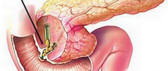
The leading role is given to surgical intervention, which solves several problems. The perforated defect is eliminated by suturing the gap, followed by suturing and sealing. During the operation, the issue of providing enteral nutrition is resolved - the probe is inserted into the stoma through the anterior abdominal wall.
The extent of surgery is determined individually; it depends on the size of the damage, the condition of the walls of the esophagus and the presence of associated factors. A good result after surgery is ensured if treatment is started immediately on the first day of the rupture.
Stimulators of esophageal regeneration can alleviate the condition.
Treatment of esophageal injury
If damage to the esophagus is suspected, it is necessary to immediately stop eating by mouth and take the victim to a surgeon. At the stage of qualified assistance, R-contrast examination of the esophagus and endoscopy are performed. Damage determined endoscopically is usually large and has crushed edges. Small lesions are often detected only by x-ray. The level of damage is determined, which determines surgical access. It is necessary to find out whether the patient has eaten, whether there is an increase in body temperature, how much time has passed since the injury, and whether there are any signs of mediastinitis. It is important to know whether before the injury there were any difficulties with swallowing due to cardiospasm, diverticulum, or stenosis.
- A conservative treatment method is used for small-sized (up to 1 cm) injuries located above the aortic arch and early diagnosed injuries. Such injuries heal in 7–10 days without surgery if treated in a timely manner.
- Surgical method. For injuries in the upper and middle third of the esophagus, a right-sided thoracotomy is performed; for low injuries, a left-sided thoracotomy is performed. In this case, a mandatory stage of the operation is the placement of a gastrostomy, since after the operation parenteral nutrition requires a lot of effort and expense. In case of cancerous perforation, perforation of a diverticulum, or extensive damage to the wall, the question arises of primary resection of the damaged esophagus. If mediastinitis develops, it is necessary to perform a cervical esophagostomy and completely exclude the esophagus from the passage.
Upon admission six or more hours after perforation, with the development of signs of mediastinitis, mediastinotomy and mediastinal drainage are indicated.
Essential drugs
There are contraindications. Specialist consultation is required.
- Cefotaxime (an antibiotic of the third generation cephalosporin group). Dosage regimen: IM 0.5 g of the drug in 2 ml (respectively 1 g in 4 ml) of sterile water for injection, injected deep into the gluteal muscle. 1% lidocaine is also used as a solvent for intramuscular administration. For intravenous administration, 0.5-1 g of Cefotaxime is dissolved in 10 ml of sterile water for injection. Inject slowly over 3–5 minutes. For drip administration (over 50-60 minutes), 2 g of the drug is dissolved in 100 ml of isotonic sodium chloride solution or 5% glucose solution.
- Cefepime (IV generation cephalosporin antibiotic). Dosage regimen: adults and children weighing more than 40 kg with normal renal function 0.5-1 g intravenously (for severe infections up to 2 g) or deep intramuscularly at intervals of 12 hours (for severe infections - after 8 hours ).
- Amoxiclav (broad-spectrum bactericidal antibacterial agent). Dosage regimen: intravenously, adults and children over 12 years of age or weighing more than 40 kg - 1.2 g of the drug (1000 + 200 mg) with an interval of 8 hours, in case of severe infection - with an interval of 6 hours.
Forecast
The prognosis for esophageal rupture varies. If the patient has uncomplicated forms of this disorder, which are quickly stopped, then complete recovery occurs.
The prognosis worsens under such circumstances as:
- late visit to the clinic;
- wait-and-see tactics;
- development of complications.
The mortality rate from ruptures of the esophagus is quite high - according to various sources, out of ten patients, from two to eight die with such a diagnosis.
Kovtonyuk Oksana Vladimirovna, medical observer, surgeon, consultant doctor
1, total, today
( 45 votes, average: 4.62 out of 5)
Biliary sludge: causes, signs, treatment, prevention
Dysbacteriosis in infants: why does it develop and should it be treated?
Related Posts
Incidence (per 100,000 people)
| Men | Women | |||||||||||||
| Age, years | 0-1 | 1-3 | 3-14 | 14-25 | 25-40 | 40-60 | 60 + | 0-1 | 1-3 | 3-14 | 14-25 | 25-40 | 40-60 | 60 + |
| Number of sick people | 0 | 0 | 4 | 21 | 21 | 21 | 21 | 0 | 0 | 4 | 21 | 21 | 21 | 21 |
How is the treatment carried out?
Patients with a similar diagnosis are taken to the hospital. They are examined by a surgeon and sent for surgery. Conservative therapy will be appropriate only for minor tears. In this case, antibiotics and dietary nutrition will be prescribed. The patient will be hospitalized in any case, since any drug for such a pathology is administered through a tube.
In most cases, to overcome the disease, the doctor resorts to surgery. The purpose of the operation will be:
- suturing the rupture site;
- prevention of peritonitis (drainage of pustules, if any);
- temporary “disconnection” of the esophagus from digestive processes.
At the end of the manipulation, the patient will be prohibited from eating for 2-3 days in his usual way - through the mouth. Nutrients will be administered through a gastrostomy tube. When the condition improves, you can eat in the usual way.
In the first days, the diet should include the following dishes:
- porridge;
- boiled meat, fish;
- baked vegetables and fruits;
- casseroles made from meat or cottage cheese;
- soups.
Those foods that can irritate the mucous membranes should be removed from the usual diet. These are, first of all, fatty foods, fried, sour, spicy. You will also have to give up flour products.
All dishes must first be pureed and heated (or cooled) to room temperature before eating.
Read: can you drink milk if you have pancreatitis?
Find out how Ursosan acts on the digestive organs.
We advise you to find out what symptoms indicate pancreatic cancer.
Prognosis and prevention
Death is most often observed after mechanical damage to the esophagus, when the upper respiratory tract is also affected. And this despite timely medical assistance. In such cases, 50-60% of patients die.
Important information! If surgery is performed within the first 48 hours after the onset of symptoms, the prognosis will be better. About 10% of all cases are fatal.
Preventive measures consist of following simple recommendations:
- eat only fresh foods to avoid frequent poisoning;
- take antiemetic drugs on time if you have a history of illnesses that cause constant severe vomiting;
- in case of gastroenterological diseases, you should adhere to the correct diet;
- avoid sudden physical activity and heavy lifting;
- try not to overeat.
As for children, you need to ensure that the child does not accidentally swallow foreign objects (batteries, nails, needles). In addition, you need to teach children not to rush and not talk while eating.
Symptoms
| Occurrence (how often a symptom occurs in a given disease) | |
| Increased salivation (hypersalivation, excessive salivation, drooling) | 70% |
| A sore throat | 40% |
| Difficulty swallowing (dysphagia, difficulty swallowing, swallowing disorder) | 40% |
| Belching and vomiting blood | 30% |
| Chest pain without connection with physical or psycho-emotional stress | 10% |
Prognosis and prevention
As in the presence of a rupture of the esophagus, Mallory-Weiss syndrome, the prognosis for recovery largely depends on the time interval between the start of treatment and the time of damage to the esophagus. Complications that accompany the pathology, the location and size of the rupture, the general condition of the patient, and chronic diseases also play an important role.
Preventive measures in this case play a secondary role. However, eliminating certain factors will prevent the development of the disease. You should avoid iatrogenic damage, prevent your body from reaching a state of bulimia, and undergo a timely medical examination.
Some rules must be followed to minimize the risk of perforation. Teach children to always eat food slowly and chew it thoroughly. Often perforation occurs due to swallowing a large piece of food. Do not forget about the saying “when I eat, I am deaf and dumb.” You should avoid strenuous physical activity and heavy lifting. The diet should be balanced and correct; you should not abuse alcoholic beverages.