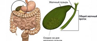
What is epithelium?
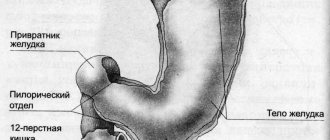
The gastric epithelium is a collection of columnar cells. They are called single-layer columnar epithelium. Against the background of prolonged irritation of the mucous membrane of the esophagus by gastric juice, for example, with gastroesophageal reflux, when the contents of the stomach are thrown into the esophagus, the risk of developing the gastric variant of esophageal metaplasia increases.
The essence of the disease is the replacement of the stratified squamous epithelium of the esophagus with a single-layer cylindrical epithelium, characteristic of the stomach. The part of the esophagus in which the mucous membrane of the organ passes into the stomach is most susceptible to tissue replacement. It is in the area of connection of the two organs that the most intense irritation with hydrochloric acid occurs against the background of gastroesophageal reflux.
Barrett's esophagus and prevention of esophageal adenocarcinoma
Transcript of the report by Prof. Sheptulina A.A. at the II International Internet Congress of Internal Medicine Specialists.
Professor Erdes S.I.: – Dear listeners, dear colleagues, we continue our conversation about cancer prevention of the esophagus. And we move on to consider issues directly related to the features of such a formidable pathology as Barrett’s esophagus. Let me give the floor for the next report to Professor Arkady Aleksandrovich Sheptulin for the message “Barrett’s Esophagus and Prevention of Esophageal Adenocarcinoma.”
Professor Sheptulin A.A.: – Good afternoon, dear colleagues.
(00:35) Screensaver: Barrett's esophagus and prevention of esophageal adenocarcinoma.
Professor Sheptulin A.A.: – The topic of my short lecture: “Barrett’s esophagus and prevention of esophageal adenocarcinoma.” I will start by demonstrating a clinical example - an example of a patient who was in our clinic not long ago.
The patient is 49 years old. He was admitted with a fairly classic, often encountered set of complaints: nausea, vomiting in the morning, discomfort in the epigastric region, unpleasant taste in the mouth and general weakness. The patient himself considers himself as such for three years, when, against the background of heartburn, he developed nausea and vomiting, and gastroscopy revealed a picture of erosive gastritis. He took proton pump inhibitors with good effect and continued to work. But a more thorough questioning showed that the patient first noticed heartburn at the age of 30, that is, 20 years ago. This is very significant, because most patients who experience heartburn are treated independently on the advice of friends and acquaintances - the so-called “telephone reflux”. Meanwhile, gastroesophageal reflux disease progresses significantly during this time. And he had been deteriorating since August 2010 (this case dates back to that year) - this was literally two months before admission. Gastroscopy revealed an esophageal ulcer, and proton pump inhibitors were again prescribed.
What can you say about your life history? The patient, although he was admitted to a reputable clinic, does not belong to the highest strata of society, to put it mildly. He works as an electrician, is divorced, and abuses alcohol. The amount that was indicated is 150-200 ml, most likely an underestimate, and a daily amount. Smokes 20 cigarettes a day. The fact that he didn’t have a single tooth in his mouth, and he didn’t even bother to get dentures, speaks volumes about his social status and its level. This is how he ate, muttering with his toothless mouth. However, based on the results of the previous examination, we diagnosed him with stage IV reflux esophagitis according to the Savary-Miller classification, since he had a peptic ulcer of the esophagus. And since the patient is a heavy smoker, he had manifestations of chronic obstructive bronchitis and pulmonary emphysema.
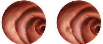
In a general blood test, as is typical for patients with obstructive bronchitis, he had an increased level of hemoglobin - secondary erythrocytosis. He had a high color index, and the size of red blood cells was increased - this reflects a hidden deficiency of vitamin B12 and folic acid, and is considered one of the indirect markers of chronic alcoholism. There is nothing special in the urine test, the stool test is negative. The biochemical blood test is surprising - the ideal state of liver enzymes and GGT (then prove that alcohol affects the liver). An ultrasound examination revealed nothing special about the liver, pancreas or gallbladder. But when a gastroscopy was done, characteristic “flames” were discovered - this is what is characteristic of Barrett’s esophagus: restructuring of the epithelium according to the intestinal type. We were also able to observe a peptic ulcer and convergence of folds, but most likely this ulcer will heal fairly quickly. And our very experienced endoscopist assessed this picture as erosive and ulcerative reflux esophagitis, complicated by the development of Barrett’s esophagus. We know what Barrett's esophagus can lead to. The endoscopist took seven fragments of their edges and the bottom of the ulcer.
The most intensive treatment of esophagitis was prescribed, which we use for complicated forms: twice-daily use of proton pump inhibitors, in particular Pariet, which has a number of advantages; combination with an H2 blocker (evening dose), which is considered to give the most powerful effect. And as symptomatic therapy - Almagel and Motilium. Quite quickly, this peptic ulcer of the abdominal part of the esophagus healed, and the symptoms of reflux esophagitis persisted. The patient felt great and insisted on being discharged, assuring that his friends had been waiting for him for a long time. Nevertheless, we told him that he needed to wait for the histological report. And we saw that the presence of Barrett’s esophagus with intestinal type restructuring of the epithelium was indeed confirmed, a picture of classic erosive-ulcerative esophagitis. But what’s sad is that against the background of this esophagitis, atypical cells were identified. And the morphologist interpreted this picture as mucinous adenocarcinoma. Accordingly, we began to investigate further.
We took an x-ray and identified a filling defect. And the clinical diagnosis was changed to cancer of the abdominal esophagus - an infiltrative-ulcerative form. And gastroesophageal reflux disease was indicated as the background disease: cardia insufficiency and Barrett's esophagus. This example is indicative in that the patient, over 20 years of illness for which he did not apply or receive treatment, went through all the stages that gastroesophageal reflux disease goes through: the stage of erosive esophagitis, the stage of Barrett’s esophagus and ended with the development of adenocarcinoma. Referring to the recommendations of the American College of Gastroenterologists, we can define what “Barrett's esophagus” is - the replacement of the squamous epithelium of the distal esophagus with columnar epithelium, which is detected by endoscopy and further confirmed by histological examination by the detection of intestinal metaplasia in this area. Data on the epidemiology, incidence and prevalence of Barrett's esophagus vary: 6% among all patients with symptoms of dyspepsia; 8% among all patients with heartburn. But the data is more plausible - 10-15% of all patients with gastroesophageal reflux disease (we just emphasize) are of the erosive form. For all patients, including non-erosive forms, Barrett's esophagus accounts for approximately 3%. It is very significant that the incidence of Barrett's esophagus is increasing significantly, in parallel with the increasing prevalence of gastroesophageal reflux disease.
And it is especially significant that the prevalence of Barrett’s esophagus according to sectional data is 5 times higher than according to clinical data. That is, out of five patients, four patients with Barrett's esophagus walk and do not know that they have this complication. Risk factors that predispose to the development of Barrett's esophagus: older age of patients, male gender, Caucasian race, long-term (more than 13 years) history of GERD symptoms - although the development of symptoms of this complication should be feared in cases where the duration of erosive esophagitis exceeds five years - high secretion of hydrochloric acid and high content of bile acids in refluxate.
Metabolic syndrome is also a risk factor for Barrett's esophagus. The presence of this syndrome increases the risk of developing Barrett's esophagus by 2 times, because obesity leads to an increase in the content of periesophageal adipose tissue. And due to the release of cytokines and chemokines from it, it promotes the development of inflammatory changes in the mucous membrane of the esophagus. And this explains the rather low incidence of Barrett's esophagus in Japan. Unlike, say, the United States, it is difficult to find an obese Japanese person in Japan. The prevalence of obesity in the United States is about 60% overweight, and in Japan it is 5%.
I would like to draw attention to the risk of malignancy in Barrett's esophagus. In general, it is low - a meta-analysis of 25 studies showed that the risk of malignancy is 0.5% per year. But this is a general risk and may vary from patient to patient. First of all, the risk of malignancy differs in the presence of dysplasia and in its absence. In particular, the risk of developing adenocarcinoma in patients without dysplasia is low, but depends on the length: short segment - less than 3 cm, and long segment - restructuring of Barrett's esophagus. We see that with a larger extent of the lesion, the risk of malignancy is higher. Also, the risk of malignancy depends on the presence of dysplasia. It is relatively low with low-grade dysplasia, and increases significantly if we are already faced with high-grade dysplasia. The annual risk of developing high-grade dysplasia in Barrett's esophagus is 1%. Indeed, almost 8 thousand patients with Barrett's esophagus were examined and followed up over a period of 25 years. And 1% per year during this time, 20-25% of patients developed high-grade dysplasia. The risk of developing high-grade dysplasia depends on the length of the segment of Barrett's esophagus. In those patients who developed high-grade dysplasia or adenocarcinoma over several years, the length of Barrett's segment was 6 cm; in those who did not progress, it was only 3 cm. Also, the risk of malignancy depends on the number of foci of dysplasia. In the presence of multifocal dysplasia, that is, many foci, the risk of developing adenocarcinoma is 3 times higher than in cases where there is one focus of restructuring.
The “gold standard” for diagnosing Barrett's esophagus is esophagogastroduodenoscopy with multiple biopsies, which are done every 2 cm, thus taking up to eight biopsies. As for additional methods, they play a purely auxiliary role. There are difficulties in screening patients with Barrett's esophagus for the following reasons.
Firstly, if there are clinical symptoms, they do not differ at all from the clinical symptoms of other forms of GERD, and half of patients with Barrett's esophagus have no clinical symptoms at all. And even the characteristic endoscopic picture is “tongues of flame”; they are not found in all patients. The tactics for managing such patients are all determined by the presence or absence of dysplasia. If it is just Barrett's esophagus, but there is no epithelial dysplasia, then such patients need to have a biopsy twice within a year.
And if there is no dysplasia, they are then subjected to dynamic monitoring with endoscopic examination every three years. If patients are diagnosed with low-grade dysplasia, a repeat examination is carried out after six months. The diagnosis is confirmed by an independent expert pathologist. And if low-grade dysplasia persists, dynamic observation with repeated endoscopic examination with an interval of one year is indicated. And finally, if patients have high-grade dysplasia, then multiple biopsies are taken over time over the first three months to rule out adenocarcinoma. And if the diagnosis of “high-grade dysplasia” is confirmed, the issue of using endoscopic or surgical research methods is decided.
Such a mutilating operation - esophagectomy, which was previously performed for Barrett's esophagus and high dysplasia, is no longer considered. Endoscopic and surgical methods for treating high-grade dysplasia are primarily photodynamic therapy, often in combination with proton pump inhibitors. It is effective in almost 80% of patients and reduces the risk of developing adenocarcinoma by 50%. Thermoablation using argon plasma coagulation is also effective in more than 80% of patients. Treatment methods include radiofrequency ablation, although in the late period this may lead to the development of esophageal strictures, and cryoablation. However, as recommended by expert consensus, endoscopic resection of the esophageal mucosa is currently the best treatment for patients with Barrett's esophagus with high-grade dysplasia. It can be combined with radiofrequency ablation or cryoablation.
What is the benefit of taking additional proton pump inhibitors for Barrett's esophagus? First of all, if patients with GERD constantly take proton pump inhibitors for a long time, then the risk of developing Barrett's esophagus itself is reduced by 2 times. In addition, materials were obtained from the use of Pariet, which showed that long-term use helps prevent further progression of GERD and the development of its relapses. In addition, long-term use of proton pump inhibitors for more than 10 years reduces the risk of developing high-grade dysplasia and adenocarcinoma of the esophagus. And interestingly, although there is indeed no complete restoration of the normal structure, a review of nine cohort studies with duration of use of proton pump inhibitors up to 13 years showed that in more than half of the patients, individual islands of normal stratified squamous epithelium are restored. Thus, the length of the metaplastic mucous membrane of the esophagus decreases.
Finally, active antisecretory therapy with proton pump inhibitors promotes better re-epithelialization of the esophageal mucosa after radiofrequency therapy. Well, unfortunately, the “fly in the ointment” is the work that was carried out in Northern Ireland. Almost 500 patients with adenocarcinoma were examined. And it turned out that only 7% had adenocarcinoma detected during follow-up of patients with early Barrett's esophagus. The remaining 90% were the same as the patient I was talking about. They have already reached the stage of adenocarcinoma of the esophagus. And this once again emphasizes the need for clinical monitoring of patients, especially with erosive forms of reflux esophagitis, in order to prevent the development of Barrett's esophagus, and then monitor such patients.
In conclusion, I can say that the presence of Barrett's esophagus should be excluded in any patient with a long history of gastroesophageal reflux disease. The risk of developing adenocarcinoma appears to be highest in patients with a long segment of Barrett's esophagus and high-grade dysplasia. The management tactics for such patients depend on the presence or absence of epithelial dysplasia and its degree and involve dynamic follow-up, repeated endoscopic examinations with biopsies, long-term use of proton pump inhibitors as monotherapy or in combination with other treatment methods.
Risk factors
Cylindrical cell metaplasia of the esophagus occurs under the influence of the same provoking factors that lead to the appearance of gastroesophageal reflux disease. Experts identify the following risk factors:
- Decreased tone of the esophageal sphincter in the lower part. This occurs due to insufficient innervation, when the sphincter does not compress tightly and gastric juice is thrown back into the esophagus.
- Changes in the structure of the congenital type. Under the influence of genetic factors, the diameter of the lower part of the esophageal sphincter increases or decreases.
- Previously experienced inflammatory processes. Localized in the lower part of the esophagus, such processes provoke the appearance of scars in the organ and lead to incomplete compression of the sphincter.
- Varicose veins in severe form. In this case, it is the nodes formed during varicose veins that prevent the sphincter from completely closing.
- Disorder of the motor-evacuation function of the stomach and esophagus. Against the background of such a disorder, antiperistalsis is observed, when the walls of the organs begin to move, leading to the movement of food masses into the upper parts of the esophagus.
The listed factors can provoke the appearance of gastroesophageal reflux, which in turn can cause erosive type gastritis, which later develops into gastric and intestinal metaplasia of the esophagus.
Causes of esophageal metaplasia and risk factors
As already stated, it would be logical to consider Barrett's esophagus as a complication of reflux esophagitis . Essentially, the body is trying to protect the esophageal epithelium from erosion by gastric juice by replacing normal cells with cells typical of the duodenum, which are able to withstand high acidity. But it turns out that there are quite a few patients with Barrett's esophagus who have never suffered from heartburn or reflux. Therefore, the true cause of Barrett's esophagus is currently unknown.
However, a number of factors are well known that increase the risk of its development:
- Presence of gastroesophageal reflux.
- Transition to 60 years of age. Barrett's esophagus is rare in young people.
- Male gender. Men are more likely to develop the disease (twice as likely as women).
- Belonging to the Caucasian race.
- Consuming excessive amounts of alcohol.
- Smoking tobacco.
- Obesity. It increases the risk of reflux disease and therefore the formation of Barrett's esophagus.
Symptoms
Many people are interested in how the disease manifests itself?
If we are talking about focal metaplasia of the esophagus, then clinical manifestations of this disease, as a rule, are not observed. Suspicion of metaplasia arises from clinical manifestations of gastroesophageal reflux. Therefore, we can say that the signs of the diseases coincide and are as follows:
- Frequent occurrence of heartburn. A strong burning sensation occurs in the retrosternal region, provoked by irritation of the epithelial layer of the esophagus with the contents of the stomach during the reverse reflux.
- Sour belching. Most often it is a precursor to heartburn. Belching appears after eating or while bending the body.
- Pain in the chest area. The intensity of the pain syndrome increases on an empty stomach and tends to radiate to the area of the neck, lower jaw and left half of the chest, as well as to the space between the shoulder blades.
- Extraesophageal signs. This may include shortness of breath, cough, dryness and sore throat, hoarseness, feeling as if the stomach is full even after a small amount of food.
When these signs appear, you should be examined for the presence of foci of metaplasia of the gastric mucosa into the esophagus and determine their extent and severity.
Classification and symptoms
The progression of Barrett's syndrome can be divided into two stages:
- The first stage is distinguished by its moderation in changes in cell structure;
- The second stage is characterized by complete replacement of epithelial cells.
Moving on to listing the complex of symptoms, it should be noted that in 25% of cases, esophageal metaplasia occurs quietly, that is, asymptomatic. In other patients it is not expressed too clearly:
- regular heartburn - burning in the esophagus and retrosternal area;
- belching with a sour taste - manifests itself after eating;
- nausea and vomiting after eating, in the last stage with blood;
- erosion of tooth enamel (rare);
- sore throat when bending forward and taking a horizontal position;
- impaired swallowing reflex;
- chest pain radiating to the jaw or neck.
In some cases, intraesophageal symptoms are observed (hoarseness, sore and dry throat and a gradual increase in cough).
Complications
Replacing esophageal tissue with single-layer columnar epithelium can cause a number of serious complications, including:
- Transformation of foci of metaplasia into malignant neoplasms. The most common form of esophageal cancer is adenocarcinoma.
- Hemorrhagic syndrome localized in foci of metaplasia and damage to the esophagus.
- The appearance of peptic-type strictures. These are adhesions of connective tissue that narrow the lumen of the esophagus at their location and provoke the development of dysphagia, which consists in disrupting the process of swallowing food.
To prevent such complications, it is important to diagnose metaplasia in time and receive appropriate treatment aimed at reducing the size of the lesions and their number.
How to detect esophageal metaplasia?
It is impossible to make a diagnosis based on a person's complaints. Changes in the mucosa are detected during endoscopic examination (FEGDS). The final diagnosis is made on the basis of histological examination of a piece of tissue. It is important to establish the type of metaplasia. It can be intestinal and gastric. If columnar epithelium with goblet cells is detected, this indicates Barrett's esophagus.
The following studies are needed:
- esophagogastroscopy;
- biopsy;
- histological examination;
- pH-metry;
- biochemical and general blood tests;
- examination of feces for occult blood;
- acid production test;
- contrast radiography;
- manometry (assessment of motor function of the esophagus);
- bacteriological examination;
- Ultrasound of the abdominal organs.
Diagnostics
The diagnosis of columnar cell metaplasia of the esophagus is made only on the basis of an instrumental examination of the patient. Modern medicine uses several methods to visualize the mucous membrane of the esophagus, including:
- Esophagoscopy. An instrument called an endoscope, which is a fiber optic tube equipped with lighting and a camera, is inserted into the lumen of the organ. The doctor can assess the condition of the esophageal mucosa by receiving an image from an endoscope on a special monitor. To obtain a more detailed image of metaplasia, a contrast agent is used to highlight foci of the pathological process. In this way, it is possible to establish the presence of an oncological neoplasm and identify metaplasia at the initial stage of development.
- Targeted biopsy. It is carried out along with the previous diagnostic method. Esophagoscopy determines the exact location of the foci of metaplasia, which allows tissue to be taken for histological examination specifically from the affected area. A specialist examines the resulting sample in a laboratory setting, determining the presence of single-layer columnar epithelium in the esophageal tissue.
- X-ray examination. Helps to visualize the structures of the walls of the esophagus and assess its motor-evacuation ability. This method also uses a contrast agent injected into the lumen of the esophagus.
- Manometry. It is carried out by measuring pressure in the esophageal lumen.
- Impedancemetry. Study of the motor-evacuation ability of all parts of the esophagus.
After a thorough examination and clarification of the diagnosis, the doctor prescribes treatment for metaplasia.
Diagnosis and treatment
Before treating esophageal metaplasia, it is necessary to diagnose it. There are several diagnostic methods that help determine the type of disease depending on its location.
The mucous membrane of the esophagus can be visualized using the following methods:
- Targeted biopsy is one of the types of biopsy, which consists of histological examination of tissue;
- Esophagoscopy - examination of the surface of the esophagus from the inside using an endoscope and methylene blue, which is used to stain the esophagus, for more accurate visualization;
- X-ray of the esophagus - pictures are taken using a barium mixture, which gives better contrast to the walls of the esophagus in the image;
- Esophagomanometry – is performed to assess the contractile activity of the esophagus;
- Impendansometry is a study of the esophagus in case of impaired motor-evacuation function.
As for treatment, it can also be carried out in several ways, the use of which depends on the degree of development of the disease. Therapeutic measures promote regression of foci of columnar epithelial cells.
Drug treatment
Drug treatment is characterized by the use of drugs from certain pharmacological groups that prevent the reflux of gastric juice.
| Pharmacological group | Impact | Medications |
| Antacids | It is a neutralizer of the chloride component of gastric juice. Used in the treatment of gastrointestinal diseases (gastric ulcers, gastritis, duodenitis). Therapy for Barrett's esophagus is carried out with drugs containing algetdarate and magnesium hydroxide; | "Phosphalugel"; "Maalox"; "Gastal"; "Omeprazole"; |
| PPIs (Proton Pump Blockers/Inhibitors) | Antisecretory drugs that are used to treat diseases of the digestive system. Reduces the secretion of hydrochloric acid. Application requires consultation with your doctor, as information about the level of acidity of gastric juice is required; | "Pantoprazole"; "Famotidine"; "Alfogel"; "Betomax"; "Gastracid"; |
| Prokinetics | Drugs that help normalize motor activity and gastrointestinal motility and block the reflux of food into the upper digestive tract. | "Ganaton"; "Motilium"; "Motilak". |
Table 1
Treatment with pharmaceutical drugs is used at the first stage of development of foci of metaplasia, as well as a course before surgery or for preventive purposes.
Surgical removal
It is not always possible to eliminate such a disease using conservative methods. Sometimes they resort to surgical removal of foci of Barrett's syndrome. However, even this radical method is divided into several techniques.
- Laser removal. Using a low-frequency laser beam to destroy columnar epithelium in the first stages;
- Photodynamic therapy (PDT). The destruction of the pathology of metaplasia occurs under the influence of light of a certain wavelength, as well as with the help of photosensitive elements - photosensitizers;
- Fundoplication involves suturing the fundus of the stomach to the diaphragm to create an acute angle with the esophagus. This treatment eliminates the possibility of gastric juice escaping into the esophagus;
- Free surgery. During the operation, the affected area of the esophagus is performed. Resection is performed with maximum and irreversible progression of the disease.
Note! After surgery, it is also necessary to continue the course of medication and adhere to a special diet, which will help prevent the development of complications.
Nutrition adjustments
Properly adjusted nutrition is the key to the success of therapeutic therapy. There are several rules that must be followed when choosing foods and dishes for the daily diet. They are based on exception:
- fermented milk products;
- alcoholic and carbonated drinks;
- coffee and tea;
- various spices and seasonings;
- fatty foods;
- citrus fruits;
- sweet;
- milk and goose meat.
Eating during metaplasia should be done in small portions (small portions 5-6 times a day). You should add more fruits (not citrus), vegetables (with fiber) and different types of legumes to your diet. Preventive measures for Barrett's esophagus are aimed at reducing the patient's weight category, getting rid of bad habits and addictions, which can provoke irritation of the epithelial tissue of the esophagus. You should also limit heavy lifting and physical overexertion.
Remember! Diet and preventative measures are not the primary method of therapy and should be combined with other appropriate treatments for this disease.
Treatment: general principles
The basic principles of treatment of esophageal metaplasia are the elimination and reduction of the size of foci of a single-layer columnar epithelial layer in the esophageal mucosa. Therapy is carried out comprehensively and includes both conservative treatment and surgical methods, and general recommendations on nutrition and lifestyle.
Experts believe that an important stage in the treatment of gastric metaplasia of the esophagus is the implementation of general recommendations that allow you to relieve the symptoms of the pathology. In addition, following the rules listed below increases the effectiveness of other treatment methods:
- Maintaining a balanced diet. Hot and cold dishes, as well as fried, fatty, smoked foods, marinades, pickles and spices are excluded.
- Supporting proper nutrition. You should eat at least five times a day in small portions. The final meal should be at least two hours before going to bed. You should try not to lie down after eating.
- Bringing body weight back to normal, subject to its deficiency or excess.
- Quitting bad habits, including alcohol and smoking. Such habits contribute to even greater irritation of the mucous membrane of the esophagus and increase the risk of increasing lesions of the disease.
- Physical activity should be moderate. Lifting heavy objects should be avoided as this increases the risk of developing pressure within the peritoneum.
The listed rules will increase the effectiveness of the treatment and prevent surgical intervention by eliminating small foci of gastric metaplasia of the esophageal mucosa.
Prevention and lifestyle recommendations
There is no specific prevention of metaplasia of the esophageal mucosa. Prevention of pathology consists of:
- In the correct and timely treatment of esophageal diseases associated with the development of this pathology (GERD, chronic gastritis, Helicobacter pylori infection).
- It is necessary to regularly and timely follow all doctor's instructions.
- It is necessary to strictly observe meal times, eat in small portions, avoiding gluttony.
- Get rid of nicotine and alcohol addiction.
Sports are contraindicated for patients with metaplasia (as it is associated with large muscle strains, including the abdominal muscles), but physical activity should not be neglected.
Rest should be complete. If possible, exclude all traumatic situations.
Drug treatment
Conservative treatment of esophageal metaplasia involves taking medications whose action is aimed at reducing the amount of gastric juice reflux into the esophagus. For this purpose, specialists prescribe the following groups of medications to patients:
- Antacids. The effect of these drugs is to reduce the acidic qualities of gastric juice. Antacids include Maalox, Almagel, Phosphalugel, etc.
- Proton pump blockers. These drugs inhibit the production of hydrochloric acid. The most popular drug in this group for esophageal metaplasia is Omeprazole.
- H1-histamine receptor blockers. Also used to reduce the amount of acid produced. The most commonly prescribed drug is Famotidine.
- Prokinetics. These drugs have a stimulating effect on the peristalsis of the stomach and esophagus, which prevents the reflux of food mass into the upper part of the digestive organs. One of the brightest representatives of this group of medicines is Motilium.
The blockers mentioned above are prescribed only after checking the acidity of the gastric juice, as well as determining the intensity of its peristalsis. Only small foci of metaplasia of the esophageal epithelium can be treated with medication.
Metaplasia of the esophagus: causes, symptoms, treatment
Cylindrical cell metaplasia of the esophagus is a common disease that has many other medical names, such as Barrett's syndrome or Barrett's esophagus. This spasm of the esophagus is a pathological phenomenon of the digestive system, which is characterized by the replacement of squamous epithelium with columnar cells. After this, they tend to collapse under the influence of highly acidic juice secreted by the stomach.
Surgery
This is a radical treatment option and is considered a last resort. Surgeons use several techniques to remove esophageal metaplasia, including:
- Open access surgery. This is a traumatic manipulation when the chest is dissected and the affected part of the esophagus is removed. The operation is prescribed in cases of extensive damage to the esophagus by foci of metaplasia.
- Fundoplication. This operation is also performed with an open approach, but during fundoplication the gastric fundus is sutured to the diaphragm. In this way, it is possible to form an acute angle with the esophagus and reduce the reflux of stomach juice into the organ.
- Laser therapy. This is a more modern method than abdominal surgery. Using a low-frequency laser, the single-layer cylindrical epithelial layer in the area of metaplasia formation is destroyed.
- Photodynamic therapy. Foci of metaplasia are destroyed by introducing a special photochemical element into them. Next, the substance is exposed to light of a certain wavelength, which leads to the death of unnecessary cells.