Baby safety from birth
The life of the adults surrounding the baby changes with his birth. Young parents and grandparents consider this moment the happiest in their lives.
At first, a newborn child is almost constantly in his crib; adults protect him and provide him with conditions that will be safe for his life.
Foreign body in the nose in a child or adult - first aid at home and see a doctor
But the baby is growing. Now he is already crawling, taking the first steps in his life, and it is becoming increasingly difficult for the surrounding relatives to monitor the more active child. The baby cannot be responsible for his actions, actions on the path of understanding the world, not yet knowing the danger.
Young children are curious, they get to know the world around them through touch, taking objects into their mouths and tasting them. Because of this, children's curiosity sometimes leads to disastrous results.
Video: What to do if a child swallows something
What do they choke on and is it dangerous?
There are a wide variety of objects that a child may swallow or inhale, and the severity of the situation must be assessed based on what exactly the baby has swallowed. It is clear that a small and smooth cherry pit that gets into the digestive tract will not cause any harm to the baby. You don’t have to worry - after a while the baby successfully goes to the toilet and the same cherry pit will be found in the feces. The same can be said about situations in which a child suddenly swallows gum.
Therefore, parents should evaluate the nature of the surface of the swallowed object, as well as its size.
Even if a child swallows a plastic part from a construction set, one should talk about danger only if this part has sharp, uneven edges, which theoretically could injure the esophagus or intestinal walls.
The most dangerous objects that children often swallow are:
- button batteries and regular AA batteries;
- screws;
- small nails;
- pins;
- coins;
- large buttons;
- glass;
- pits from plums, fish, chicken, dates.
In this case, parents should definitely contact a medical facility, even if the child looks good and does not show any negative symptoms. Signs may appear later, and it is important to prevent this.
However, a foreign body that enters the respiratory tract rarely “behaves” without symptoms. And such an incident often requires emergency assistance. Indeed, a swallowed foreign object itself, even if it is paper, a napkin, or if a baby chokes on food, may well harm the child, but much more often he is harmed by the unreasonable and incorrect actions of colicky parents.
Parents should at least have a rough idea of not only the size and texture of what they swallow, but also the volume.
A harmless cherry pit will not cause harm if there is one, maximum two or three. But just a handful of these seeds can cause intestinal obstruction.
Why children swallow foreign objects - the main reasons
Pediatric surgeons often discover foreign bodies in toddlers, and doctors add to the collections of their own museums by removing them. Statistics show that almost every fourth child from one to six years old has swallowed small objects, causing their parents to worry a lot.
Children between one and three years of age are more likely to swallow objects.
Exploring the world, the child goes through the “oral stage ,” and at this stage of development he studies various objects according to their shape, tastes, and properties. And parents need to monitor the safety of this knowledge, constantly watch the child’s play, what he picks up and then puts into his mouth. For your baby to play, you need to choose large objects that have a safe surface.
But there are situations when it is difficult to keep track of a child, or adults are distracted and forgetful.
While playing, the baby may become interested in some foreign object that catches his eye. But objects differ in shape, size, and surface type - this affects whether they pose a danger to children.
What objects are usually considered foreign bodies?
Small, beautiful, shiny objects attract children's attention and can easily become a foreign body.
Young children commonly swallow food (meat, nuts, seeds, candy, fruit pits, and popcorn). Coins, marbles, pins, buttons, pencil pieces, erasers, pieces of paper, stones and beads can also become foreign bodies.
Children can easily swallow small batteries. They are dangerous due to their toxicity.
Toothpicks and razors are also dangerous.
Also dangerous are small parts, such as screws, eyes, noses and other parts of toys.
What kind of objects or substances can a baby put in his mouth, why is swallowing them dangerous?
Objects that are found in a child’s body (digestion or respiratory tract) are conventionally divided into two types:
- Threatening to health.
- Non-harmful.
The first group includes:
- Pins and needles, push pins, and paper clips that are considered sharp.
- Objects larger than 3 cm in size are considered long for children under one year old, and more than 5 cm for children aged one year and older.
- Batteries.
- Magnets - when a baby swallows 2 magnets, sticking of the intestine may occur (in the area where the magnets stick together, necrosis of a section of the intestine is possible, which causes an inflammatory process in the abdominal cavity, subsequently leading to peritonitis and sepsis).
- Toxic substances and poisonous compounds.
If a child swallows the items or substances described above, help is needed and can be provided in a medical facility.
When an item from the above, or a dangerous substance, enters the body for a sufficiently long period of time, this sometimes leads to serious consequences!
Small fruit seeds, buttons, beads and other very small objects are considered non-hazardous ingested items
If non-hazardous objects enter a child’s body, the baby must be closely monitored to see if his well-being changes. If the baby's condition is good, he plays, there are no unpleasant sensations - then there is no need to worry.
Small objects can leave the body on their own - they can be found with feces in the pot.
But quite often there are times when an object swallowed by a baby cannot pass through the esophagus or intestines. And quite large objects with a complex shape can also get stuck in the stomach.
Video: If a child swallows something...
Results and discussion
Collecting anamnesis was made difficult by the small age of the patients, the presence of children without supervision, and a negative emotional reaction. Anamnestic evidence of MIT ingestion by children was present in 5 cases and was confirmed by plain radiography of the abdominal organs. In 7 cases, an acute abdomen was present and during the examination, ultrasound examination (US) and plain radiography of the abdominal organs were performed, which made it possible to determine the presence of MIT in various parts of the intestine. In 2 children in serious condition, ultrasound visualization of diffuse peritonitis excluded preoperative radiography of the abdominal cavity, and MITs were an intraoperative finding. Of the 7 cases with single MIT, spontaneous evacuation occurred in 5, but 2 children had complaints of dysphagia and abdominal pain when magnets were retained in the stomach for more than a day, which required their immediate removal using FEGDS.
We present clinical observations of children with multiple MITs.
1. Patient L
., 3.5 years. Upon admission to the Central District Hospital, the child had a history of ingesting several magnetic elements. A chest x-ray shows entangled foreign bodies in the lower third of the esophagus. An attempt to extract them was unsuccessful. After 6 hours, during hospitalization at Emergency Hospital No. 2 (EMS No. 2), complaints of pain in the epigastrium were noted, and MIT were identified on the X-ray image in the projection of the stomach (Fig. 1, a).
Rice.
1. Patient L., 3 years 5 months. When radiography is performed, 2 MITs are visualized in the projection of the lower third of the esophagus; b — the extracted magnet after esophagoscopy has a cylindrical shape; c — dynamics of MIT progress (x-ray control). During esophagoscopy, a non-displaceable foreign body in the form of a metal cylinder with a diameter of 8 mm was discovered in the cardiac part of the esophagus; it was concluded that another magnet is mutually attracted from the side of the stomach. After several attempts, the magnet of the cardiac part of the esophagus was captured and forcefully removed (see Fig. 1, b). A pronounced hematoma is visualized on the gastric mucosa in the area of fixation of the second magnet. To reduce the manipulation time in the absence of danger, the second single magnet is left for independent evacuation from the gastrointestinal tract. The control radiography of the abdominal cavity confirmed the migration of the magnetic object through the intestines (see Fig. 1, c). The magnet came out on its own. 2. Patient G
., 4.5 years. Received on 08/31/11 with complaints about a “black” stool that appeared on 08/30/11. Upon examination, moderate tension in the muscles of the anterior abdominal wall and pain on palpation in the epigastrium were revealed. An abdominal radiograph showed 28 round foreign bodies. During an additional history taking, the mother remembered that on August 26, 2011 the child swallowed an unknown amount of magnets from a construction set and had no complaints. FEGDS was performed: moderate hyperemia of the mucous membrane of the cardial part of the esophagus, the gastric mucosa was hyperemic, with hemorrhages, with small ulcerations, a chain of round magnets was found extending into the pyloric part of the intestine. 21 MITs were removed, the remaining chain of MITs was tightly fixed to the posteroinferior wall of the stomach. Mutual attraction of MIT from the stomach and duodenum with perforation of the walls is assumed. Conversion was performed: laparotomy, the peritoneum was hyperemic, serous effusion up to 100 ml, gastrotomy along the greater curvature, 8 MITs were removed. A through perforation of the posteroinferior wall of the stomach, perforation of the mesentery of the transverse colon, and perforation of the small intestine with a diameter of up to 4 mm with compacted edges (distance from the ligament of Treitz 1 meter) were found. The perforations are sutured with double-row sutures with a strand of omentum brought to the wall of the stomach. The abdominal cavity is drained. Control radiography of the abdominal cavity: there are no MITs. The postoperative period was without complications; he was discharged in satisfactory condition after 12 days.
3. Patient F
., 2 years 3 months. She was admitted to the emergency room due to the ingestion of an unknown number of round magnets, about 2 days ago, according to her mother. Upon examination, the child had no complaints or anxiety; the abdomen was soft and painless on palpation in all parts. On a survey radiograph of the abdominal cavity, a chain of rounded radiopaque shadows (75 pieces) is visualized in the projection of the stomach and intestines (Fig. 2, a).
Rice.
2. Patient F., 2 years 3 months. An X-ray of the abdominal cavity shows a chain of magnets in the projection of the stomach; b - a chain of 79 magnets removed from the stomach. FEGDS was performed and part of the MIT was removed. To remove the remaining chain of magnets fixed by the magnetic field through the walls of the stomach and duodenum, an upper-median laparotomy was performed: the stomach in which the objects were palpated was brought out into the wound, 78 magnetic balls were removed using gastrotomy (see Fig. 2, b). A double-row suture was placed on the wall of the stomach. X-ray control of the abdominal cavity: no MIT. The postoperative period proceeded smoothly, and she was discharged after 13 days. 4. Patient G
., 10 years. He independently went to the emergency room with complaints of abdominal pain that arose 36 hours ago, vomiting, and loose stools. Taking into account complaints, anamnesis, and clinical picture, a diagnosis was made: acute appendicitis, peritonitis. After preoperative preparation, laparoscopy was performed using video assistance: the appendix was not changed, intestinal volvulus was detected around a tight local junction - the anastomosis of two loops of the small intestine, caused by round MIT with a diameter of 4 mm, located in the intestinal lumen (Fig. 3).
Rice.
3. Patient G., 10 years old. A developing anastomosis of two loops of the small intestine was found (indicated by an arrow), formed by the mutual attraction of two groups of MIT. A conversion was performed: laparotomy, during which 9 magnetic balls were removed, the removed perforations (upper 2 m, lower 1.5 m from the ileocecal angle) were sutured with double-row sutures, the abdominal cavity was drained. X-ray control: no MIT in the abdominal cavity. He was discharged in satisfactory condition on the 11th day. 5. Patient M
., 4 years old. She was admitted by ambulance with complaints of abdominal pain for 25 hours and repeated vomiting. On examination, the abdomen is soft, painful in the epigastrium and right iliac region, symptoms of peritoneal irritation are negative. Ultrasound of the abdominal cavity revealed that the appendix with a diameter of 6 mm was not changed, but a conglomerate of intestinal loops was detected, which did not change its shape and size during polypositional examination, and had no changes in the intestinal wall. There was no intestinal peristalsis. On an x-ray of the abdominal cavity in the projection of the small pelvis on the right, contrasting shadows are determined in the form of a chain of 7 balls with a diameter of 0.47 cm (Fig. 4, a). The loci of X-ray and ultrasound navigation of the intestinal MIT coincided. After preoperative preparation, surgical intervention was performed (pararectal midline incision on the right): upon opening the peritoneum, a serous transparent effusion in a volume of up to 100 ml was revealed; during inspection of the intestine, two perforations were found (the dome of the ileocecal angle and the base of the appendix) with the presence of MIT in their lumen (Fig. 4, b) and the third perforation in the small intestine (distance up to 50 cm from the ileocecal angle), covered with fibrin and with the leakage of intestinal contents (Fig. 4, c). 7 MITs were removed from the perforation hole at the base of the appendix. An appendectomy was performed, the perforations were sutured with double-row sutures, and the abdominal cavity was drained. X-ray control of the abdominal cavity: no MIT. The postoperative period was smooth, she was discharged in satisfactory condition after 16 days.
Rice. 4. Patient M., 4 years old. a - on an X-ray of the abdominal cavity, a chain of 7 MITs is determined in the projection of the intestine; b — perforation was detected at the base of the appendix with the presence of a round magnet in the lumen (indicated by the arrow); c - perforation in the small intestine with leakage of intestinal contents.
Rice.
4. (end) Patient M., 4 years old. a - on an X-ray of the abdominal cavity, a chain of 7 MITs is determined in the projection of the intestine; b — perforation was detected at the base of the appendix with the presence of a round magnet in the lumen (indicated by the arrow); c - perforation in the small intestine with leakage of intestinal contents. 6. Patient B
., 7 years. The patient was delivered by emergency medical care one day after complaints of abdominal pain and vomiting occurred. On examination, the abdomen is moderately swollen, soft, painful in all parts, there are no peritoneal symptoms. According to abdominal ultrasound: intestinal motility is reduced, the appendix with a diameter of 4.3 mm is unchanged. On a survey radiograph of the abdominal cavity in the projection of the small intestine, an irregularly shaped body measuring 4.0×2.5 cm of metal density and many contrasting shadows of different densities with a diameter of 0.2 cm are visualized (Fig. 5, a).
Rice.
5. Patient B., 7 years old. On a plain radiography of the abdominal cavity dated October 18, 2013, in the projection of the small intestine, an irregularly shaped body measuring 4.0 × 2.5 cm of metal density is visualized; b — X-ray control of the abdominal cavity: the position of foreign bodies remains in the small intestine; c - composition of a foreign conglomerate extracted from the small intestine: a chain of small metal balls, batteries (3 pieces), self-tapping screws and bolts (5 pieces), metal balls (6 pieces), nails (8 pieces), nuts (2 pieces), button, construction cartridge and 2 magnets (indicated by arrows). After the examination, the child remembered that about 3 days ago he had swallowed various metal objects, including magnets. It was collectively decided to carry out conservative therapy in order to achieve spontaneous release of foreign bodies out under ultrasound and radiation control of the abdominal cavity. No reliable movement of foreign bodies was detected in the dynamics (see Fig. 5, b). A planned surgical intervention was performed: lower median laparotomy, 2 tightly fused hyperemic loops of jejunum were found in the small pelvis over a distance of 1 m from each other (the distance of the nearest loop from the ligament of Treitz is approximately 1.5 m). Fixed foreign bodies of metallic density were detected in the lumen of both loops. The walls of the two loops are tightly fused over a distance of 5 cm with the presence of 3 separate covered perforation anastomoses at a distance of 1 cm from each other. A gentle resection of the perforated and fused sections of the intestine was performed, 2 end-to-end anastomoses were performed with a double-row suture, and the abdominal cavity was drained. BMIs are represented by 35 metal parts with two strong magnets (see Fig. 5, c). X-ray examination of the abdominal cavity: no foreign bodies. The postoperative period was smooth, 13 days after the operation he was discharged in satisfactory condition. 7. Sick T
., 3 years 2 months. He was admitted as an emergency medical aid. From the anamnesis it is known that, against the background of complete health, 4 days ago periodic abdominal pain, hyperthermia up to 37.5 ° C, two loose stools, and vomiting appeared. The circumstances surrounding the child's swallowing of magnetic objects remain unknown. The local pediatrician suspected pneumonia. During antibiotic therapy, abdominal pain, hyperthermia up to 37.6 °C, and repeated vomiting persisted. Ultrasound of the abdominal organs: echo signs of peritonitis, the appendix is not visualized, there is no free fluid, intestinal motility is sluggish. A diagnosis was made: acute appendicitis, complicated by diffuse peritonitis. A median laparotomy was performed, and during inspection of the intestine, 9 perforations with a diameter of up to 0.5 cm were found (2 on the dome of the cecum and 7 on the jejunum, of which 2 were 10 cm from the ligament of Treitz and 5 were located over 30-60 cm from the Treitz ligament), 9 magnetic objects were removed (Fig. 6).
Rice. 6. Patient T., 3 years old. Surgical finding: 7 MIT during surgery for diffuse appendiceal peritonitis. Perforations of the jejunum are sutured with double-row sutures, resection of the ileocecal angle, enterocecostomy, appendectomy, sanitation of liquid pus and intestinal contents, drainage of the abdominal cavity, laparostomy. The postoperative period was complicated by the development of severe sepsis. On the 3rd day, the course of ongoing peritonitis was determined, surgical intervention was performed: revision and sanitation of the abdominal cavity (liquid pus in the lower parts on the right and left), there is no intestinal contents in the abdominal cavity, the sites of intestinal perforations are sealed, intestinal paresis, enterocecostoma is functioning sluggishly, laparostomy abandoned. Opening of the pelvic abscess on the 6th day. Planned suturing of the laparostomy on the 7th day. He was discharged on the 35th day with a functioning enterocecostomy. After 3 months, a planned resection of the enterocecostomy was performed and an end-to-side antireflux ileoascendoanastomosis was performed. There were no complications. The child was discharged in satisfactory condition.
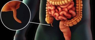
Foreign body in a child’s esophagus - main symptoms and risks
A foreign object swallowed by a child may become stuck in the esophagus. This case is very dangerous due to the increased sensitivity of the esophagus and its vulnerability.
The esophagus is equipped with muscle groups in which spasm may occur due to irritation of these muscles by the edges of a foreign object, which can cause complications.
Therefore, you should carefully monitor the symptoms that can alert you when monitoring your child and his well-being:
- When a child swallows, he experiences pain that is localized in the chest area, behind the sternum.
- He experiences discomfort when swallowing saliva, and solid food will not be swallowed at all.
Dangerous symptoms include nausea, frequent vomiting, and coughing. If such symptoms occur, the child must be examined urgently!
If these symptoms appear, delay threatens esophageal perforation and further bleeding. Food can get inside the chest, which will threaten the health and life of the baby.
Signs that a child has swallowed a foreign object
If a child managed to swallow a small object, and the parents did not notice this and did nothing, after some time certain signs may appear:
- Copious secretion of saliva.
- Sharp cough and difficulty breathing.
- Temperature increase.
- Bloating and severe abdominal pain.
- The appearance of bloody discharge in the stool.
- Nausea or vomiting.
- Chest pain.
If a child begins to have a sharp cough, he begins to choke and turns pale, then most likely a foreign object has entered the respiratory tract; you need to take the baby to the hospital as quickly as possible.
First aid for a child if he has swallowed a foreign body - what to do in such cases
The material of the object found in the body, its shape and size will influence the child’s behavior and the symptoms that manifest themselves in the current situation.
The baby must be kept in a state of physical rest if there is a foreign body inside it.
If a foreign object gets into the baby’s digestive tract, you need to urgently call an ambulance so that an examination can be carried out in the hospital.
Before the ambulance team arrives, you should not take out the object yourself or push it further - this can harm the child. Do not give your baby food or drink until examined by a doctor.
Usually the situation ends favorably, although there may be dangers. If the baby is calm when a foreign object is inside the body, then most likely it has already moved into the stomach and can leave the body on its own.
To check this, you need to wait one or two days from the moment of this event - and examine the children's feces. The swallowed object usually comes out within four days. But, if it does not leave the body, then an urgent fluoroscopy in a medical institution.
When you are sure that the swallowed object is dangerous, call emergency services !
What is not recommended to do?
- Parents make a mistake when they give their child several enemas and use laxatives to quickly remove the object. A foreign body entering the digestive system causes stress in it, and attempts to speed up peristalsis can cause traumatic damage to the digestive organs by the protruding edges of the object - it gets stuck in the intestine, leading to intestinal obstruction.
- If a foreign body is in a child’s body, you cannot use laxatives , induce vomiting, or give an enema . This may have a negative impact on your health.
- Do not attempt to remove it, so as not to further injure the baby.
- It is not recommended to shake out the object or try to push it further after chewing the crust of the bread.
- Do not feed or give water to the baby, because the object may be large and have sharp edges.
eating bran, fruits, and vegetables will help get rid of the object in the body .
If you are not sure that the baby has swallowed any object, then you must first observe the child’s condition and behavior for three days, and if alarming symptoms arise, consult a doctor immediately!
Clinical picture, help with getting into the throat
The clinical picture of a foreign body in a child’s throat leaves no doubt - it is often visible to the naked eye. If large objects get in, you can try to remove them. if these are small beads and parts, then you shouldn’t even try to provide help in this way.
Foreign bodies in the pharynx are quite common and enter it mainly with food.
The most common objects removed from the throats of children are fish and chicken bones, seeds and husks from them, and various “inedible” little things. The clinical picture is so pronounced that there is usually no doubt: a sensation of a foreign object and a stabbing pain in the throat, aggravated by swallowing, a paroxysmal cough and gagging, with the help of which the body seeks to remove the foreign body. Increasing pain when swallowing forces the child to stop swallowing saliva, and the baby leaks it from the mouth, and the older child spits it out.
These signs indicate the presence of a foreign body in the pharynx or tonsil tissue, and parents have no choice but to consult an otolaryngologist. Removing a foreign body from the pharynx is not difficult for a specialist, and after a few seconds your child is cheerful and healthy again.
The popularly recommended method is to swallow the dry crust - sometimes it can help, but it can drive the sharp bone deeper and make it more difficult to remove. Before contacting a doctor, you can try to remove a foreign body located on the surface by gargling. But this procedure can be performed by a schoolchild, but it is not suitable for children.
Medical care for a foreign body in a child’s digestive system
At the medical clinic, the child will be examined by experienced pediatricians and surgeons, and if necessary, an x-ray, additional ultrasound or endoscopic examination will be prescribed.
Based on the results of these studies, it is possible to establish an accurate diagnosis and determine the location of the foreign body.
- To remove it from the body, a laxative .
- If a foreign body that has entered the child’s body moves with difficulty, then the attending physician will prescribe an endoscopy .
- If endoscopy is not practical, surgery or laparoscopy , but this is too traumatic for the baby and often causes complications.
Prevention of foreign bodies entering the child’s digestive organs
If you did not see how the baby swallowed any object, but only scattered small objects and held them in his mouth, then you need to carefully inspect the place for playing - maybe the object rolled under the sofa or under the closet.
Babies need constant attention!
When they begin to actively crawl and then walk, they open the drawers and look into them, reaching for the shelves.
Parents need to remember that knowledge of the world occurs through the mouth and hands. The baby is able to put some small object in his mouth to find out its taste, and then accidentally inhale or swallow it.
A foreign body can pose a threat to the health and life of the baby. If parents are careless and do not look after the child very carefully, such cases happen often.
Therefore, adults should know what symptoms this manifests, what threat it poses, and what needs to be done.
To avoid getting into such situations...
- You cannot leave a child alone , without the supervision of adult relatives.
- The baby's play area must be cleared of small objects that are dangerous.
- Do not allow your child to take small objects to play with.
- Adult relatives need to carefully select children's toys, which need to be sorted by the size of the parts - and based on the age of the baby.
- When you need to leave the room, check the toys and their condition, or take your baby with you. Let the playing baby be in your visibility zone, under constant and careful control!
Rate
—
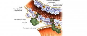
Classification of foreign bodies of the stomach
By size, foreign bodies can be:
- small - <1 cm in diameter or up to 5 cm in length;
- medium - up to 2 cm in diameter or up to 10 cm in length;
- large - > 2 cm in diameter or > 10 cm in length.
By form:
- rounded;
- piercing;
- cutting;
- cylindrical;
- irregular shape, etc.
By origin, foreign bodies can be organic and inorganic. Among organic foreign bodies, trichobezoars occupy a special place - a consequence of the habit of sucking strands of hair or tearing out and swallowing one's own hair. Phytobezoars are also common - rounded foreign bodies made of indigestible plant fiber.