Esophageal atresia is considered a severe congenital defect, incompatible with life without emergency surgical intervention.
With this defect, the esophagus becomes infected at a distance of about 12 cm from the oral cavity. The pathology usually forms at the level of the tracheal bifurcation. With chromosomal abnormalities, esophageal atresia in newborns is observed in 5% of cases. According to statistics, 2-3 babies and their mothers per 10,000 births have to face the problem and find out what esophageal atresia is. Up to 30-40% of children with this pathology are born premature and with developmental delays.
Reasons for the formation of a defect
The appearance of atresia occurs through disruption of the formation of the esophagus during intrauterine development of the fetus. The trachea and esophagus grow from the foregut and are initially united; their separation occurs at the end of the 4th week of fetal development and ends towards the beginning of the fourth month of pregnancy. Under the influence of specific conditions during this period of time, esophageal atresia occurs.
Factors that most often cause violations:
- X-ray exposure in the first 12 weeks of pregnancy;
- taking medications that are prohibited during pregnancy;
- environmental factors;
- influence of chemicals;
- bad habits;
- first pregnancy after 35 years.
Classification
The main forms of this defect in the development of the esophagus are:
- Esophageal atresia with inferior fistula (85-90% of cases). With this pathology, its upper section ends blindly, and the lower section is combined with the trachea.
- Isolated atresia (8%). It is characterized by closure of both parts of the esophagus.
- Isolated esophageal fistula (3-4%). The upper and lower sections of the esophagus are attached to the trachea in one place, and a fistula can be located on any of them. Its diameter is usually up to 4 mm.
- Atresia with superior fistula (0.5%). The lower part of the esophagus is closed, and the upper part is connected to the trachea.
- With two fistulas (1%). Both parts of the esophagus are united in different places with the trachea.
The absence of the esophagus or agenesis is diagnosed extremely rarely, and is often combined with other severe defects. Most newborns in this situation die.
Description
Esophageal atresia is a severe malformation. With pathology, the lower (proximal) section of the esophagus is completely closed, and the upper (distal) is connected to the trachea through the fistula canal. An anomaly occurs during intrauterine development in the first three months of pregnancy. Atresia is formed due to cellular degeneration. Often the speed and direction of growth of the trachea does not correspond to the same parameters of development of the esophagus, resulting in the formation of atresia. Predisposing factors from the pregnant woman's medical history are polyhydramnios and the threat of miscarriage in the early stages.
The frequency of recorded cases is 1 in 5000 newborns. If esophageal atresia is not detected in a timely manner and without surgical intervention, death occurs. The disease is often accompanied by defects in other organs and systems, such as the heart, gastrointestinal tract, genitourinary and musculoskeletal systems, and pathologies of the craniofacial part.
Atresia of the esophageal tract with lower tracheoesophageal fistula is one of the most common pathologies of this type.
Symptoms
Esophageal atresia in babies is detected at the first attempt at feeding: it does not work - the milk or formula flows out uncurdled. Additionally, the presence of a defect is indicated by:
- discharge of foamy, viscous mucus from the mouth and nose;
- cough;
- dyspnea;
- bluish skin;
- flatulence;
- vomit.
With this pathology, dehydration of the body quickly occurs, fever, and aspiration pneumonia develop. As a result, without timely surgical intervention, everything ends in death.
Treatment
Esophageal atresia is an indication for emergency surgery: the operation must be performed within the first 36 hours of the newborn's life. Preoperative preparation begins in the maternity hospital. It includes:
- giving the baby a position that prevents acidic gastric contents from entering the respiratory tract through the distal tracheoesophageal fistula;
- avoiding oral feeding;
- repeated and frequent aspiration of mucus from the respiratory tract;
- continuous inhalation of humidified oxygen;
- antibacterial therapy;
- correction of water and electrolyte disorders.
Diagnosis of pathology
Early diagnosis of the disease is very important, so examination of the esophagus is prescribed for all newborns with feeding problems. The diagnosis is confirmed by obstruction of the nasogastric tube into the stomach. He hits an obstacle. The presence of air in the stomach and intestines is noted if there is an esophageal fistula.
Prenatal diagnosis
Determination of a defect during pregnancy is possible using an ultrasound examination (gives 40-50% reliability). Warning signs during this procedure:
- polyhydramnios, which occurs due to the child’s inability to swallow amniotic fluid;
- problems with visualization of the gastrointestinal tract during several ultrasounds;
- discrepancy between the size of the stomach and the gestational age.
But similar deviations also occur with other defects.
Diagnosis of a newborn
The baby is examined using several methods:
- Intranasal sounding. It is carried out by inserting a catheter with a rounded end through the nose. With atresia, it, plunging up to 12 cm from the edge of the gums, rests on the blind process and comes back out.
- Elephant sample. A little air is injected into the catheter, lowered into the esophagus, using a syringe. With atresia, it begins to whistle, and noisily returns through the patient’s mouth and nose.
- Esophagoscopy with bronchoscopy: an endoscope is inserted into the esophagus to examine it from the inside.
- Contrast radiography. This method helps clarify the diagnosis in difficult situations. Most often it is done using iodolipol.
Differentiation of pathology is carried out:
- to separate other options from atresia with esophageal fistula;
- to exclude other gastrointestinal defects.
Prenatal diagnosis of laryngeal atresia
Ultrasound machine HM70A
Expert class at an affordable price.
Monocrystal sensors, full-screen display mode, elastography, 3D/4D in a laptop case. Flexible transformation into a stationary scanner with a cart.
Introduction
Congenital obstructive upper airway disease (COLD) is a rare life-threatening condition that results from a variety of fetal anomalies, including laryngeal and/or tracheal atresia, laryngeal cysts, and oropharyngeal or neck tumors. The most common cause of OPVDP is laryngeal atresia, but the incidence of this defect is not precisely known; the ratio of boys to girls is 1:1 [1].
There are 3 types of laryngeal atresia according to classification II Smith, AD Bain [2].
Type I - complete atresia of the larynx above and below the glottis as a result of the fusion of the arytenoid cartilages and the paired intrinsic laryngeal muscles in the midline.
Type II - laryngeal atresia under the glottis. In this case, the arytenoid cartilages, vocal cords and vestibule have a normal shape. Occlusion of the lumen of the larynx is achieved due to the presence of a dome-shaped cricoid cartilage.
Type III - laryngeal atresia at the level of the vocal cords due to the presence of a diaphragm consisting of connective and muscle tissue in the anterior sections and cartilaginous tissue in the posterior sections. The cartilaginous tissue of the diaphragm is formed by interconnected arytenoid cartilages and the corresponding vocal cords. The vestibule and cricoid cartilage have a normal shape.
Typical ultrasound findings in the prenatal diagnosis of laryngeal atresia are sharp enlargement and increased echogenicity of the lungs, dilatation of the trachea and bronchi, ascites and a flattened or inverted diaphragm. The lungs are affected bilaterally, and the contours of the lungs are smooth. First of all, attention is drawn to the visualization features of a four-chamber section of the heart [3]. An increase in the size of the lungs leads to compression of the heart and large veins, which complicates venous return to the heart, contributes to compression of the pulmonary vessels, the development of cardiovascular failure and, as a consequence, ascites [4]. Changes in the lungs and dilation of the bronchi and trachea are caused by the accumulation of fluid secreted by the alveolar epithelium and disruption of its outflow from the fetal respiratory tract. For the same reason, flattening and then inversion of the diaphragm occurs. These signs are clearly visible when studying parasagittal and coronal scans. Very often, laryngeal atresia is accompanied by polyhydramnios due to impaired swallowing due to compression of the esophagus by hyperplastic lungs [5]. However, cases of laryngeal atresia in combination with oligohydramnios in the fetus have been described. This is explained by insufficient replenishment of the amniotic fluid volume due to the pulmonary component [6].
The prognosis for laryngeal atresia is considered unfavorable. This is due both to the severity of the defect itself and to the presence of associated anomalies. If laryngeal atresia is detected, especially in combination with pathology of other organs, before the period of fetal viability is reached, the issue of termination of pregnancy should be discussed with the parents. When prolonging pregnancy, two tactics for providing emergency care to the newborn are possible: 1) rapid delivery and immediate tracheotomy; 2) carrying out the EXIT procedure (Ex Utero Intrapartum Treatment), which is currently widely used in foreign countries. EXIT is performed during a cesarean section after removal of the fetal head and neck from the uterine cavity and before cutting the umbilical cord and consists of performing intubation or tracheotomy. After this, the umbilical cord is cut and the newborn is removed [7]. Despite the fairly successful use of this procedure in the presence of tumors or cysts that contribute to the formation of OPVDP, since 1993, no more than 10 newborns with laryngeal atresia who survived after EXIT have been described in the literature. At the same time, the quality of life of these children remains reduced [8].
Material and methods
Patient P., 40 years old, underwent a routine screening test at 19-20 weeks of pregnancy at her place of residence, after which she was sent to the medical genetics department of MONIIAG with a diagnosis of fetal ascites. This pregnancy is from a second marriage, the sixth in a row. Both spouses are healthy and have no occupational hazards. Heredity is not burdened. There are five normal births from the first and second marriage, all children are healthy. The patient was registered with the antenatal clinic after 18 weeks of pregnancy; she did not undergo screening in the first trimester. The pregnancy proceeded without complications.
The study was carried out using an ultrasound machine WS80A (Samsung Medison).
results
An ultrasound examination in the medical genetics department of MONIIAG revealed one living fetus. The gestational age was 20.3 weeks. No pathological changes in the placenta were detected. The size of the fetus was disproportionate and did not allow us to judge the gestational age. The fetal life span corresponded to a period of 22.3 weeks, the length of the fetus was 21.6 weeks, and the length of the femur was 18.4 weeks. The abdominal circumference was sharply increased due to pronounced ascites and corresponded to a period of 30.5 weeks.
Evaluation of the chest at the level of the four-chamber section of the heart revealed enlarged, hyperechoic lungs and a compressed heart that occupied less than 1/6 of the cross-section of the chest. An anechoic avascular round formation was detected in the tracheal area (Fig. 1).
Rice. 1.
Cross section of the chest at the level of the four-chamber section of the heart. Enlarged hyperechoic lungs of the fetus, compressed, reduced in size heart and trachea, defined as a round anechoic formation anterior to the spine.
Also noteworthy were pronounced ascites and inversion of the dome of the diaphragm (Fig. 2, 3).
Rice. 2.
Transverse scan. Severe ascites.
Rice. 3.
Parasagittal scan. Marked ascites and inversion of the dome of the diaphragm (arrows).
A thorough assessment of the lung tissue revealed a trachea dilated throughout its entire length (Fig. 4) and dilated bronchi (Fig. 5).
Rice. 4.
Parasagittal scan. Trachea dilated to the level of obstruction.
Rice. 5.
Transverse scan. Expanded bronchial tree.
Additionally, an abnormal position of the fetal feet was identified, which, apparently, was a consequence of oligohydramnios: the amniotic fluid index was 90 mm (
The diagnosis was made: “Pregnancy 20.3 weeks. MFPR of the fetus: laryngeal atresia, abnormal placement of the feet. Low water." Medical genetic counseling and prenatal consultation were conducted. At the request of the parents, the pregnancy was terminated and the diagnosis was confirmed at autopsy.
Discussion
It is believed that obstructive lesions of the upper respiratory tract can be diagnosed no earlier than 17 weeks, since before this period the respiratory bronchioles, alveolar ducts and alveoli have not yet been formed, which means that a sufficiently large amount of pulmonary secretion is not formed, causing the development of large hyperechoic lungs [1]. Nevertheless, a case of a diagnosis of laryngeal atresia as early as 15 weeks of pregnancy has been described [9].
The differential diagnosis of laryngeal atresia should first of all be carried out with cystic adenomatous lung malformation (CAPL) and pulmonary sequestration. A distinctive feature of APVDP is bilateral damage, dilated bronchi and trachea, and the absence of fluid movement in the trachea during respiratory movements of the fetus. In CAPRL, the lesion is usually unilateral and most often affects one of the lobes of the lung. Pulmonary sequestration is characterized by the presence of a feeding vessel originating from the aorta.
Laryngeal atresia may be associated with a number of other malformations (esophageal or duodenal atresia, horseshoe kidney, renal agenesis, malformations of the hands and feet) and minor malformations (persistent superior vena cava, single umbilical artery). The combination of laryngeal atresia with tracheoesophageal fistula (TEF) significantly complicates the diagnosis, since the echogenicity of the lungs may be normal or slightly increased. This is explained by the fact that with TPF, the fluid produced by the alveolar epithelium enters the esophagus and does not accumulate in the lungs, and therefore their size and echogenicity do not change. The table presents an algorithm for the differential diagnosis of isolated laryngeal atresia, proposed by ME Furness et al. [10].
Table
. Algorithm for differential diagnosis of isolated laryngeal atresia [10].
| Ultrasound sign | Isolated laryngeal atresia | Laryngeal atresia with TPF | Tracheal agenesis |
| Fluid in the trachea | Yes | — | — |
| Enlarged lungs | Yes | — | Yes |
| Echogenic lungs | Yes | Yes | Yes |
Chromosomal abnormalities in laryngeal atresia are extremely rare, and therefore, when an isolated abnormality is detected, invasive diagnostics and prenatal karyotyping are not advisable. Although associations of this defect with partial trisomy of chromosomes 9 and 16, deletion of the short arm of chromosome 5, and deletion of 22q11.2 have been described [11].
At the same time, the risk of combined non-chromosomal syndromes in fetuses with laryngeal atresia is considered high, which is of fundamental importance in medical genetic counseling of the family. The most common among them is Fraser syndrome, an autosomal recessive disease characterized by a combination of laryngeal atresia, cleft lip/palate, heart disease, syndactyly, microophthalmia, abnormal auricular structure, and bilateral renal agenesis (OMIM #219000) [7].
Additionally, laryngeal atresia may be part of such syndromes as Opitz - Frias, Di Georges, Marshall - Smith, Laurence - Moon - Bardet - Biedl, short ribs - polydactyly, type II, Pallister - Hall, Schinzel - Giedion, Goldenhar and Pfeiffer [ 1].
Thus, when identifying ultrasound signs of laryngeal atresia, a thorough assessment of the entire ultrasound anatomy of the fetus is necessary with an assessment of all organs and systems to exclude concomitant pathology. When ascites is detected in a fetus, a thorough assessment of the entire ultrasound anatomy of the fetus is also necessary, since this condition can not only be associated with the presence of obstetric complications of pregnancy (Rhesus conflict, infectious or viral diseases), but also be a consequence of congenital diseases or malformations. The range of congenital pathological conditions accompanied by ascites is extremely wide: laryngeal atresia, meconium peritonitis [12], large-sized CAPRL [13], some heart defects, cloacal dysgenesis [14], obstructive lesions of the urinary tract [15], agenesis of the ductus venosus, anomaly development of the portal system [16], tumors, anemia, metabolic diseases, etc. It is important to remember that some of the developmental defects, in particular laryngeal atresia, may be part of non-chromosomal syndromes, which further determines the tactics of adequate prenatal counseling and the reproductive prognosis for the family .
Literature
- Medvedev M.V. Prenatal echography. Differential diagnosis and prognosis. 3rd ed., additional, revised. M.: Real Time. 2012.
- Smith II, Bain AD Congenital atresia of the larynx. A report of nine cases // The Annals of Otology, Rhinology, and Laryngology. 1965. V. 74. P. 338-349.
- Aslan H., Ekiz A., Acar DK, Aydiner B., Kaya B., Sezer S. Prenatal diagnosis of congenital high airway obstruction syndrome (CHAOS). Five case report // Med Ultrason. 2015. V. 1. P. 115-118.
- Andreeva E.N., Bortnovskaya N.P., Krivolapov O.V., Nikolaeva Yu.Z., Volkov A.E., Mishin A.V., Podolsky A.L., Chuchvaga S.M. Prenatal diagnosis of rare congenital defects and syndromes. XXIV. Laryngeal atresia, type III // Prenatal diagnosis. 2008. T. 7(4). 283-286.
- Garg M. Case report: antenatal diagnosis of congenital high airway obstruction syndrome. Laryngeal atresia // Indian Journal of Radiology and Imaging. 2008. V. 18(4). P. 350-351.
- Yudina E.V. Prenatal ultrasound diagnosis of congenital and hereditary defects in the second half of pregnancy. Lungs // Prenatal echography / Ed. Medvedeva M.V. M.: Real Time, 2005. pp. 341-370.
- Paladini D., Volpe P. Ultrasound of congenital fetal anomalies. Differential diagnosis and prognostic indicators. 2nd edition. CRS Press. 2014. P. 233-267.
- Saadai P., Jelin EB, Nijagal A., Schecter SC, Hirose S., MacKenzie TC, Rand L., Goldstein R., Farrell J., Harrison M., Lee H. Long-term outcomes after fetal therapy for congenital high airway obstructive syndrome // J Pediatr Surg. 2012. V. 47(6). P. 1095-1100.
- Gilboa Y., Achiron R., Katorza E., Bronshtein M. Early sonographic diagnosis of congenital high-airway obstruction syndrome // Ultrasound Obstet Gynecol. 2009. V. 33(6). P. 731-733.
- Furness ME, Donnelley BW, Lipsett J. Larynx atresia. 1991-03-11-21 // www.thefetus.net
- Chaemsaithong P., Chansoon T., Chanrachakul B., Worawichawong S., Wongwaisayawan S., Promsonthi P. Prenatal Diagnosis and Pathology of Laryngeal Atresia in Congenital High Airway Obstruction Syndrome // Case Reports in Radiology. V. 2012.
- Tsai MH, Chu SM, Lien R., Huan HR, Luo CC Clinical manifestations in infants with symptom atic meconium peritonitis // Pediatr Neonatol. 2009. V. 50(2). P. 59-64.
- Suk HJ, Won HS, Lee EJ, Lee MY An unusual prenatal manifestation of a huge congenital cystic adenomatoid malformation with favorable perinatal outcome // Obstet Gynecol Sci. 2014. V. 57(1). P. 73-76.
- Chen CP, Liu YP, Chang TY, Tsai FJ, Chen CY, Wu PC, Chen TH, Wang W. Prenatal diagnosis of persistent cloaca with hydrometrocolpos and ascites by magnetic resonance imaging in one fetus of a dizygotic twin pregnancy // Taiwan J Obstet Gynecol. 2010. V. 49(3). P. 385-386.
- Guyot A., Rosenblatt J., Bidat L., Bensman A., Benifla JL, Jouannic JM Impact of fetal urinary ascites on serum beta 2 microglobuline in obstructive uropathies: a case report // J Gynecol Obstet Biol Reprod (Paris). 2010. V. 39(5). P. 418-421.
- Achiron R., Gindes L., Kivilevitch Z., Kuint J., Kidron D., Boyanover Y., Yakobson J., Heggesh J. Prenatal diagnosis of congenital agenesis of the fetal portal venous system // Ultrasound Obstet Gynecol. 2009. V. 34(6). P. 643-652.
Ultrasound machine HM70A
Expert class at an affordable price.
Monocrystal sensors, full-screen display mode, elastography, 3D/4D in a laptop case. Flexible transformation into a stationary scanner with a cart.
Treatment of the disease
Preoperative preparation
When a pathology is diagnosed, the child is transferred to the surgical department of the hospital. Preoperative preparation is prescribed, which consists of:
- fixing the baby in a special elevated position;
- feeding only through a tube;
- aspiration of the contents of the mouth and nasopharynx every half hour;
- prescribing antibiotics and infusion solutions to maintain the stability of the condition;
- the use of oxygen inhalation in case of hypoxia development.
Basic assistance begins after an accurate diagnosis and when the child weighs more than 2 kg.
Carrying out the operation
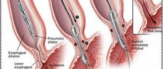
Treatment is exclusively surgical. If children are born full-term and do not have any malformations of the esophagus, then atresia with inferior fistula is performed by thoracotomy, dividing the process. If the diastasis of the esophageal segments is no more than 2 cm, then a direct anastomosis is performed. In more complex cases, the cervical esophagostomy method is used. This is also done with the non-fistula form of atresia.
If the operation has a significant risk, then it begins with the introduction of two gastrostomies into the stomach (to normalize intragastric pressure) and the duodenum (to facilitate feeding). Their outer ends are secured to the skin. After a few days, esophagoplasty is performed.
Postoperative period
In the absence of complications, removal of a special tube from the trachea (extubation) is allowed. If there are serious breathing problems, the child receives artificial ventilation. The baby's neck is firmly fixed for about a week to reduce the risk of sutures coming apart. Analgesics are prescribed to relieve pain. Antibiotic therapy is also carried out.
For 2 weeks after surgery, the sutures are carefully taken care of and ultraviolet irradiation is prescribed. The child is fed through a tube. After 5-7 days, the suture is checked for consistency, contrast radiography is performed, and based on its results, they begin to give food to the baby through the mouth. After 3 weeks, a control examination is done with an endoscope. Usually the site of the anastomosis remains unchanged, but if it narrows, bougienage is performed.
To prevent complications, up to a year the baby is recommended to be fed only crushed, homogeneous food. After the end of the recovery period and provided there are no other reasons that could cause health problems, children live a full life.
Rehabilitation
Treatment tactics for a child in the postoperative period are selected according to certain criteria:
- degree of prematurity;
- level of respiratory dysfunction;
- type of combined pathologies.
In the absence of complications and the above parameters, extubation is allowed, that is, removal of the breathing tube from the child’s trachea. If there is severe respiratory instability, the baby is connected to a mechanical ventilation device, which is turned off only when the ability to breathe independently is restored.
If there is weakness in the walls of the trachea, which is typical with a fistulous connection of the trachea and esophagus, the baby undergoes tracheopexy or tracheostomy.
In the postoperative period, the baby’s neck is securely fixed for 3-7 days in order to minimize the risk of tension on the anastomosis and suture divergence. To relieve pain, opioid analgesics are prescribed through an IV. On days 3-5 it is possible to switch to injections. Antibacterial therapy is performed with metronidazole in the amount of 15 mg/(kg*day). In the future, the appointment depends on the condition of the baby.
Possible complications
A variant of the norm is hoarseness, which can last up to a year. Appears due to damage to the laryngeal nerve during surgery.
Other possible complications in the postoperative period:
- pneumonia;
- failure of sutures;
- mediastinitis;
- laryngotracheomalacia;
- gastroesophageal reflux;
- difficulty swallowing food;
- anemia.
If a child experiences at least one of the above consequences, an esophagoscopy is prescribed.
Forecast
The prognosis for esophageal atresia depends on the type of pathology and the impact of conditions aggravating the treatment process. In the isolated form, the survival rate is about 90-100%, in the case of severe congenital defects and varying degrees of prematurity - 30-50%.
For about a year after treatment, the baby is under the supervision of pediatric doctors: a surgeon and a gastroenterologist.
Diagnostics
In the presence of the above-described clinical picture, diagnosis should be carried out in a short time, since the risk of deterioration in the child’s well-being is present even when symptoms are relieved.
In this case, diagnostic measures are divided into two categories - perinatal and after the birth of the child. The first group should include the ultrasound procedure. The presence of pathology will be indicated by the following:
- small size of the stomach or its complete absence on the image;
- polyhydramnios - this factor is due to the fact that the child does not swallow amniotic fluid due to intestinal obstruction.
To confirm pathology in a newborn child, the following diagnostic measures can be carried out:
- physical examination;
- checking intestinal patency using a special catheter - if esophageal atresia is present, the tube extends 8–10 centimeters, after which it rests or is tucked in and exits back through the mouth;
- esophagoscopy;
- bronchoscopy;
- X-ray of the esophagus with contrast agent.
Pathology examination
During the examination, additional consultation with a surgeon and endocrinologist may be necessary. Laboratory research methods are not carried out, as they do not have diagnostic value.
In some cases, differential diagnosis may be necessary to confirm or exclude the presence of the following pathological processes:
- esophageal stenosis;
- congenital pyloric stenosis;
- cleft larynx;
- esophagospasm.
If the diagnosis of esophageal atresia is confirmed, then urgent surgical intervention is required.
Prevention
Prevention is possible only during pregnancy:
- healthy lifestyle;
- absence of bad habits;
- avoiding contact with pesticides and x-ray radiation (especially in the first trimester of pregnancy);
- limiting the use of medications that may adversely affect the fetus;
- accommodation in an environmentally safe area.
Although esophageal atresia in children is a severe pathology of the gastrointestinal tract that can end the life of a child, if it is diagnosed in a timely manner and emergency surgery is performed, the chances of a full recovery are very high. And the child himself will subsequently be no different from his peers.
What is esophageal atresia
Esophageal atresia is a congenital pathology in which part of the esophagus is missing in a newborn, which leads to esophageal obstruction. The only treatment for this disease is surgery. It should be noted that this kind of pathological process occurs in both boys and girls. In the absence of early surgical intervention, this pathology leads to the death of the newborn.
- Etiology
- Classification
- Symptoms
- Diagnostics
- Treatment
- Complications
- Prevention
According to the International Classification of Diseases, Tenth Revision, this disease is designated as “esophageal atresia with tracheal-esophageal fistula.” ICD-10 code – Q 39.1.