Gastric papillitis - what is it? How to treat such a disease? Before answering these questions, it should be recalled that any neoplasms that appear on human skin (face, neck, limbs) or its mucous membranes clearly indicate activation of the activity of papillomaviruses. What is the danger of their presence in the body? Doctors say that if external papillomas are detected, there is a high probability of their occurrence on internal organs. Moreover, it is almost impossible to find out about their existence without a special examination.
What it is?
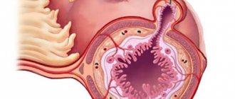
What is gastric papillitis? In medical practice, this term is used to refer to benign formations that arise on the mucous layer of the mentioned organ. This disease is caused by an inflammatory process that, for one reason or another, develops on the walls of the stomach.
Timely detection of such formations, as well as their careful treatment, guarantees the absence of serious consequences. After all, it is no secret that papilloma viruses can easily develop into cancer. That is why, after discovering gastric papillitis, doctors try to quickly determine the type of virus, which helps to learn about all the risks.
How dangerous is the disease?
Any benign formations on the mucous membranes are dangerous due to the risk of cells degenerating into malignant ones. If papillitis is not treated, there is a possibility of the disease progressing to stomach cancer.
In advanced cases, papillitis is characterized by the formation of multiple gastric papillomas. Inflammation of polyps is accompanied by the formation of erosions. If there are many lesions, the walls and mucous membrane of the stomach suffer, and acute pain and indigestion become constant companions of the disease.
The chronic form of papillitis is accompanied by anemia and negatively affects the functioning of all body systems. The development of chronic papillitis is due to the protracted course of the disease without taking the necessary therapeutic measures.
The appearance of blood in the stool indicates gastric bleeding. This condition is often diagnosed in the chronic form of papillitis and requires timely treatment, and in some cases, urgent hospitalization of the patient.
It is important to remember that papillomas are caused by the action of a virus that is very contagious. A person with gastric papillitis poses a threat to his sexual partner, since HPV is transmitted through sexual contact. There is also a high probability of infecting others if personal hygiene rules are not followed and someone else’s towels are used.
Types of formations
Gastric papillitis, which should be treated immediately after its detection, may vary. In medicine, it is usually divided into two groups:
- Single or, conversely, multiple growths that have peculiar legs and rise above the gastric mucosa.
- Weakly defined papillae, accumulating in small groups and resembling the convolutions of the brain in shape.
Typically, the diameter of such formations reaches 3 cm. They can be either pink or bright red.
Traditional methods of treatment
If papillomas appear in the stomach, treatment with folk remedies is possible only after examination and confirmation of the diagnosis. Before starting to use traditional medicine, you must consult your doctor.
To prepare a home remedy, you can use one of the recommendations given.
- Pour 5 g of dried celandine into 500 ml of boiling water and simmer over low heat for 10 minutes. Without straining, wrap in a terry towel and leave for 5 hours for the medicine to infuse. The infusion is taken in a large spoon 3-4 times a day, the course of treatment is 4 weeks.
- 5 g (small spoon) of a mixture of celandine and calendula is poured into two glasses of boiling water and left for 4 hours. The mixture is taken according to the same scheme as in the first recipe.
- Pour 2 large spoons of burdock rhizome with two glasses of hot water and boil for several minutes. The decoction is taken throughout the day instead of water. The permissible dosage is 3 glasses per day. The course of treatment is 6-8 weeks.
- The color of lilac will help relieve inflammation. Two tablespoons of flowers are poured with 6 cups of boiling water, then the medicine is infused for 6 hours. The infusion is taken half a glass three times a day. The course of treatment does not exceed a month. It is recommended to give preference to white flowers.
An infusion of lilac flowers will help with stomach papillomas
For stomach polyps, treatment is supplemented by taking propolis tincture with butter or water. Every day you need to drink a tablespoon of this remedy. It is recommended to include honey in the diet, as it has antiseptic properties and accelerates the healing of erosions.
Folk remedies should be used only after consultation with a specialist. Celandine is poisonous and can lead to poisoning. It is important to remember that home-made herbal medicines can cause an allergic reaction.
Forms of development
Gastric papillitis has such forms of development as:
- Erosive. This form is characterized by the presence of erosion directly on the tumor, as well as a strong inflammatory process.
- Chronic form. This disease usually occurs due to a constant irritant. It is characterized by long healing of existing ulcers and their severe bleeding.
There is also catarrhal papillitis of the stomach. Its distinctive feature is that benign formations arise only in a certain section of the main digestive organ (catarrhal).
Features of the therapeutic course
To begin treating papillitis, you need to see a doctor and get examined. The type of treatment is selected based on the information received regarding the severity of the pathology and its origin. As for benign education, the course of therapy consists of a strict diet, elimination of bad habits, proper rest and work regimen. As a preventative measure, it is necessary to undergo fibrogastroscopy twice a year.
If large papillomas are found, they must be removed through surgery. The complication occurs due to dysfunction of the cardiovascular system. Medium-sized neoplasms on knives are treated using an endoscope. The essence of the therapy is to remove them with a metal loop. This form of therapy helps to prevent organ rupture and also significantly reduce the risk of injury to the walls. The tissues heal quickly, and stomach deformation is prevented.
Numerous papillomas deserve special attention. They are removed using a special laser. This method is only suitable for flat tumors that do not have legs. The end of treatment involves the use of electrocoagulation to stop hemorrhages. Before the operation, appropriate preparation is required, consisting of a gentle diet, fractional meals, and no breakfast.
Papillitis cannot be ignored. It is important to start timely treatment. Otherwise, there is a risk of developing complications that are difficult to treat.
Symptoms of the disease
What signs indicate the development of catarrhal or erosive papillitis of the stomach? According to doctors, in the early stages, such a disease does not cause any serious symptoms that would force the patient to consult a specialist. A person usually attributes mild discomfort in the form of heartburn, leukoplakia, heaviness in the epigastric region, as well as lack of appetite to banal fatigue, consumption of stale or low-quality products, etc. And only when all these signs begin to actively intensify and become acute in nature, does the patient contact See a doctor for help.
Gastric papillitis has the following symptoms:
- bowel dysfunction;
- aching and nagging pain in the epigastric region, which often extends to the lumbar region or shoulder blades (usually after eating);
- putrid odor from the mouth;
- active salivation;
- frequent manifestations of gas formation;
- vomiting and nausea (arise due to hyperesthesia of stomach tissue);
- bleeding in the stomach (usually caused by large polyps).
Stenosing duodenal papillitis: etiology, diagnosis, treatment.
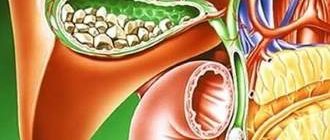
BDS stenosis is a benign disease caused by inflammatory changes and cicatricial narrowing of the papilla, which cause obstruction of the bile and pancreatic ducts and associated pathological processes in the bile ducts and pancreas. In clinical practice, the term “stenosing duodenal papillitis” means: stenosis of the papilla of Vater, stenosis of the duodenal papilla, stenosis of the terminal part of the common bile duct, stenotic odditis, fibrosis of the sphincter of Oddi, stenosis of the hepatopancreatic ampulla, that is, narrowing of the ampulla of the BDS or the hepatopancreatic sphincter ampulla, as well as the adjacent section of the common bile duct. BDS is often called the Oddi space (zone). Narrowing of the space of Oddi occurs primarily due to inflammatory and fibrosing processes. It is known that the structure of the BDS can be modified taking into account age characteristics. According to V.V. Pushkarsky (2004), with cholelithiasis in old and senile age, the atrophic-sclerotic form of chronic papillitis predominates (up to 54% of cases); at the age of up to 60 years, hyperplastic (adenomatous, adenomyomatous) changes in the BDS predominate.
The increased attention to acute and chronic inflammatory changes in the BDS is not accidental. According to A.I. Edemsky (2002), acute and chronic papillitis is observed in 100% of patients suffering from cholelithiasis, and in 89.6% of patients with recurrent pancreatitis. There are 3 forms of chronic pathological changes in the papilla: chronic adenomatous, adenomyomatous and atrophic-sclerotic chronic papillitis.
The BDS is located on the border of two (common bile duct and duodenum), and sometimes three (when the large pancreatic duct flows into the ampulla of the papilla) hollow systems. Pathogenic microflora, fluctuations in pressure and pH, stagnation in these two or three cavities contribute to the development of pathological changes in the BDS. Undoubtedly, the passage of dense structures, primarily the migration of stones along the common bile duct, also traumatizes it. The length of the BDS usually does not exceed 5–10 mm. Inside the papilla, in approximately 85% of cases, there is an extension of the common bile duct, which is designated as the ampulla of the papilla. The terminal part of the common bile duct adjacent to the papilla, with an average length of about 1 cm (0.6–3 cm), is located inside the wall of the duodenum and is called the intramural segment of the duct. Physiologically, this segment forms a single whole with the BDS. The cavity of the BDS, together with the terminal part of the common bile duct, is designated as the space of Oddi.
The locking apparatus of the BDS - the sphincter of Oddi - consists of: 1) the sphincter of the duodenal papilla itself, the so-called Westphal sphincter, which is a group of ring-shaped and longitudinal fibers reaching the apex of the duodenal papilla; when contracting, the Westphalian sphincter delimits the cavity of the papilla from the cavity of the duodenum; 2) the sphincter of the common bile duct - apparently the most powerful of this group of sphincters - the sphincter of Oddi, reaching a width of 8–12 mm; its proximal part often extends beyond the wall of the duodenum; during its contraction, it delimits the cavity of the common bile duct (and sometimes the pancreatic duct) from the cavity of the BDS; 3) the sphincter of the large pancreatic duct, usually poorly developed and sometimes completely absent. Stenosing duodenal papillitis affects not only the Westphal sphincter zone and the ampulla of the papilla, but also often the sphincter zone of the common bile duct, i.e. the entire zone of Oddi. Thus, stenotic duodenal papillitis is, to some extent, a collective concept that covers at least two pathological processes: 1) stenosis of the duct in the area of the ampulla of the ampulla; 2) stenosis of the terminal (mainly intramural) part of the common bile duct itself.
A significant part of long-term dyskinesias of the sphincter of Oddi essentially represents the initial stage of stenotic duodenal papillitis. With direct transduodenal endoscopy with thin (diameter 2.0–2.1 mm) probes, scar changes in the area of the space of Oddi are detected in many such patients. The close anatomical and topographic relationship of the BDS with the biliary system and pancreas, as well as the dependence of the function of the BDS on the state of the organs of the biliopancreaticoduodenal zone and the pathological processes developing in them, significantly influence the state of the BDS. This leads to the fact that specific symptoms characteristic of the disease itself are quite difficult to identify. For this reason, the pathology of BDS is often not diagnosed. Nevertheless, the main symptom in which the doctor should think about a possible pathological process in the BDS is biliary or pancreatic hypertension (the occurrence of jaundice or the pain syndrome characteristic of pancreatitis).
BDS diseases can be divided into primary and secondary. Primary diseases include pathological processes localized in the BDS itself: inflammatory diseases (papillitis), benign and malignant tumors. Secondary diseases of the BDS include stones of the ampulla of the BDS, stenosis of the BDS (as a consequence of cholelithiasis), as well as compression of the BDS due to a pathological process localized in the head of the pancreas with pancreatitis or a tumor. Secondary diseases of the BDS include dysfunction of the sphincter apparatus of the BDS that occurs against the background of duodenal ulcer and duodenostasis. If the pathological process in the BDS develops against the background of diseases of the biliary system, the clinical picture is manifested by symptoms characteristic of cholelithiasis. In cases where the pathological process in the BDS is the cause of the development of inflammation of the pancreas, this is accompanied by clinical signs of pancreatitis. The appearance of jaundice may indicate that the pathological process is localized in the BDS. In this case, changes in the color of feces (gray, discolored) and urine (beer-colored urine) are noted. Violation of the outflow of bile into the duodenum may be accompanied by an increase in the patient’s body temperature, which is associated with the development of acute cholangitis. Stenosing duodenal papillitis is a disease that is often asymptomatic and sometimes asymptomatic. Very often, the symptoms of narrowing of the BDS and the terminal part of the common bile duct are mistakenly associated with other pathological processes, primarily with manifestations of cholelithiasis itself (stone of the common bile duct, etc.). Perhaps, due to these circumstances and difficulties in recognition, a sometimes quite formidable disease did not attract the attention it deserved for a long time. Stenosing duodenal papillitis was described only at the end of the 19th century. as cicatricial stenosis of the papilla caused by an impacted stone. In 1926, D. Dell Vail and R. Donovan reported stenosing papillitis not associated with cholelithiasis, calling it scleroretractile odditis. As in Langebuch's time, stenotic duodenal papillitis continued to be considered a rare casuistic disease. Only in the 1950s and 60s did the situation change. The use of intravenous and operative cholangiography, manometry and radiometric studies allowed P. Mallet-Guy, J. Caroli, N. Hess and other researchers to identify the widespread prevalence of this disease, especially in cholelithiasis. Thus, W. Hess, out of 1220 cases of diseases of the gallbladder and biliary tract, noted stenosis of the BDS in 29%. With acalculous cholecystitis, stenotic duodenal papillitis was observed in 13%, with cholecystolithiasis - in 20%, with choledocholithiasis - in 50% of patients. In the last two decades, since the widespread use of endoscopic examinations, and in particular endoscopic papillosphincterotomy, the incidence and clinical significance of this disease have become abundantly clear. There is a need to clearly distinguish between stenotic and non-stenotic (catarrhal) duodenal papillitis.
The development of stenotic duodenal papillitis is most often associated with cholelithiasis, primarily with choledocholithiasis. Injury to the papilla during the passage of the stone, an active infectious process in the folds and valve apparatus of the ampulla cause further development of fibrous tissue and stenosis of various parts of the ampulla of the BDS or the part of the common bile duct immediately adjacent to it, i.e., the zone of Oddi.
With calculous cholecystitis and especially acalculous cholecystitis, the development of this disease is associated with a chronic infection that spreads through the lymphatic tract. P. Mallet-Guy suggested that in the pathogenesis of papillitis, an important role belongs to the following mechanisms: hypertension of the sphincter of Oddi, delayed evacuation of bile into the duodenum, activation of the infectious process in the area of the urinary tract, and the development of inflammatory fibrosis. Inflammatory-fibrosing processes in the BDS often occur in patients with parafateral diverticulum, some forms of duodenitis, and duodenal ulcer. In case of peptic ulcer with localization of the ulcer in the duodenum and partly with duodenitis, the peptic factor plays a certain role in the development of stenotic duodenal papillitis. When alkalization processes in the vertical part of the duodenum were disrupted, which was confirmed by the method of multichannel pH-metry, traumatization of the BDS with hydrochloric acid was detected. It is the peptic component that in many cases is the cause of pain in people suffering from stenotic duodenal papillitis, which explains the analgesic effect of antacids and H2-blockers. The injured mucous membrane of the abdominal cavity, including the ampoule, is subsequently easily subject to bacterial invasion, and an infectious-inflammatory process develops.
As indicated, stenotic duodenal papillitis in many cases is a secondary process, in which cholelithiasis is considered as the root cause of the disease. Primary stenotic papillitis, in which there are no traditional causes (cholelithiasis, parafateral diverticula, etc.), appears to be less common. According to J. Caroli, this development of the disease is observed in 2–8% of patients. In recent years, the frequency of primary forms of stenotic duodenal papillitis has increased to 12–20%. The histological picture of the primary forms of the disease is identical to the secondary ones. The etiology of primary stenoses remains unclear. Based on morphological characteristics, three main forms of stenosis of the abdominal joint can be distinguished:
- inflammatory-sclerotic, characterized by varying degrees of fibrosis; in the early stages - hypertrophy and degenerative changes in the muscle fibers of the valve apparatus of the BDS with the presence of round cell infiltrates, as well as fibrous tissue; in advanced cases, fibrous tissue is almost exclusively determined;
- fibrocystic form, in which, along with the phenomena of fibrosis, a large number of tiny cysts are detected, often representing sharply expanded pericanalicular glands, compressed by hypertrophied muscle fibers;
- adenomyomatous form, characterized by adenomatous hyperplasia of the pericanalicular glands, hypertrophy of smooth muscle fibers, proliferation of fibrous fibers (fibroadenomyomatosis), is often observed in older people.
Normally, the pressure in the common bile duct does not exceed 150 mm of water. Art. With stenotic duodenal papillitis, it increases to 180–220 mm of water. Art. and more. With a rapid increase in pressure to 280–320 mm water. Art. An attack of hepatic colic may develop. In the duodenum, normal pressure is up to 6–109 mm of water. Art. , under pathological conditions it can increase to 250–300 mm of water. Art. In the pancreatic ducts under conditions of secretory rest, the pressure is 96–370 mm of water. Art. At the height of secretin stimulation in the distal part of the main pancreatic duct, the pressure can reach 550–600 mm of water. Art. In recent years, special catheters with a diameter of 1.7 mm (for example, Wilson-Cook, USA) are used to measure pressure, inserted into the duodenal papilla through an endoscope. The obtained data is recorded in the form of various curves.
The clinical picture of the disease is determined by the degree of narrowing of the bile and pancreatic ducts, biliary and pancreatic hypertension, infection, and secondary damage to the liver and pancreas. It is still unclear why in patients with almost identical anatomical changes in the obdominus and the terminal part of the common bile duct, in some cases constant excruciating pain is observed daily, in others - only with errors in the diet, and in others - only minor episodic pain and heartburn are noted.
The most common symptom of stenotic duodenal papillitis is pain. Usually the pain is localized to the right and above the navel, sometimes in the epigastric region, especially in its right half. In a small proportion of patients, it migrates between the right hypochondrium and the epigastric region. Several types of pain can be distinguished: 1) duodenal type, when the patient is bothered by “hungry” or late pain, often quite long-lasting and monotonous; 2) sphincteric - short-term cramping, sometimes occurring with the first sips of food, especially when drinking cold fizzy drinks and fortified wines; 3) choledocheal itself in the form of severe monotonous pain that appears 30–45 minutes after eating, especially heavy or rich in fat. In severe cases, the pain is persistent, long-lasting, and is often accompanied by nausea and vomiting. The most pronounced pain syndrome is more often observed in patients with a relatively insignificant expansion of the common bile duct to 10–11 mm. In rare cases of sudden dilatation of the bile duct (up to 20 mm or more), the pain syndrome is much less pronounced. It has already been indicated that pain occurs and intensifies after a rich, fatty meal. Refractory fats (pork, lamb, beef lard, sturgeon fat) are dangerous in this regard. The combination of fat and dough is especially dangerous - pies, goose pies, pancakes with sour cream; It is they who often provoke a sharp exacerbation of the disease. Cold fizzy drinks are intolerable to most patients. In some patients, warm bread causes increased pain.
More than half of the patients experience various manifestations of dyspeptic syndrome: nausea, vomiting, bad breath and heartburn. In some patients, frequent vomiting is the most painful manifestation of the disease. After endoscopic papillosphincterotomy, the previously observed vomiting, as a rule, stops, while pain in the upper abdomen only decreases. Vomiting is considered a characteristic symptom of stenotic duodenal papillitis. In contrast to the latter, vomiting is very rarely observed in uncomplicated forms of BDS cancer. A common concomitant disease, cholangitis, is associated with complaints such as chills, malaise, and low-grade fever. Stunning chills with paroxysmal rise in temperature are less common than in persons with a common bile duct stone. Mild short-term jaundice is observed in a third of patients. Bright, long-term jaundice in the absence of concomitant diseases (common bile duct stone, parafateral diverticulum, etc.) is rare. Progressive weight loss is also rare. A slight weight loss of 2–3 kg is often noted. Palpation of the epigastric region in most patients gives an uncertain result. Only in 40–45% of patients it is possible to identify an area of local (usually low-intensity) pain 4–6 cm above the navel and 2–5 cm to the right of the midline, approximately corresponding to the Shoffar area. Peripheral blood in most patients is not changed; only 20–30%, during exacerbation of the disease, experience slight leukocytosis and, even more rarely, a moderate increase in erythrocyte sedimentation rate (ESR).
The addition of cholangitis, especially purulent one, entails the appearance of leukocytosis with a band shift and a significant increase in ESR. Similar changes are observed with the development of acute pancreatitis in patients with stenosing duodenal papillitis. Delayed movement of bile through the common bile duct and the major duodenal papilla is an important diagnostic sign of the disease. Two methods help in this regard. In case of short-term (0.5–3 days) disturbances in the outflow of bile, occurring after the consumption of significant doses of alcohol or errors in the diet and, probably associated with increased edema in the area of the ampulla of the papilla, a short-term but significant (5–20 times ) increased activity of glutamate dehydrogenase, aminotransferases and serum amylase. These changes are recorded especially clearly in the first 4–8 hours of increased pain. Such exacerbations of the disease often occur in the afternoon or at night. A simultaneous moderate increase in serum bilirubin levels with such short-term disturbances in the outflow of bile is rarely observed. With a single emergency blood draw performed in the first hours of a sharp increase in abdominal pain, an increase in enzyme activity is detected in 50–60% of those examined. In a double study of this type, severe hyperenzymemia is detected in 70–75% of those examined.
For long-term stable disorders of bile outflow, radionuclide methods are quite effective. When performing isotope hepatography in 50–60% of patients, a slowdown in the entry of radionuclide into the duodenum is detected. When conducting cholescintigraphy using acetic acid derivatives (drugs HIDA, IDA, etc.), a moderate slowdown in the entry of radionuclide into the duodenum is observed in 65–70% of those examined; in 7–10%, a paradoxical phenomenon is detected - accelerated entry of small portions of the drug into the intestine, apparently associated with the weakness of the sphincter system of the abdominal joint. In general, a repeated emergency study of enzyme activity at the onset of a sharp increase in pain and planned cholescintigraphy can identify in 80–90% of patients with stenosing duodenal papillitis symptoms of delayed flow of bile into the duodenum (in essence, symptoms of acute and chronic biliary hypertension).
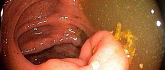
An important place in the diagnosis of the disease is occupied by the endoscopic method and combined endoscopic-radiological (x-ray) research methods. With catarrhal and stenotic papillitis, the papilla is often enlarged, reaching 1.5 cm. The mucous membrane is hyperemic and edematous. An inflammatory whitish coating is often visible at the top of the papilla. A characteristic sign of stenosing papillitis is flattening of the papilla. A flattened, wrinkled papilla is characteristic of a long-term process.
Intravenous cholegraphy data often play a major role in distinguishing between catarrhal and stenotic papillitis. With a stenotic process in 50–60% of patients, as a rule, a moderate (10–12 mm) expansion of the common bile duct is determined. The contrast agent is retained in the common bile duct. In some patients, it is also possible to detect a funnel-shaped narrowing of the terminal part of the common bile duct. Sometimes this narrowing looks peculiar - in the form of a writing pen, an inverted meniscus, etc. Occasionally, an expansion of the ampulla of the BDS is detected. Important examination results can be obtained during laparotomy. Operative cholangiography, often performed through the stump of the cystic duct, brings results close to those of endoscopic retrograde cholangiopancreatography (ERCP). It is often performed in two steps. First, 1/3 of the contrast volume is injected and an image is taken. It usually shows the stones of the common bile duct quite clearly. Then a second, larger portion of contrast is injected. The image shows tight filling of the common bile duct, its narrowing and delayed emptying are visible. Small stones in the duct are often invisible when filled tightly. Manometric examination of the common bile duct is mainly performed during surgery, although in recent years special probes adapted for manometry have been manufactured that are inserted transduodenally.
Bougienage of the OBD for diagnostic purposes during surgery finds some application. Normally, a probe with a diameter of 3 mm passes relatively freely through the Oddi zone into the duodenum. The possibility of inserting a probe of a smaller diameter (2 mm or only 1 mm) indicates stenosing duodenal papillitis. The bougienage procedure itself is quite traumatic. Sometimes it causes severe injury to the area of the duodenal papilla. Not all surgeons are willing to undertake this study.
ERCP plays an important role in recognizing stenotic duodenal papillitis. When performing catheterization of the BDS, difficulties often arise. Some of them, when a catheter is inserted into the common bile duct, may indicate stenosing duodenal papillitis. Moreover, when inserting a catheter, the endoscopist sometimes quite accurately determines the length of the narrowing area. Varying degrees of narrowing of the space of Oddi are observed in 70–90% of patients. The delay in emptying of contrast plays a certain diagnostic role. If the delay is more than 45 minutes, we can talk about stenosis or prolonged spasm of the Oddi area. Differential diagnosis. First of all, the issue of the possible presence of stones in the common bile duct should be resolved. The results of ultrasound and intravenous cholegraphy when identifying stones in large bile ducts are often not reliable enough, so in such a situation, ERCP is necessary. Occasionally, in cases of persistent jaundice, it is necessary to resort to percutaneous cholangiography. Among other diseases of the common bile duct and adjacent organs with relatively similar symptoms, one should keep in mind: 1) parafateral diverticulum; 2) indurative pancreatitis; 3) proximally located narrowing of the common bile duct, primarily in the area of confluence of the cystic duct; 4) cancer of the head of the pancreas; 5) cancer of the common bile duct; 6) primary sclerosing cholangitis.
Both primary and secondary sclerosing cholangitis are usually characterized by the fact that the extrahepatic and less intrahepatic ducts are affected, which is recorded by ERCP in the form of alternating narrowings and dilations of the common bile duct. In doubtful cases (and this is not so rare), ERCP has to be repeated, and the second targeted study, as a rule, gives a definite, almost unambiguous result. As you can see, ERCP in combination with ultrasound and CT plays a major role in differential diagnosis.
Clinical significance of stenotic duodenal papillitis. In most cases, this disease is, as it were, in the shadow of another pathology, which is considered as the main one. First of all, such a basic disease is choledocholithiasis, somewhat less often - cholecystolithiasis. It is not so rare that stenotic duodenal papillitis appears in the shadow of chronic acalculous cholecystitis and parafateral diverticulum. To a certain extent, these four different diseases are united by the low effectiveness of treatment when patients also have papillitis. Removal of stones from the gallbladder and common bile duct, rehabilitation of acalculous cholecystitis and parafateral diverticulum often turn out to be ineffective, i.e., they do not reduce the manifestations of clinical symptoms when papillitis is ignored therapeutically. In more than half of patients with postcholecystectomy syndrome, symptoms are mainly or largely associated with stenotic duodenal papillitis, which was either not recognized or was not eliminated during the period of cholecystectomy. Of the two diseases that the patient suffered from, cholecystectomy resolved the issue with only one of them. It is not surprising that after cholecystectomy, papillitis is often more severe than before the operation. Before a planned cholecystectomy, the danger of viewing papillitis and choledocholithiasis makes it necessary to perform duodenoscopy and intravenous cholegraphy.
Treatment. Patients with the most severe forms of stenotic duodenal papillitis, occurring with persistent pain, vomiting, repeated jaundice, and weight loss, are subject to endoscopic or surgical treatment. As a rule, they perform endoscopic papillosphincterotomy. Only in cases where the stenosis extends beyond the space of Oddi, transduodenal papillosphincterotomy with plasty is performed.
For milder forms, conservative therapy is prescribed, which includes diet No. 5, antacid therapy, and for particularly persistent pain, H2 blockers are used; anticholinergic therapy - atropine, platifillin, metacin, aeron, gastrocepin; antibacterial therapy.
Endoscopic papillosphincterotomy is especially indicated for dilation of the common bile duct. Its effectiveness varies. More often the pain syndrome decreases. Vomiting usually stops. Jaundice does not recur. In patients with a preserved gallbladder, acute cholecystitis develops relatively often (up to 10%) immediately after the intervention and during the first month. This pattern seems to emphasize the connection between pathological processes in the area of the obstructive system and the gallbladder.
During the period of remission, patients with papillitis are recommended a special diet, which can be regarded as maintenance therapy. Patients are also recommended to walk at least 5–6 km daily, do morning exercises without jumping and do abdominal exercises. Swimming is recommended. Nutrition should not be excessive, you should monitor the stability of body weight. Meals should be frequent: at least 4 times a day. It is advisable to enrich the diet with vegetables and vegetable oil. Refractory fats, cold fizzy drinks, hot seasonings, and fried foods are prohibited. Large meals at night are especially undesirable. If there is a slight increase in dull pain in the right hypochondrium, nausea, or heartburn, a course of treatment with choleretic drugs is recommended.
Thus, the pathology of BDS often leads to severe complications, often requiring emergency surgical treatment. At the same time, qualified morphological diagnosis of the pathological process of a given anatomical formation is extremely important, which subsequently plays a leading role in the choice of treatment tactics and the extent of surgical intervention.
Development risk
Which category of people most often develops a disease such as gastric papillitis? As is known, such a formation arises when infected with the corresponding virus. Due to the fact that the transmission rate of papillomavirus is very fast, its carrier can infect a healthy person even through normal contact.
Thus, people with reduced immunity, various diseases of the gastrointestinal tract, and also a certain age category are at risk. By the way, the presence of chronic diseases of the stomach causes fibrous changes in its tissues, which greatly complicates the diagnosis of such formations. People who smoke and drink are also at risk. The fetus in the womb of an infected woman can also become infected with papillomavirus.
Diagnostics
How is gastric papillitis (papillae) diagnosed? Reviews from patients with this diagnosis contain a lot of information on how to suspect the development of this disease. However, experts do not recommend self-diagnosis, much less self-treatment based on personal assumptions.
To identify such a disease, you should contact a general practitioner, who should subsequently refer the patient to a specialist - a gastroenterologist. After examination and questioning, the doctor recommends undergoing a full examination. As a rule, it includes procedures such as submitting biomaterial for laboratory analysis.
Also, to make an accurate diagnosis and identify papillitis, a specialist may recommend undergoing one of the following examinations:
- X-ray of the stomach;
- Ultrasound performed with a special composition, which is injected directly into the digestive organ;
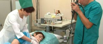
- fibrogastroscopy;
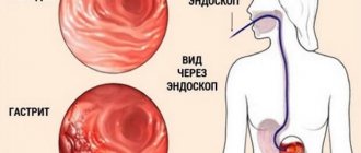
- endoscopy, that is, a detailed examination of the stomach from the inside, which is carried out using a special device that displays images on a monitor screen.
The use of one or more research methods will necessarily confirm or refute the presence of papillomas on the mucosa of the main digestive organ.
Danger of papillitis
Any benign neoplasm, especially papilloma, can, under certain conditions, develop into malignant. This is the main danger of the disease. This condition becomes especially threatening in the absence of clinical signs or late diagnosis.
In the acute erosive form, there is a high risk of gastric bleeding, followed by anemia, iron deficiency and oxygen starvation of the body. The human papillomavirus can be transmitted from mother to child, through sexual contact, through hygiene items, even by touch.
Gastric papillitis: how to treat?
Diagnosis and prevention of this disease should be carried out in a timely manner, since in the future such formations can cause the development of a cancerous tumor. As for treatment, it should only be prescribed by a gastroenterologist. This is done on the basis of the collected material and test results, which show not only the picture of the disease, but also help to build a strategy for further therapy. Typically, the initial stage of the disease in question is treated without surgery. In this case, the patient is strongly recommended to follow a number of rules, thanks to which there will be no further exacerbation:
- Drug therapy aimed at eliminating the inflammatory process and suppressing the activity of the virus in the body.
- Proper nutrition in fractional mode.
- Complete elimination of bad habits, including drinking alcohol.
- Constant rest, avoidance of stress and good sleep.
- Annual complete examination of the gastrointestinal tract.
If a systematic check reveals the growth of formations, then doctors recommend other methods, namely the removal of papillomas.
It should be noted that the choice of type of operation for this disease depends on the size of the formations and their shape:
- For medium-sized papilloma, which is elongated on a stalk, endoscopic intervention is used. Its essence lies in the fact that the formation is removed using a special metal loop. At the same time, the wounds resulting from the surgical procedure heal very quickly.
- The most difficult operation is performed on large papillomas. Very often, concomitant cardiovascular diseases interfere with its implementation. This operation involves the use of a scalpel, although it is also possible to use high-frequency current, which serves as an alternative to a laser.
- As for laser removal of benign growths, this procedure is applied to multiple formations. However, they must be flat and have no legs. To stop bleeding from the wound, electrocoagulation is used.
It is important to know that after any operation the patient must be observed by a doctor for at least two weeks. In this case, the patient is prescribed strict bed rest, any physical overload is prohibited and a certain diet is recommended.
Reasons for the formation of papillomas in the stomach
The main reason for the development of the pathological process is the penetration of papillomavirus into the body through tactile or sexual contact. But the disease may not manifest itself for a long time, and develops only when there are favorable conditions for the activity of the virus. The following factors contribute to this process:
- violations in the diet - consumption of significant amounts of protein foods, alcohol abuse;
- chronic infections of the body's digestive system;
- long-term drug therapy with antibiotics or other potent drugs;
- decreased immunity due to chronic stress or concomitant diseases of internal organs;
- metabolic disease.
Once in the body on fertile soil, the virus begins its destructive effect, which later manifests itself in clinical symptoms.
Preventive measures
How to prevent the development of gastric papillitis? Reviews from people with this disease are full of regrets that they never thought about preventing papillomavirus. After all, it is quite possible to avoid the development of this unpleasant disease with serious consequences.
To do this, you just need to follow a few simple rules:
- avoid unprotected sexual intercourse;
- when visiting bath complexes and swimming pools, be sure to use slippers or flip flops;
- remember to wash your hands regularly, especially after a seemingly ordinary handshake or visiting a public toilet;
- use only personal hygiene products and utensils;
- do not wear other people's things and underwear.
Symptoms and signs of papillitis
When papillitis occurs in the rectum, the clinical picture is similar to hemorrhoids and anal fissure; the patient complains of the following unpleasant symptoms:
- pain in the anus, which may occur when visiting the toilet or at rest;
- false urge to go to the toilet due to the sensation of a foreign object in the anus;
- bleeding from the anus;
- mucus secretion;
- swelling of the anal folds;
- itching and burning of the anus;
- leakage of rectal contents.
Since the symptoms of the disease do not directly indicate papillitis, differential diagnosis will be required, that is, an examination during which diseases that have a similar clinical picture will be excluded.