Intestinal metaplasia - what is it?
Intestinal metaplasia is a disease in which the tissues of the gastric mucosa are replaced by intestinal cells.
Intestinal metaplasia is a disease in which the tissues of the gastric mucosa are replaced by intestinal cells. The disease was first described by Professor Kupfer more than 100 years ago.
Elderly people are most often affected. According to statistics, 80% of those infected are diagnosed with chronic gastritis, as well as duodenal ulcer.
In a healthy state, the tissues that cover the walls of the stomach are constantly renewed. When damaged, cell division increases, which leads to increased migration and restoration of cellular renewal. In patients diagnosed with chronic gastritis, this process is disrupted, resulting in the inability of the gastric glands to perform their functions, which leads to metaplasia.
Causes
The reasons that cause intestinal metaplasia of the mucosal surface in the stomach have not been fully studied and there is no clear answer. But provoking factors have been identified, a combination of which or prolonged exposure to one of them can give impetus to the development of the disease. Among the common unfavorable factors, prolonged negative psycho-emotional stress, stress or depressive states have a negative impact.
Other causes of metaplasia:
- frequent consumption of alcohol, spicy, fatty, fried foods, which cause irritation of the inner surface of the stomach walls;
- chronic inflammation and ulceration of the epithelium;
- throwing intestinal contents into the stomach cavity;
- decreasing the acidity level of gastric juice;
- penetration of the bacterium Helicobacter pylori into the stomach.
During the examination, patients are found to have low acidity of gastric juice; in such an environment, the microflora necessary for normal gastric function dies. The bactericidal properties of the juice are reduced and favorable conditions are created for the development of pathogenic intestinal bacteria, including the dangerous microbe Helicobacter pylori. Its vital activity is accompanied by the release of enzymes, which, as a result of complex chemical transformations, form nitro compounds with carcinogenic properties.
What are the features of the disease
There are two types of gastric metaplasia: small intestinal (complete) and large intestinal (incomplete). Although now such a clear distinction is called into question. Small intestinal metaplasia is not a precancerous disease, while colonic metaplasia is considered a precancer. The reason is that small intestinal metaplasia is characterized by the restructuring of the organ to a different type of secretion while maintaining the function of the metaplastic cells. With colonic metaplasia, the processes of cell differentiation are disrupted and the process is similar to dysplasia. This indicates malignancy.
The danger is that there are no specific symptoms for this disease. It all depends on the cause of the disease and its manifestations. With chronic gastritis with high acidity, pain on an empty stomach, frequent heartburn, and feeling of hunger at night will be observed. With a peptic ulcer, pain of a certain localization will bother you in a hungry state, with exacerbations in spring and autumn. If the cause of metaplasia is the reflux of intestinal contents into the stomach, then this condition will manifest itself as a feeling of bitterness in the mouth, pain, and often vomiting. If the reasons are the reflux of stomach contents into the esophagus, then the symptoms will be heartburn and sour belching.
Very often, the disease as such does not manifest itself and symptoms such as heartburn, belching, and minor pain may not be given much importance, which is extremely dangerous. In any case, if a person experiences symptoms such as lack of appetite, nausea, vomiting, discomfort or pain in the epigastric region, it is necessary to consult a doctor for examination and diagnosis.
Features for different parts of the stomach: antrum, pyloric region
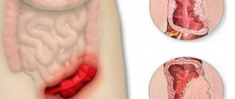
Metaplastic changes can occur in any part of the stomach, occupying only part of the mucosa or the entire thickness. The lesions are located in the membrane of the body, fundus or pyloric region, less often in the antrum.
The stomach is a rather complex organ
Important! Pathological changes in the gastric mucosa are considered a precancerous condition, so the disease is classified as dangerous. Studies conducted by many scientists have confirmed the presence of metaplasia in 94% of patients suffering from stomach cancer.
Observations over the past two decades have confirmed that tissue changes in intestinal metaplasia and intestinal type gastric cancer (Lauren classification) are completely identical.
Doctors believe that intestinal cancer occurs under the influence of external carcinogenic factors (substances that cause cancer). Most often occurs in the body of the stomach. Studies have shown that the disease develops in complex epidemiological areas.
Characteristic symptoms
The danger of the pathology, first of all, lies in the fact that it can be practically asymptomatic. At an early stage of development, with successful treatment, the disease can manifest itself only as mild discomfort, to which patients, as a rule, do not pay any attention. If metaplasia occurs due to the development of a chronic form of gastritis or gastric ulcer, then in addition to the symptoms of existing pathologies, new ones arise.
Main symptoms of metaplasia
The most common symptoms of gastric metaplasia include:
- sudden weight loss;
- deterioration of appetite or complete loss of it;
- painful sensations in the right or left side;
- a feeling of bitterness in the mouth, an attack of heartburn;
- frequent attacks of hunger;
- severe burning sensation in the stomach area;
- attacks of nausea and vomiting.
According to statistics, the symptoms of the disease appear mildly and irregularly, which significantly complicates the diagnostic process . Incorrect diagnosis of pathology ultimately leads to its serious progress. Therefore, at the first suspicious symptoms, you should immediately seek help from a doctor.
Stomach cancer
Gastric metaplasia: what is this pathology, causes and treatment
Gastric metaplasia is a change in the structure of the tissues of the gastric mucosa. Under the influence of unfavorable factors, they become similar to the membranes of the small and large intestines, due to which the basic functions of the digestive tract are disrupted.
This is not a separate disease, but a very alarming symptom of problems in the body. In addition, with progressive metaplasia, cells begin to degenerate, leading to cancer and death.
Many factors lead to metaplasia, the main ones being regular dietary errors, bad habits and systemic use of medications. Under this influence, the cells of the gastric mucosa cease to perform their function, and an inflammatory process develops with a further change in the structure of the lining.
- Small intestinal complete metaplasia is most often caused by the presence of chronic gastritis and leads to the formation of foci of changes. A characteristic sign of complete metaplasia is the detection of Paneth nuclei (cells) in the epithelium of the gastric mucosa. The altered tissues not only form the walls of the small intestine, but also begin to function in the same way.
- Colonic metaplasia is even more dangerous. In this pathology, the epithelium of the stomach consists almost entirely of cells of the large intestine. The normal process of formation of the gastric glands, as well as its digestive function, is disrupted. This condition is a precancerous threat, because in more than 94% of patients with stomach tumors the process began this way.
The development of such changes occurs at different speeds. It is believed that older people (especially those over 70 years of age) are prone to metaplasia, but the disease often occurs in younger patients. The degree is determined according to the volume of tissue implanted and the area of the lesion.
There are three types of changes:
- Mild metaplasia, in which destruction of the digestive tract does not exceed 5%.
- The average degree characterizes changes of up to 20%.
- Severe form, with a degree of damage over 25%.
Kinds
Classification by type can be defined as small intestinal (complete) and large intestinal (incomplete). Both conditions are quite dangerous, caused by various reasons with mandatory medical correction.
- Small intestinal complete metaplasia is most often caused by the presence of chronic gastritis and leads to the formation of foci of changes. A characteristic sign of complete metaplasia is the detection of Paneth nuclei (cells) in the epithelium of the gastric mucosa. The altered tissues not only form the walls of the small intestine, but also begin to function in the same way.
- Colonic metaplasia is even more dangerous. In this pathology, the epithelium of the stomach consists almost entirely of cells of the large intestine. The normal process of formation of the gastric glands, as well as its digestive function, is disrupted. This condition is a precancerous threat, because in more than 94% of patients with stomach tumors the process began this way.
Main varieties
Small intestinal complete metaplasia is most often caused by the presence of chronic gastritis and leads to the formation of foci of changes.
Metaplasia also has a gradation according to the types of changes in tissues. It is carried out at the location of the lesions and comes in the following types.
Classification by types of lesions:
Focal and diffuse forms of metaplasia most often occur in the antrum of the stomach.
Complete metaplasia
Complete metaplasia is characterized by the presence of all the cells characteristic of the small intestine, which usually line regular tubular formations. Goblet cells, as a rule, do not lie in a continuous layer, but are interspersed, as in the small intestine, with bordered absorptive enterocytes that do not secrete mucus. The deep sections of the pits are identical to the intestinal crypts. They are lined with borderless basophilic epithelium and contain Paneth cells. The presence of Paneth cells is the most important sign of complete intestinal metaplasia.
Incomplete metaplasia
In incomplete (colic) metaplasia, goblet cells are located among tall prismatic cells resembling colonocytes. Paneth cells are not detected. Nuclear polymorphism is expressed in the epithelium, and nuclear-cytoplasmic ratios are significantly increased.
With incomplete metaplasia, in contrast to complete metaplasia, the superficial parts of the glands are almost no different from the deep ones, which indicates a violation of maturation and differentiation. Incomplete metaplasia is clearly distinguished from complete metaplasia by its histochemical characteristics. In case of incomplete intestinal metaplasia, special staining reveals sialomucins and sulfamucins, which are absent in complete metaplasia.
Most often in chronic gastritis, complete metaplasia occurs; incomplete metaplasia is found in only 11% of patients with all benign diseases of the stomach. At the same time, with stomach cancer it is observed in 94% of patients. This allows us to classify it as a precancerous change and requires an indispensable differentiated assessment of intestinal metaplasia.
Volgograd
Atrophy of the mucous membrane is characterized by a decrease in the number of normal glands. This definition is reasonable and simple, and it would seem that, based on it, it would be easy to diagnose atrophy, but this is far from the case, especially in the antrum.
This is due, first of all, to the lack of understanding of the normal antrum.
In addition, diffuse inflammatory infiltration can push the glands apart, resulting in the impression of atrophy, since there are actually fewer glands in the field of view, although their absolute number remains the same.
Lymphoid follicles, often found in Helicobacter gastritis, located in place of the glands, can also imitate atrophy. True, such pictures are unlikely to confuse an experienced pathologist, but from a “normal” point of view, one can assume that since the number of glands in the specimen is reduced, this is atrophy.
In Helicobacter pylori gastritis, it is optimally possible to distinguish true atrophy from false atrophy on the basis of dynamic observation after successful antimicrobial therapy. If the inflammatory infiltration has disappeared or significantly decreased and the glands have become closer to each other, the diagnosis of atrophy is removed. If at the site of infiltration the connective tissue layers and glands are still moved apart, we can talk about true atrophy. It should, of course, be borne in mind that a decrease in lymphoplasmacytic infiltration can be expected no earlier than after several months.
It is necessary to distinguish true from false atrophy not only to ensure the “purity of the diagnosis”, but also based on practical significance. Only true atrophy can be considered a precancerous change. In addition, the lack of a clear definition of the term atrophy of the gastric mucosa appears to be associated with occasional reports of the reverse development of atrophy after successful treatment of Helicobacter pylori infection and the development of atrophic fundic gastritis with long-term use of proton pump blockers. Since the term "atrophic gastritis" is associated with the risk of stomach cancer, it may seem that the use of powerful antisecretory drugs can lead to cancer.
Regardless of the mechanisms of damage, the gastric mucosa can regenerate and restore its original structure or undergo adaptive regenerative changes. If damaged glands cannot regenerate, their place is taken by the so-called extracellular matrix or fibroblasts. Such patterns of irreversible loss of functional structures represent true atrophy.
Another way of development of atrophy is the formation of specialized metaplastic epithelial cells in place.
Based on these considerations, R. Genta (1997) proposed a “precise definition” of atrophy. Atrophy should be considered the irreversible loss of gastric glands with their replacement by metaplastic epithelium or fibrous tissue. If these signs are absent, but there are few biopsies against the background of inflammation of the glands, the presence or absence of atrophy can be judged by the results of a repeat biopsy and not earlier than after 6 months. after eradication of H. pylori or discontinuation of NSAIDs.
If changes are detected in the preparations that suggest atrophy, one should act in the same way as when detecting dysplasia.
Intestinal metaplasia.
Intestinal metaplasia was described by Kupfer more than 100 years ago, but is still actively studied, mainly because of its possible connection with gastric cancer.
As the name indicates, we are talking about replacing the gastric epithelium with intestinal epithelium.
This type of metaplasia occurs quite often. In old age, it is also found in practically healthy people, but especially often in chronic gastritis.
With atrophic gastritis and stomach cancer, it is observed in almost 100%, with stomach ulcers - in 81-100%, with duodenal ulcers - in 47-54%.
Intestinal metaplasia is diagnosed not only by histological examination, but also macroscopically, by staining the mucous membrane with methylene blue, which selectively binds to the intestinal epithelium.
This technique can be applied not only to the resected stomach, but also used during endoscopic examination.
In this case, one can judge the volume of metaplasia:
- weak metaplasia occupies up to 5% of the surface of the stomach,
- moderate up to 20%,
- and pronounced - over 20%.
Until relatively recently, it was assumed that with intestinal metaplasia, the gastric epithelium is replaced by an epithelium similar to the epithelium of the small intestine. In this regard, there was a synonym for intestinal metaplasia - enterolization.
Currently, it is customary to distinguish two types of metaplasia:
- full, resembling a small intestine,
- incomplete - fat.
These types differ not in volume (complete or incomplete replacement of the mucous membrane in area and depth), but in the ratio of cellular elements and some of their histochemical properties.
Complete metaplasia (synonym: mature, small intestinal, type 1 metaplasia).
Complete metaplasia is characterized by the presence of all the cells characteristic of the small intestine, which usually line regular tubular formations.
The presence of Paneth cells is the most important feature of complete intestinal metaplasia.
Complete intestinal metaplasia is similar to the small intestine not only structurally, but also functionally and morphologically.
Incomplete metaplasia (synonyms: immature, colonic, type 2 metaplasia).
In incomplete metaplasia, goblet cells are located among tall prismatic cells resembling colonocytes. Paneth cells are not detected.
Most often in chronic gastritis, complete metaplasia occurs.
Incomplete metaplasia is found in only 11% of patients with all benign diseases of the stomach.
At the same time, with stomach cancer it is observed in 94% of patients. This allows us to classify it as a precancerous change and requires an indispensable differentiated assessment of intestinal metaplasia. Therefore, the morphologist’s conclusion cannot say: “atrophic gastritis with intestinal metaplasia.” You must also specify its type.
The fact that complete intestinal metaplasia is much more common than incomplete intestinal metaplasia, and that they are often combined, suggests that incomplete metaplasia develops from complete metaplasia.
There may be two types of combination of these types of intestinal metaplasia. More often they are visible in adjacent areas of the mucous membrane, less often - within the same gland.
Pyloric metaplasia.
Pyloric metaplasia is characterized by the formation in place of the main mucous glands, reminiscent of pyloric glands.
Such glands were described at the beginning of the century and were called pseudopyloric glands of Sterk.
Pyloric metaplasia can be:
- diffuse,
- focal.
With a diffuse zone of the pyloric glands, the zone of the pyloric glands shifts in the oral direction to the territory of the fundus of the stomach.
Focal metaplasia is manifested by the replacement of individual fundic glands against the background of inflammation and impaired cellular renewal.
By analogy with intestinal metaplasia (L.I. Aruin et al. (1986) and M. Takematsu (1988)), two types of pyloric metaplasia are distinguished:
- full
- and incomplete.
Ciliated metaplasia.
Ciliated cells are not found in the digestive tract, so their appearance may be associated with metaplasia.
It has been noted that ciliated metaplasia occurs in gastric adenocarcinomas, in which ciliated cells are found, however, it is difficult to talk about its precancerous potential, since it has always been combined with intestinal metaplasia.
Pancreatic metaplasia.
The term “pancreatic metaplasia” was proposed relatively recently, in 1993 (C. Doglioni et al).
We were talking about the appearance in the gastric mucosa of cells with acidophilic fine-grained cytoplasm in the apical part and with basophilic cytoplasm at the opposite pole.
Pancreatic metaplasia was described: in the intrahepatic bile ducts of humans and experimental animals, in chronic cholecystitis, in Meckel's diverticulum, at the same time, pyloric metaplasia was found in the pancreas.
Dysplasia with gastritis
Dysplasia, which should also be considered as precancerous changes, is divided into two degrees:
- low-grade dysplasia is characterized by elongation of pits, increased diameter and hyperchromatosis of nuclei, increased nuclear-cytoplasmic ratios, and a predominance of diploid cells;
- with high-grade dysplasia, the mucous membrane thickens and foci of dysplasia can rise above the surface of the mucous membrane. Cellular atypia with anisokaryosis and nuclear hyperchromatosis is observed. The average DNA content and the number of cells in the DNA synthesis phase are sharply increased. Cellular anaplasia can be so severe that differential diagnosis from carcinoma becomes very difficult. The most important feature of severe dysplasia is p53 expression.
A. Kalinin, etc.
“Metaplasia with gastritis” and other articles from the section
Intestinal metaplasia, its types and the risks of developing benign neoplasms have not been fully studied. In their work, doctors base their work on disorders of cellular metabolism that provoke intestinal-type development.
Causes
Despite numerous studies in this area, the mechanism of occurrence of such pathologies is not fully understood.
It is believed that the root cause of metaplasia is the activity of Helicobacter pylori, which also causes some types of gastritis and ulcers. In some cases it was not detected, so it is very difficult to establish a direct relationship.
Other causes of metaplasia:
In addition, the disease can develop due to intestinal dysfunction, when duodenal and gastric contents are refluxed into the esophagus. The normal microflora of the stomach is destroyed, and pathological processes occur in the upper layers of epithelial tissue.
Types of pathology
In medicine, there are two types of gastric metaplasia: incomplete colonic and complete small intestinal. Taking into account the area of modifications and the degree of distribution of the lesion, the following forms can be distinguished: mild (5% of lesions), moderate (20%), severe (more than 20%). As for the severity of atrophy of the glandular layer, there are such varieties as: complete (C), intermediate (B), minor (A).
Small intestinal metaplasia, unlike the large intestinal form, should not be classified as a precancerous disease. This is due to the fact that small intestinal metaplasia of the stomach is characterized by the transformation of one organ to another form of secretion, but the functions of the cells are preserved. Colonic metaplasia is accompanied by a disturbance in the processes of cell differentiation, therefore these transformations have all the signs of dysplasia, which indicates malignancy.
According to the nature of the lesion, ciliated, antral and pancreatic forms are distinguished. Another interesting thing is that you can read about polyps of the antrum of the stomach in an article on the topic. The danger of the pathology lies in the fact that there are no specific signs. The disease can be detected based on the manifestations of the underlying disease that caused pathological processes in the gastrointestinal tract.
With gastritis with high acidity, painful sensations on an empty stomach, hunger at night and constant heartburn predominate. As for peptic ulcers, the pain is localized in the stomach on an empty stomach, exacerbations occur in spring and autumn. If the cause of metaplasia is a reflux disease, then the symptoms will be as follows: vomiting, pain, bitterness in the mouth, heartburn, belching.
Main symptoms
The situation is worsened by the fact that metaplasia usually develops against the background of chronic gastritis or ulcers, so patients mistake the signs for symptoms of existing diseases.
One of the main threats of this disease is its practically asymptomatic course. In the early stages, when treatment is particularly successful, the disease manifests itself as minor discomfort, which usually no one pays attention to.
The situation is worsened by the fact that metaplasia usually develops against the background of chronic gastritis or ulcers, so patients mistake the signs for symptoms of existing diseases.
The main signs of gastric metaplasia:
In most patients, symptoms appeared irregularly and without great intensity. This significantly complicates the diagnostic task and also leads to significant progress of the disease.
Known types of metaplasia
Today, there are two main types of gastric damage:
- Complete or small intestinal metaplasia. It occurs much more often than the second type of pathology. The disease is similar to dysplasia (improper development of organs or tissues). It is characterized by the fact that the gastric epithelium is replaced by cells of the small intestine, as a result of which it acquires intestinal functional properties. Complete metaplasia has the most favorable treatment outcome, since it is considered the initial stage of the disease and biologically cannot be a precancerous condition;
- Pathology of the second type (colon) is called incomplete or immature. This is a dangerous disease that is a precursor to a malignant process. Cells from both the large and small intestines are found in the stomach. Most often, oncology is diagnosed in patients whose epithelium is affected to a large extent, rather than by small lesions. Precancerous changes require early initiation of therapy, since a person who does not receive treatment will face death.
There are divisions of types of pathology based on other criteria. So, metaplasia happens:
Diseases of the gastrointestinal tract
- Focal. The pathology is characterized by individual small areas of damage formed as a result of damage to the gastric mucosa. The main location is the antrum of the stomach (the place where eaten food ends up). During the process of destruction, the affected cells lose their ability to renew and atrophy over time;
- Diffuse. Replacement progresses (also in the antrum of the digestive organ) without the death of tissue-forming cells, and the deep epithelial layers are not affected;
- Pancreatic. A distinctive feature of the pathological process is the replacement of healthy matter with cells that have acidophilic endings. Typically, this pathology is observed against the background of chronic gastritis;
- Eyelash. The replacement of epithelial cells in this form of the disease resembles microscopic cilia (similar to cancer metastases). Such destructive development is considered a harbinger of malignant degeneration, for example, gastric adenocarcinoma.
Intestinal metaplasia is also classified according to the extent of the epithelial lesions. It happens:
- weak (about 5% of general lesions of gastric tissue);
- moderate (up to 20% of altered epithelium);
- pronounced (more than 20% replacement of stomach cells with intestinal ones).
According to the degree of gland atrophy, metaplasia is divided into:
- minor (A);
- intermediate (B);
- full (C).
Diagnostic methods
The main study that can detect or suspect pathological changes in the gastric epithelium is endoscopy.
What can show metaplasia:
When contacting a gastroenterologist, special examinations may be prescribed, and the endoscopy procedure will need to be completed several times. Detection of the root cause of the disease is important in further therapy.
The diagnostic standard is considered to be histological examination, which reveals the form of the disease. During the procedure, small pieces of tissue taken from the human body are examined. The method of collecting cells or epithelium is called a biopsy. This is a mandatory way to confirm the diagnosis if the formation of malignant tumors is suspected.
To determine the extent of damage, an additional examination of the gastrointestinal tract is carried out using endoscopic equipment with cell staining. Suspected pathological tissues are tinted with a special paint - methylene blue, which is absolutely safe for human health. Damaged cells acquire a special color and become visible under a microscope.
A combination of methods allows you to diagnose the disease more accurately. In addition, the degree of identification of the bacterium that causes chronic gastritis is increasing, and the need to identify it in intestinal metaplasia in order to prevent a precancerous condition is increasing.
Diagnostics
Since metaplasia is practically invisible in the early stages, the disease can be discovered incidentally during endoscopy performed for other diseases. The main method is FGDS with sampling of material for histological studies.
When performing endoscopy, a targeted biopsy is performed, that is, cells are taken from altered areas of the mucosa. A histological examination not only reveals the presence of metaplasia, but also determines its type. The following types are distinguished:
- Small intestinal. In most cases, this pathology develops against the background of chronic gastritis. This type of pathology occurs most often; it has a favorable prognosis if treatment is started on time.
- Colonic. This is a more dangerous type of pathology; it is a precancerous condition. The course of the disease is unfavorable, complete recovery is observed only in 5% of cases. It is characterized by the fact that the epithelial cells of the stomach are replaced by tissues that are characteristic of the large intestine.
Advice! There are also intermediate states in which cells characteristic of the small and large intestines are found in the stomach.
In addition, during the endoscopic examination, the area and extent of the lesion are assessed. Based on the area of damage, they are distinguished:
- mild degree, in which up to 15% of the mucosa is affected;
- moderate – with damage to up to a quarter of the entire mucosal area;
- severe, in which a third or more of the mucous membrane is affected.
According to the degree of tissue atrophy, incomplete, intermediate and complete stages are distinguished. During diagnosis, the location of the lesions is identified. Most often, focal metaplasia of the antrum occurs. At the same time, the lesions are small. In addition, ciliated metaplasia is distinguished, in which ciliary epithelial cells appear. Pathology is a precancerous condition.
Chronic gastritis with focal metaplasia
Chronic gastritis - inflammation of the mucous tissues of the stomach, impaired formation of gastric acids of increased, decreased fermentation.
Chronic gastritis - inflammation of the mucous tissues of the stomach, impaired formation of gastric acids of increased, decreased fermentation. Low acidity forms the most dangerous forms of the disease, leading to the death of glandular cells, often leading to a malignant process; if you do not undergo medical examination and treatment on time, it can lead to death.
Chronic gastritis is considered a disease of civilization. Eating heavy, unhealthy foods, abuse of alcohol, carbonated drinks and other harmful substances leads to consequences such as the development of various pathologies of the digestive system.
The working classification of chronic gastritis according to morphological characteristics identifies one of its pathological varieties - gastric metaplasia. Chronic gastritis with focal metaplasia is a pathology in which the cells of the gastric tissue, under the influence of unfavorable causes, are damaged, in place of which cells appear that are similar to another organ: the large or small intestine.
Subsequently, these cells perform their physiologically established function, stopping the function characteristic of the stomach. This disease is rarely diagnosed due to the specific changes in the gastric mucosa.
The period of exacerbation of metaplasia is capable of malignant degeneration.
Treatment of intestinal metaplasia of the gastric mucosa
Patients with gastric metaplasia should be prepared for long-term treatment. The specialist will select a comprehensive therapy, including medications, traditional medicine recipes, diet and adjustments to the usual daily routine.
The following categories of drugs are used:
Drug therapy requires careful adherence, monitoring progress in treating the disease and adjusting drugs and doses if treatment is ineffective.
The radical solution to the problem is surgery. It is carried out after a full examination, assessment of the risks and chances of success and the patient’s condition.
Surgical intervention is also recommended if the effectiveness of medications is low, in order to prevent the degeneration of pathogenic epithelial tissues into a malignant tumor.
Folk remedies
Dietary nutrition is of great importance in case of pathologies of the gastrointestinal tract.
At the same time, even specialists recognize the effectiveness of some home methods, so it would be useful to adopt some recipes.
Traditional medicine recipes for metaplasia:
You should definitely discuss the medications you take with your doctor. Only a specialist can approve and authorize the use of traditional medicine, which many of us are accustomed to consider completely safe.
If you self-medicate and ignore drug therapy, the consequences can be dire, even death.
Dietary nutrition is of great importance for pathologies of the gastrointestinal tract. You should change your usual lifestyle once and for all, give up bad habits and devote more time to your health.
Metaplasia cannot be treated overnight, especially since the risk of relapse is high. That is why it is worth remembering the principles of dietary nutrition, which will help keep the gastrointestinal tract healthy and reduce the risk of tumors.
The main points of the diet for metaplasia:
Prevention
There are no special measures to prevent the development of metaplasia. These are the principles of healthy eating, giving up bad habits and self-medication (including folk recipes), as well as feasible physical activity.
All these recommendations will help not only avoid the development of this terrible pathology, but also significantly improve the quality of life. Gastric metaplasia is a serious disease that is difficult to treat. Many factors lead to the development of the disease, some of which are difficult to prevent.
If symptoms of gastrointestinal problems are detected, you should definitely visit a doctor for further examination. The danger of metaplasia lies in its asymptomatic onset and rapid progression to irreversible changes.
What factors can trigger the disease, what are the main symptoms and methods of diagnosing the disease, as well as suitable types of treatment - all the information of interest on the topic is in our article.
Prognosis and prevention
The prognosis for gastric metaplasia depends on the timeliness of the patient's contact with a gastroenterologist. In the early stages, the pathology is reversible; in advanced cases, the prognosis worsens significantly. Measures to prevent metaplasia differ little from preventive measures to prevent other stomach diseases. Recommended:
- avoid stressful situations, stress provokes many diseases, including stomach diseases;
- carefully observe the rules of personal hygiene, this will help prevent the likelihood of contracting Helicobacter pylori infection;
- eat rationally, giving preference to natural products. You need to avoid foods that are too fatty, use a frying pan less often, and don’t get carried away with smoked foods;
- give up or minimize bad habits, this applies to both alcohol and smoking.
All of the above recommendations will help maintain a healthy digestive system. And if you experience discomfort in the stomach area, you should not postpone a visit to a gastroenterologist.
So, gastric metaplasia is a dangerous pathology, which can be the initial stage of a precancerous condition. But if the patient consults a gastroenterologist in time and undergoes a course of treatment, the prognosis is quite favorable. Changes at this stage are reversible, so full recovery is possible. The main thing is not to start the process, avoiding the development of complications.
Reviews from patients
My father died at the age of 40 from stomach cancer, so when I heard the news from the doctor about pathological changes in the mucous membrane, I was simply shocked. Fortunately, medicine has advanced a lot during this time, and new techniques have emerged. After the examination, the situation improved a little, since I had an “uncomplicated” stage of metaplasia, amenable to drug treatment. Now I’m taking the full course, judging by the tests, the progress is positive. The family had to completely reconsider all views on nutrition, give up alcohol and quit smoking. In general, I’m setting myself up for success; just in case, I’m looking for a good surgeon abroad.
Practical recommendations
Treatment of metaplasia should begin as early as possible, and all recommendations must be strictly followed.
Treatment of metaplasia should begin as early as possible, and all recommendations must be strictly followed. It is necessary to establish a daily routine and wakefulness. Sleep duration should be 7-8 hours. Proper nutrition is important. You need to eat in small portions 4-5 times a day, avoiding long breaks. It is worth excluding fried, spicy, and salty milk from your diet. Coffee, strongly brewed teas, carbonated drinks, and alcohol are excluded. The diet should contain a sufficient amount of fruits and vegetables. Different types of cereals are useful. The last meal should be at least 2 hours before bedtime. Following this diet is extremely important. As with any other intestinal pathology, it makes up a significant part of the treatment.
The main thing is to treat the underlying disease. In the case of chronic gastritis and detection of carriage of the Helicobacter bacterium, eradication therapy is necessary with mandatory monitoring of cure. For this purpose, established first- or second-line treatment regimens are used.
For gastroesophageal reflux disease, drugs are used to correct gastric secretion:
- antacid drugs (phosphalugel, magnagel, maalox);
- H2-histamine blockers (ranitidine, cimetidine, famotidine);
- proton pump inhibitors (omeprozole, rabeprozole).
Prokinetics (domperidone) and reparants (sucralfate, solcoseryl) are used. Nonspecific anti-inflammatory drugs are also used. But their effect has not been fully studied.
They also use traditional medicine methods and make various herbal decoctions. For example, a decoction of chamomile, yarrow, calendula, marshmallow root. Take a teaspoon of each herb and pour boiling water over it. Infuse for an hour and consume 20-30 ml 3-4 times a day half an hour before meals. However, it is necessary to understand that traditional medicine methods are used as additional ones and only after consulting a doctor.
Usually, with proper treatment and compliance with all recommendations, the process of metaplasia slows down. The question of a complete cure is now being debated. It is believed that atrophy of stomach tissue can be completely cured, but areas of metaplasia are not completely cured, but only stop developing. Therefore, all patients should constantly monitor the condition of the gastric mucosa.
All those who are found to have gastric metaplasia are registered at the dispensary. Usually observation is carried out by a gastroenterologist at the place of residence. In his absence, this is done by a general practitioner. Once every 1-2 years, it is necessary to conduct a diagnostic fibroesogastroduodenoscopy to control the disease and prevent the development of cancer.
https://fitsmed.ru/atrophic-hyperplastic-gastritis-with-intestinal-metaplasia-intestinal-metaplasia-of-the-stomach-is-a-common-severe-disease/
https://gastritinform.ru/yazva.bezgastritov.ru/yazva/ochagovaya-kishechnaya-metaplaziya-zheludka-vylechivaetsya-li/
How to treat?
Focal gastric metaplasia and other types of disorders require an integrated therapeutic approach. In case of pathology, a special diet is followed and medications are taken. If the patient is not treated on time, the disease requires surgical intervention, in which the damaged areas are removed with a laser or scalpel. In the early stages, it is possible to eliminate the symptoms of gastric metaplasia with the drugs presented in the table:
| Medication group | Medication |
| Proton pump inhibitors that reduce acidity | "Rabeprazole" |
| "Omeprazole" | |
| "Pantoprazole" | |
| Antacids that neutralize hydrochloric acid | "Maalox" |
| "Phosphalugel" | |
| H-2 receptor blockers | "Cimetidine" |
| "Ranitidine" | |
| Gastroprotectors | "De-Nol" |
| "Cytotech" |
Treatment with folk remedies
Therapy with the gifts of nature will eliminate the inflammatory process in the internal organ.
It is recommended to use natural ingredients in combination with other therapeutic measures. Thanks to natural substances, it is possible to eliminate the inflammatory reaction in the internal organ and relieve unpleasant symptoms. Gastric metaplasia can be treated with the following folk remedies:
- Medicinal collection. To prepare the drug, use chamomile inflorescence, yarrow, calendula, and marshmallow root. The ingredients are taken in equal quantities and poured with boiling water. Leave for 60 minutes, filter and take orally 4 times a day half an hour before meals.
- Flax-seed. A decoction is prepared from the component using 25 grams of raw materials. Cook over low heat for 5 minutes. Allow to cool and drink in small sips within 2 hours.
- St. John's wort. Prepare the tincture for 12 hours by brewing 15 grams of herb with 250 ml of boiling water. Filter and take 50 ml orally 30 minutes before meals. The duration of treatment is 14 days, after which they take a 7-day break and continue taking the medication.
Therapeutic diet
Comprehensive treatment of gastric metaplasia necessarily includes proper nutrition. The daily menu does not contain fatty, hot, spicy and other unhealthy foods. You need to eat fractionally, serving food in small portions. The diet necessarily excludes alcoholic drinks, soda, and coffee. It is recommended to eat fresh salads, fruits, and vegetables. All types of cereals are useful for illness.
With metaplasia, warm dishes are consumed, since too cold or hot food has an irritating effect on the mucous membrane.