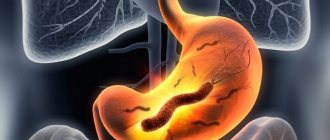
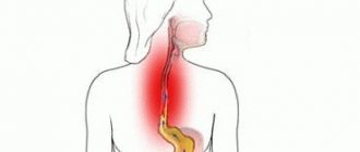
Gastric leiomyoma is absolutely benign in morphological essence, grows very slowly and in most cases does not manifest clinical symptoms. Large nodes can disrupt the normal functioning of the gastrointestinal tract and lead to dangerous complications.
- General information
- Kinds
- Reasons for appearance
- Symptoms
- Diagnostics
- Treatment
- Possible complications
- Prevention
- Forecast
Externally, leiomyoma cannot be distinguished from malignant neoplasms, so it is removed for microscopic and IHC studies, which prove the absence of signs of malignancy.
General information
Leiomyomas develop from cells of the muscular layer of the stomach, which is why their second name is “fibroids.” The tumor develops mainly in older people, but there are known cases of diagnosis of formation in childhood. In the scientific literature of the last century, a more frequent incidence of leiomyoma in women was noted; today, pathology is more often found in men from 40 to 70 years old.
Leiomyoma is a rare pathology and accounts for 3% of all organ neoplasms, but it is also the most common benign gastric tumor - it accounts for almost 45%. Since the process is asymptomatic, in the vast majority of cases, fibroids in the stomach are discovered accidentally during an examination for another reason or at an autopsy. In the last decade, the frequency of pathology has increased; it is believed that the reason for this is the greater availability of medical examinations and mandatory medical examinations.
Symptoms of gastric leiomyoma
Symptoms of leiomyoma often signal its ulceration, when the disease reaches a life-threatening stage. Until this moment, tumor growth may not show signs and not bother the patient. The timeliness of a patient’s visit to a doctor determines the duration of treatment and the prognosis of the disease, so it is imprudent to wait for relief in cases of attacks without visiting a clinic. If one of the following signs of a tumor in the stomach was noticed by the patient, an examination by a doctor should immediately follow:
- Malaise, a feeling of fatigue and dizziness are indicated when the tumor bleeds. Since the bleeding is internal, the reason for the change in health status is unclear to a person for a long time.
- The skin turns pale due to blood loss from the body and anemic iron deficiency.
- Stomach bleeding is accompanied by a change in a person's stool to a dark color.
- Even with a good appetite, a persistent decrease in body weight occurs. Substances that are already absorbed in the stomach or digested in this section pass through the intestines undigested and undigested.
- The resulting tumor reduces the size of the stomach, causing some of the acidic gastric juice to be released into the esophagus, causing frequent heartburn. The release of stomach contents with secreted hydrochloric acid into the esophagus is facilitated by relaxation of the gastroesophageal smooth muscle sphincter.
- A blood test does not cause much concern when the hemoglobin concentration is slightly reduced. A signal of internal bleeding is a significant decrease in hemoglobin level with an increase in the number of leukocytes in 1 mm3.
- “Hunger pains” that appear in the patient mainly at night. At the same time, the patient has recently noted frequent night snacks after waking up from pain. Analgesics from your home medicine cabinet are often useless when trying to relieve pain.
Such signs indicate the development of the disease in a dangerous stage, when the enlargement of the tumor interferes with the normal functioning of the stomach or its bleeding.
Kinds
A generally accepted classification of gastric leiomyoma has not been developed.
Gastric fibroids are usually divided according to the location of the node:
- submucosal or submucosal;
- inside the muscular layer of the stomach - intramural;
- subserous, growing predominantly outward, that is, into the abdominal cavity.
Some experts classify fibroids as:
- internal, growing mainly inside the stomach - endogastric;
- external - with growth directed into the abdominal cavity - exogastric.
By localization in the stomach it is designated as follows: located in the upper third or antrum; in the middle and lower third of the organ.
For gastric fibroids, a typical localization has not been established: approximately a fifth is located in the upper parts, about a third - in the middle part.
Prevention and prognosis
The prognosis for leiomyomas is positive in most patients. However, the risk of tumor degeneration cannot be excluded. In such conditions, only 25-50% of patients live more than 5 years with chemotherapy.
Specific methods of prevention have not yet been developed. This is explained by the lack of sufficient information about the causes of the development of nodular neoplasms in the gastric mucosa.
To reduce the risk of leiomyomas, it is recommended to give up bad habits and switch to proper nutrition. In addition, endocrine pathologies that provoke tumor processes should be treated in a timely manner.
Reasons for appearance
The list of reasons for the development of a tumor in the muscular layer of the stomach includes a dozen “general” assumptions, that is, all known risk factors for tumors of any nature are mentioned.
First of all, heredity is suspected - a family history of gastrointestinal neoplasms is assumed without indicating the specific genes “responsible” for initiating the process or hereditary syndromes associated with leiomyoma. The negative impact of radiation, from electromagnetic to radioactive and solar, cannot be ruled out.
Possible factors favoring tumor pathology are immunodeficiency in HIV, as well as hormonal imbalance.
The harmful effects of the environment, unhealthy lifestyle with irregular and unhealthy diet, alcohol abuse and smoking cannot be excluded. And, of course, it was not without the complicity of nervous breakdowns in the initiation of tumor growth.
In fact, none of these reasons has reliable evidence and it would be correct to state the following - the reasons for the development of leiomyomas in the stomach are unknown to science.
Causes
Unfortunately, scientists have not yet figured out the exact reasons for the formation of leiomyoma in the stomach area, but there are known factors that can contribute to the development of this pathology:
- traumatic damage to the gastric walls;
- systematic consumption of irritating dishes and products;
- constant stress;
- hereditary factor;
- the presence of gastrointestinal diseases, including chronic pancreatitis;
- diseases of the endocrine system;
- immunodeficiency states;
- viral and bacterial lesions of the organ;
- radiation, electromagnetic, ultraviolet, ionizing effects;
- living in an environmentally unfavorable area.
More often, the neoplasm occurs in females after 50 years of age.
Symptoms
The slow growth and benign course of the myomatous node with high extensibility of the gastric wall in most cases contributes to the absence of complaints of discomfort. It is clear that the severity and range of clinical manifestations are determined by the size and location of the tumor:
- Small or “miniature” leiomyomas do not manifest themselves in any way.
- Large nodes with intracavitary growth can disrupt the evacuation function of the stomach, interfering with the movement of food masses, clinically manifested by belching, vomiting and a feeling of fullness of the stomach after eating.
- Externally located large fibroids, especially on a narrow base - a pedicle, can put pressure on neighboring organs and disrupt their function, as well as twist in the pedicle and simulate an “acute abdomen” clinic with severe pain, vomiting and intoxication syndrome.
The main danger of gastric fibroids is the complicated course of very large nodes, which we will consider further.
Since there is no generally accepted classification of leiomyomas, nodules less than 2 centimeters are considered small, large ones - more than 5 centimeters; the literature contains cases of surgical removal of huge leiomyomas that were masquerading as tumors of the abdominal cavity or ovaries.
Treatment of gastric leiomyoma
Typically, the development of pathology involves surgical intervention. Therapeutic measures do not lead to recovery. They are carried out only if there are obvious contraindications to the operation, which most often are severe cardiac disease, vascular pathology or severe metabolic disorders.
As a rule, surgical treatment is carried out urgently due to the threat of severe consequences and the risk of malignant degeneration of the tumor. In the presence of small nodes, their endoscopic resection to the border of healthy tissue formation is required.
Nowadays, cryodestruction of small leiomyomas is also used.
If there is an active ulcerative process, vessels are destroyed, or there is a clear risk of developing a cancerous tumor, then the entire stomach is removed. After surgery, it is necessary to undergo a course of intensive therapy, taking proton pump blockers and antibiotics to suppress the growth of Helicobacter pylori. In addition, compliance with the general principles of prescribing therapeutic nutrition becomes a mandatory rule for restoring the body.
It is prohibited to use:
- fried;
- fat;
- canned food;
- smoked;
- pickled;
- sausages;
- liver;
- mushrooms;
- spices;
- coffee;
- strong brewed tea;
- lemonade;
- alcoholic drinks.
These products irritate the inner surface of the organ. Therefore, it is advisable to include more cereals, vegetables and fruits, lean meat and fish in your diet.
All dishes are recommended to be served boiled or baked.
Diagnostics
Mostly the tumor looks like a nodular formation with a smooth surface, but it can also consist of several nodules. An experienced endoscopist will detect it by bulging of the wall, if growth is directed into the gastric lumen, and a change in the peristaltic “wave”.
A biopsy of the submucosal node is often inconclusive due to its deep location, so in hardly a third of cases it is possible to judge the true nature of the neoplasm.
High-tech diagnostics - CT and MRI are practically powerless for pathology of the gastric wall; the most effective detection method is endoscopy or endoscopic ultrasonography combined with ultrasound, which allows you to examine the formation from the stomach cavity, determine its boundaries inside the gastric wall, and see the structure and structure of the node.
Treatment tactics
Treatment of leiomyoma involves only surgery. When leiomyoma manifests in a patient, it is important to quickly perform surgery to excise the tumor within healthy tissue. Starting treatment for esophageal leiomyoma with folk remedies and only them is contraindicated. This can be dangerous not only for the health, but also for the life of the patient. There is no alternative treatment, and even more so, grandmother’s and folk methods against leiomyoma. The lack of adequate treatment can provoke bleeding, rupture of nodes and walls of the stomach, cause peritonitis and other serious consequences. There are two tactics of surgical intervention for different developments of leiomyoma:
- uncomplicated (excision of the tumor is carried out within the unchanged tissue);
- complicated (removal of part of the stomach along with the tumor).
To determine the extent of the operation, a consultation with an endoscopist may be required. Removal of the tumor can be delayed as much as possible in the presence of severe conditions from the cardiovascular system, end-stage renal or liver failure, during the active phase of tuberculosis and other pathologies. Treatment of gastric leiomyoma is carried out if the potential risk to life outweighs the risk of exacerbation of existing ailments.
Important! Many patients may remain patients in the gastroenterology department for a long time due to other pathologies. After leiomyoma is diagnosed, patients are transferred to surgery for further treatment. In the absence of life-threatening contraindications, radical excision of the defective tissue is performed.
Treatment
There is no need to remove small tumors that do not bother the patient if there is 100% confidence in their benign nature, but with submucosal growth such confidence is excluded. Benign fibroids and malignant GISTs are visually and morphologically indistinguishable and are differentiated only by immunohistochemical analysis of tumor sections obtained surgically.
The final diagnosis and simultaneous treatment with a diagnosis of gastric leiomyoma consists of their surgical removal. The extent of intervention and access to the organ is determined by the size and location of the tumor, the presence of complications and the patient’s initial health.
In most cases, organ-preserving endoscopic interventions are resorted to, of course, with urgent histological examination during surgery. Laparoscopic resections are quite sufficient and adequate, allowing for rapid recovery of the patient.
In case of a large complicated neoplasm that has caused ulceration of the mucous membrane, perforation of the gastric wall or bleeding, classical operations with dissection of the abdominal wall and gastrectomy are resorted to.
Diagnosis of leiomyoma
Most clinics in the country do not have a gastroenterologist on staff who can check the state of the digestive system during an annual medical examination. This doctor is consulted only if there is a referral from a therapist or the patient experiences pain in the digestive tract. Only then is fibrogastroendoscopy prescribed to check the current state of the digestive organs.
Video endoscopy revealed leiomyoma
The fibrogastroendoscopy procedure is prescribed for suspected gastritis and gastric ulcers, during which leiomyoma may be accidentally discovered. Patients who have once had gastritis or a stomach ulcer must take care to prevent not only recurrence of the disease, but also the occurrence of a benign tumor. For this purpose, it is recommended that the stomach be examined at least once every six months. When fibroendoscopy or symptoms of leiomyoma indicate its presence, the doctor diagnoses the tumor using the following methods:
- the doctor collects anamnesis, asking questions about the nature of the relatives’ illnesses, recent health status, the patient’s work and lifestyle, etc.;
- physical examination of the patient, including skin color, palpation of the abdomen, checking the degree of pain and its degree in response to external sensations of the stomach;
- laboratory analysis of urine and blood, coprographic color analysis and determination of the presence of blood in stool;
- fibrogastroduodenoscopy, which helps determine the condition of the digestive tract;
- bacteriological analysis for the presence of the causative agent of ulcerative conditions - Helicobacter;
- the presence of a benign neoplasm, location and size are accurately determined by ultrasound examination of the abdominal area;
- The initial pathology of leiomyoma is clarified using computed tomography.
A set of diagnostic techniques gives a clear idea of the diagnosis and allows you to choose the right treatment regimen.
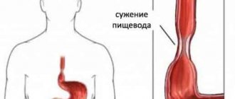
Leiomyoma of the esophagus
Possible complications
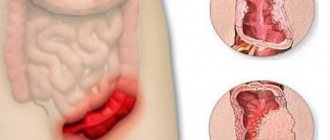
Large leiomyomas growing into the stomach cavity disrupt the structural integrity of the mucous membrane, causing its ruptures - erosion and forming ulcers. Ulcerations in clinical manifestations are indistinguishable from a banal ulcer - pain, discomfort, episodes of vomiting, changes in appetite.
And just like ordinary ulcers, they can be complicated by acute or chronic bleeding. Acute gastric bleeding is manifested by vomiting coffee grounds and black stools, decreased vascular pressure up to shock. Chronic blood loss is manifested by anemia syndrome, changes in stool, and weight loss. Insufficient nutrition of leiomyoma leads to the disintegration of its tissues with possible rupture of the gastric wall - perforation and the development of peritonitis.
The complicated course of a benign stomach tumor requires active therapy followed by long-term rehabilitation.
Possible complications and prognosis
Benign tumors differ from malignant ones in that they do not form metastases, and therefore do not disperse or grow into surrounding tissues. They usually do not affect the lives of victims.
Although they are benign, some carry risks: either they can develop into cancerous tumors, or they can become so enlarged that they affect the function of other organs. Many also cause pain when they settle in a sensitive area. Then they need to be treated.
Why tumors form is not scientifically clear. Experts suspect factors such as genetic predisposition, hormonal effects, environmental toxins, poor diet, inflammatory and stress factors.
Many benign tumors go undetected. Some are detected during routine or early cancer screening. In some cases, a person becomes aware of them because they affect body functions. And sometimes they can also be a cosmetic problem. The doctor must decide on a case-by-case basis whether it is sufficient to continue monitoring the tumor or whether it is better to remove it.
With timely treatment, the patient's life expectancy is not reduced. However, with a longer course there is a risk of developing unpleasant symptoms.
Forecast
Leiomyoma after proper removal does not recur and does not affect the patient’s life expectancy. The literature mentions the possibility of malignancy, which from an oncological point of view is an initially undiagnosed malignant slow-growing GIST; it is because of such cases that gastric leiomyoma must be removed.
A patient with suspected gastric leiomyoma should be operated on by oncological surgeons in a clinic with a powerful diagnostic service and a morphological laboratory focused on oncopathology. During surgery, it is necessary to differentiate a benign process from a malignant one in order to determine the optimal size of removal. The European Clinic is one of the few medical institutions that meet these important requirements for the patient.
Book a consultation 24 hours a day
+7+7+78
Bibliography:
- Gankov V.A., Maslikova S.A., Lazarev A.F., Oskretkov V.I./Surgical tactics for submucosal formations of the esophagus, stomach and duodenum//Bulletin of Med. Sciences No. 2 (10) 2018
- Starkov Yu.G., Solodinina E.N., Novozhilova A.V. /Submucosal neoplasms of the gastrointestinal tract in endoscopic practice// Surgery, 2010; 2.
- Firsov E.I., Vorotyntseva N.S., Novikov M.V. /Comprehensive clinical and radiological diagnosis of benign non-epithelial tumors of the stomach// Kursk scientific-practical. Bulletin "Man and his health", 2009, No. 2.
- Bucber P., Villiger P., Egger JF., et al. /Management of gastrointestinal stromal tumors: from diagnosis to treatment// Swiss. Med. wkly., 2004; 134.
- Greenson JK /Gastrointestinal stromal tumors and other mesenchymal lesions of the gut// Mod. Pathol., 2003; 16(4).
- Nishida T., Blay JY, Hirota S., et al. /The standard diagnosis, treatment, and follow-up of gastrointestinal stromal tumors based on guidelines // Gastric Cancer., 2021 Jan., 19 (1).
Diagnostic methods
Clinical diagnosis can be made using endoscopic ultrasound, which allows an accurate representation of the five-layer structure of the gastrointestinal wall and thus distinguishes hemangioma, cyst and lipoma of the stomach or intestinal wall.
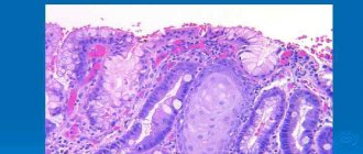
Using conventional light microscopy, distinguishing between gastric mucosa and leiomyoma can be difficult. Histological findings in hematoxylin and eosin staining cannot be reliably or specifically related to immunophenotic, immunohistochemical and molecular genetic properties.
Laboratory
To be able to distinguish between gastric leiomyoma and gastric mucosa, the histological characteristics of the various lesions are used. Histological processing of gastric stromal tumors reveals a higher cell density with consistent predominantly basophilic staining properties.
Immunohistochemical studies are available for definitive differential diagnosis. Consequently, gastric leiomyomas are positive for actin and desmin. In contrast, GMs are negative for desmin but positive for cellular markers such as CD34 and CD117.
Morphologically, leiomyoma is presented macroscopically as a defined, sometimes painful, rough nodule on the skin. Looking at the affected tissue under a microscope, the neoplasm is characterized by bundles of smooth muscle. Hyaline cartilage can be detected immunohistochemically as an additional component between these muscle bundles.
Instrumental
Symptoms such as abdominal pain or problems with bowel movements may indicate leiomyomas. To investigate this suspicion, the gastroenterologist first asks in detail about existing complaints and any previous medical history.
After collecting the medical history, an examination follows. The exact location and size of leiomyoma fibroids can be determined with help. endoscopy. Ultrasound examination can be performed through the abdominal wall.
If ultrasound does not provide an accurate diagnosis, the surgeon may perform an intra-abdominal (laparoscopic) examination. If the leiomyoma is pressing on organs, examination with ultrasound and x-rays with contrast may be necessary.
Ultrasound is a method for diagnosing gastric leiomyoma
If test results are unclear, your doctor will sometimes order a magnetic resonance imaging (MRI) scan. In addition, if necessary, a blood test is performed (if anemia is suspected) and hormone levels are measured.
The most typical radiological sign of the pathology is an oval or round filling defect with smooth edges measuring approximately 5 to 6 cm. Ulcerations are often present on the surface. The folds of the mucous membrane around the filling defect are moved apart, but not disorganized.
Endoscopy can detect benign submucosal lesions but cannot differentiate the disease. Transabdominal ultrasound and CT scans provide additional information about the type of tumor. Endoscopic ultrasound is the best way to detect the disease. Preoperative endoscopic marking of the tumor-bearing area can aid in laparoscopic identification of pathology located on the posterior wall of the stomach.
Causes of tumor formation
Leiomyoma can develop into a malignant tumor
The body never simply fails, and in order for even benign tumors to begin to appear, negative factors must influence. The main reasons for the formation of leiomyomas in the stomach include:
- Poor nutrition. As a result of the fact that a person eats incorrectly, mainly fatty and spicy foods, the mucous membrane begins to collapse, and the stomach can no longer cope with the task. In this case, a general weakening of the body occurs, which is why the smooth muscle cells on the walls of the stomach begin to grow rapidly.
- Radiation. This usually includes radiation and electromagnetic radiation, which literally affects humans every day. A huge amount of equipment, access systems - all this negatively affects the cells. People who often fly on airplanes are most susceptible to developing tumors, as they have to go through arches with X-ray radiation several times
- Polluted air, which also pollutes the lungs, and after them all organs do not receive the amount of oxygen that they so need. In addition, particles of chemical substances enter the body and settle on the bronchi.
- Bacteria and viruses that penetrate the body so quickly that a person does not always have time to react and begin to fight them. And it’s good if their presence manifests itself with obvious symptoms, but most often they destroy the body asymptomatically. Thus, the stomach is negatively affected by the Helicobacter bacterium, which, according to scientists, causes such dangerous diseases as stomach ulcers, gastritis, and sometimes cancer
- Injuries during which internal organs were damaged and the stomach was affected
- Weak immunity, as a result of which the body is most susceptible to negative factors. Due to reduced immunity, cells do not respond to changes in any way
- Problems with hormones. Hormones pose a particular danger, namely when their levels change sharply and become either too high or too low. Under the influence of hormones, tumors can form, cells can degenerate, etc.
- Inflammatory process in the stomach, namely inflammation of the mucous membrane
- Smoking and alcohol abuse. During smoking, all cells are literally saturated with nicotine, which can kill them. But alcohol gradually burns the mucous membrane
- Heredity. Heredity plays a special role. If there are people in the family (close relatives) who suffered from gastric leiomyoma, then the likelihood that it will develop increases several times
- Stress. Under the influence of stress, irreparable changes occur in the body, which most often affect the stomach and nervous system. Constant nervous tension is sometimes more dangerous than radiation
Typically, gastric leiomyoma develops in people over 50 years of age, since it is from this moment that the body becomes less resilient, and the cells gradually age.
How to treat
The treatment method depends mainly on the size of the leiomyoma, its shape and location. In most cases, doctors recommend surgical removal of the tumor. The desired effect cannot be expected from conservative methods.
If the tumor is small (up to 2-3 cm), then an endoscope is used to remove it, with possible freezing of the transformed tissues with liquid nitrogen. This allows the tumor not to grow and completely destroy diseased cells.
If the formation exceeds 3 cm in diameter, local excision is performed. This is a strip operation that requires longer rehabilitation. During this surgical intervention, 2 cm of healthy tissue is cut out from the border of the leiomyoma.
After excision of the tumor, drug therapy is prescribed to reduce the risk of complications and relieve inflammation.
- Drugs that reduce the production of HCl by the stomach.
- If Helicobacter pylori is detected, antibiotics are used to suppress growth and destroy the bacterium.