Intestinal paresis is a pathological condition that is characterized by a decrease in the level of peristalsis of the digestive system and disruption of the movement of the food bolus through the gastrointestinal tract. The disorder can occur against the background of many diseases and may indicate damage to the digestive organs and changes in the functional activity of other body systems.
Causes
Postoperative paresis
Intestinal paresis is one of the most common postoperative complications. Experts believe that this condition is triggered by trauma to the peritoneum, in response to which a protective reaction develops in the form of a decrease in peristalsis activity. Therefore, in most cases, postoperative paresis occurs only after extensive abdominal surgery.
Most often, paralysis develops after laparotomy.
Other
- Inflammatory processes in the abdominal cavity. Massive inflammation of the peritoneum is accompanied by impaired peristalsis of the digestive system, which can lead to intestinal paralysis. Such conditions include peritonitis, phlegmon of the retroperitoneal tissue.
- Impaired blood supply to the intestines. Decreased peristaltic activity may be associated with vascular compromise. The condition is one of the manifestations of acute damage to the heart and arteries, in which blood does not flow into the intestines in sufficient quantities. This disorder may be caused by an abdominal aortic aneurysm or myocardial ischemia (acute coronary syndrome). Vascular disorders can also be local. For example, acute intestinal hypoxia is observed with thrombosis of the mesenteric vessels, which supply blood to the lower parts of the digestive system.
- Violation of the nervous regulation of the intestines. The contractile activity of the digestive system is ensured by the rhythmic impulse of nerve fibers coming from the central nervous system to the intestines. Damage to any segment of the nervous tract can lead to intestinal paresis. This condition can be caused by a tumor or injury to the spinal cord, or by taking certain medications.
- Reflex disorders. Intestinal paralysis can develop in response to any acute disturbances. This may be renal colic, postoperative condition, severe intoxication, severe pneumonia.
Be sure to read: What foods cause constipation?
Combating intestinal paresis
9 May 2021, 14:25
With a gradual decrease in intestinal muscle tone with the development of paralysis due to severe diseases of the gastrointestinal tract and other body systems, intestinal paresis develops.
Symptoms of the condition are characterized by uniform bloating, nausea, ending with vomiting, acute dehydration, tachycardia, and respiratory dysfunction. Paresis is diagnosed by X-ray and ultrasound methods, tomography, colonoscopy and irrigoscopy.
Treatment involves an integrated approach, including motor stimulation, elimination of symptomatic manifestations, and intestinal decompression. More often surgery is required.
General information about pathology
Intestinal paresis is a temporary weakening of the peristaltic activity of the intestinal tract, which often develops against the background of a disorder of water and electrolyte balance due to surgery on the organ itself or in another part of the abdominal cavity. Other names for the pathology are ileus, atony, paralytic obstruction. Symptoms of the disease appear on the 2-3rd postoperative day. Atony is possible in one section of the intestine or paralysis of the entire section is possible.
When there is poor intestinal permeability due to paresis, the following appear:
- severe, widespread bloating in the abdomen;
- increased flatulence;
- diffuse, nagging pain, covering all parts of the intestine;
- constant nausea with vomiting, in the masses of which there are blood streaks, bile, gastric or intestinal contents;
- problems with stool excretion, in particular, the appearance of a small volume of liquid stool;
- soft, relaxed abdomen;
- intermittent breathing, tachycardia with a sharp decrease in blood pressure due to distension and swelling of the intestinal walls.
Disease incidence
Pathologies of internal organs lead to the development of intestinal paresis.
Intestinal paresis is a common disorder and manifestation of diseases of other organs. In 25% of patients, paralysis appears due to acute diseases of the internal organs, less often - with severe forms of pathologies of the heart and blood vessels, intoxication, and general infection.
Postoperative intestinal paresis often develops due to surgical intervention in the gastrointestinal tract, but only 0.2% of patients with this symptom underwent surgery for another reason. 70% of patients are people over 60 years old. The disease may occur in newborns and older children, in women during pregnancy (2nd, 3rd trimester) and after childbirth.
Phases of development
Intestinal paresis develops in 3 stages:
- 1st, when there is a gradual or sudden inhibition of peristalsis with the development of paresis;
- 2nd, when peristalsis stops altogether, and against the background of increased formation of gases and accumulation of fluid in the intestine, pressure rises, blood supply is disrupted;
- 3rd, when, against the background of intestinal blockage, the body is poisoned, the condition worsens, and other organs and systems become disordered.
Provocateurs of intestinal paresis
Causes can be primary or secondary. When paresis occurs as a result of surgery, abdominal trauma, or metabolic disorders, atony develops independently.
If the cause is damage to the mesentery (in particular, its artery), the occurrence of inflammation in any organ of the gastrointestinal tract, the development of other serious diseases in the abdominal cavity and pelvic organs, peritonitis (inflammation of the abdominal sheets), a secondary, symptomatic form of paresis develops.
Main reasons for development:
- side effects of opiates, calcium duct blockers and similar medications;
- hypokalemia (impaired metabolism);
- acute peritonitis;
- tumors, cysts, polyps, hematomas that provoke inflammation in the intestines;
- kidney disease;
- pathologies of the sternum (rib fractures, myocardial infarction, pleuropneumonia);
- diabetes.
After childbirth, intestinal problems arise, which soon go away.
Ileus develops in women after childbirth as a result of weakening/absence of intestinal motility in the first 24 hours after delivery. With normal recovery, the condition returns to normal within 2-3 days.
If gases do not pass on the 4th day, the doctor diagnoses paresis and prescribes treatment. In newborns, paresis develops due to congenital or acquired disorders of the nervous system.
In infants, ileus is a consequence of poor nutrition and treatment of certain infectious diseases.
Postoperative paresis: description, symptoms
Atony is a common consequence of abdominal surgery (surgeries on the internal organs of the peritoneum). In most cases, the disorder does not require treatment, as the condition resolves itself after a few days. But complications may develop.
The symptoms of the condition are determined by the degree of paresis:
- With 1st degree damage, the condition is characterized by a temporary reaction of the body to injury in the form of gastrointestinal dysfunction. With proper medical care of the patient, no treatment is required; recovery occurs on its own.
- With 2nd degree damage, the dysfunction is more profound and is manifested by a feeling of heaviness in the stomach, nausea with vomiting, and tolerable bloating. Symptoms develop against the background of stagnation of food chyme (lump), problems with the movement of food through the gastrointestinal tract. The condition requires infusion treatment aimed at regulating water-electrolyte and acid-base balance. Evacuation from the stomach to the intestines can be improved by introducing a probe or motor stimulation.
- With 3rd degree damage, intestinal paresis is accompanied by severe bloating and a constant feeling of gastric surges. In the presence of postoperative intestinal paresis, treatment regimens are used aimed at preventing complete paralysis of the intestinal muscles.
Examination methods
Methods for diagnosing diseases of the gastrointestinal tract include palpation.
Problems with intestinal motility are dealt with by a gastroenterologist and a surgeon.
Doctors perform examination, palpation and percussion (listening to the intestines) to identify intestinal obstruction and suggest the underlying causes of the condition.
Then a comprehensive examination of the patient is prescribed using the following instrumental methods:
- Survey radiography. The method allows you to confirm/refute mechanical blockage of the intestine.
- Ultrasound, MSCT, CT are specific and sensitive methods that make it possible to identify distended intestinal loops, fluid and gas levels, traumatic changes in the intestinal mucosa, and determine the intensity of ischemia of the small and large intestine.
- Irrigoscopy, colonoscopy, intended to differentiate paresis from mechanical blockage, coprostasis, infection or parasites.
Treatment methods
Treatment of intestinal paresis is carried out in intensive care. The first stage of the treatment regimen is unloading the intestines - gases are removed through outlet tubes (rectal probes).
Additionally, fasting is prescribed to reduce the load on the gastrointestinal tract, primary pathology is treated, and water-electrolyte balance and metabolism are improved.
Moderate physical activity and abdominal massage are recommended.
Newborns are treated with a regimen aimed at improving blood circulation, adjusting muscle tone, metabolism, and strengthening the nerve impulse.
Medicines
The drug improves gastrointestinal motility.
To combat postoperative intestinal paresis, conservative treatment tactics with Proserin are used, the effect of which is aimed at stimulating gastrointestinal motility.
Treatment is carried out under control in order to prevent bradycardia. If the first injection is not effective, infusion therapy with Proserin is prescribed (efficacy - 75%). This tactic is contraindicated:
- with mechanical blockage of the intestine, perforation or ischemia of the intestinal wall;
- during pregnancy (in women);
- for renal dysfunction, bronchospasm, complicated cardiac disorders.
Surgical intervention
In most cases, the tactics of minimally invasive decompression of the affected intestine are used in three different ways, selected individually:
- drainage of the colon with X-ray examination;
- puncture of the cecum, performed percutaneously;
- application of an artificial, external fistula to the cecum (cecostomy).
Surgical intervention is used for a number of indications for the procedure.
Indications for procedures:
- expansion of the large intestine by more than 10 cm in diameter;
- duration of paresis - from 3 days;
- ineffectiveness of medications after 2 days of use;
- lack of progress during treatment with Proserin or with contraindications to its use.
Colonoscopy is often done, but the procedure is prohibited in cases of peritonitis and intestinal perforation. The effect is achieved with isolated colonoscopy in ¼ of patients. An effectiveness of 90% is possible when combining colonoscopy with drainage.
Percutaneous puncture and cecostomy are performed in case of complicated course, ineffectiveness of medications and colonoscopy. Abdominal surgery is recommended in extreme cases: with peritonitis and violation of the integrity of the intestinal wall.
Open surgery involves removing the affected part of the intestine.
During surgical treatment, narcotic analgesics are prohibited - they inhibit intestinal motility.
Symptoms
- constant nausea;
- vomiting, which in the initial stages contains undigested food residues, after some time can take on a fecal character;
- severe bloating, impaired gas discharge;
- moderate pain without clear localization, which spreads over the entire surface of the abdominal wall;
- prolonged constipation, a small amount of liquid discharge is possible, but its volume is not comparable with the daily norm of feces;
- manifestation of intoxication of the body - fever, shortness of breath, weakness;
- signs of dehydration are dry skin, thirst, lack of urination, tachycardia and decreased blood pressure.
Diagnostics
- Analysis of clinical and anamnestic data. A detailed story from the patient about when the disease appeared and how it developed will help the specialist to assume the presence of intestinal paresis and determine its cause. Of great importance is the presence of concomitant pathologies - vascular disorders or innervation disorders, which can cause the development of paresis. You should definitely check if there is a history of recent abdominal surgery.
- X-ray of the abdominal cavity. The study is the primary way to diagnose paresis. Images of the abdominal cavity reveal fluid-filled loops of intestine. In this case, there should be no signs of organ blockage (mechanical obstruction).
- Ultrasound or multislice CT. The techniques are more specific and informative than x-ray diagnostics. With such an examination, it is possible to analyze the activity of peristalsis and detect increased pneumatization of intestinal loops. Research is also used to identify the causes of paresis of the digestive system.
Treatment
General events
After intestinal paresis is detected, a number of general measures are prescribed to reduce the load on the digestive system and improve the patient’s condition. These include:
- removal of intestinal gases using a rectal tube or thick gastric tube;
- food restriction to reduce enteral load;
- relief of the underlying disease that caused the paresis;
- correction of water and electrolyte balance.
Drug treatment
Severe intestinal paresis requires drug stimulation of peristalsis. For this purpose, the patient is administered neostigmine .
The drug affects blood circulation, so during its administration the doctor must monitor the patient’s condition. If there is a sharp decrease in heart rate, the patient is given atropine to normalize the heart rhythm. Be sure to read:
What foods improve intestinal motility?
If there is no effect from a single administration of the drug, massive infusion therapy with neostigmine is carried out until the effect is achieved (at least 24 hours). The drug has a number of strict contraindications, so a thorough examination of the patient is required before its administration.
Non-surgical decompression
With severe intestinal paresis, the diameter of the organ increases significantly, the pressure in the digestive system increases, which is fraught with a number of complications. Therefore, if conservative therapy is ineffective, the patient undergoes decompression, which helps reduce pressure. It can be carried out using the following methods:
- Insertion of a thick probe into the intestine under X-ray control.
- Evacuation of contents during colonoscopy with the introduction of drainage.
- Percutaneous cecostomy - bringing the cecum to the surface of the abdominal cavity and evacuating its contents.
Colonoscopy is most often used for decompression, since its use has a minimal likelihood of complications.
Surgery
If decompression fails, radical surgery is required. Its volume is determined individually for each patient. In some cases, you can get by with an open cecostomy. In the most severe cases, resection is performed - removal of the affected part of the intestine and anastomosis.
How does intestinal paresis manifest?
In gastroenterological practice, a pathological condition such as intestinal paresis is often encountered. This is not a separate disease, but a complication that develops against the background of various pathologies. Paresis must be distinguished from mechanical intestinal obstruction. In the latter case, stagnation of feces occurs due to an obstruction. These can be tumors, parasites and foreign objects.
Development of intestinal paresis
Intestinal paresis is a condition characterized by low smooth muscle tone. This leads to disruption of the movement of feces. In severe cases, surgical treatment may be necessary.
This pathology develops after surgery, against the background of diseases of the lungs, heart and digestive tract. In surgical practice, intestinal paresis accounts for up to 0.2% of all diseases.
The risk group includes older people over 60 years of age. This is due to the presence of chronic diseases and frequent operations. Paresis is often diagnosed in newborns and pregnant women. This condition results in paralytic ileus when half-digested food stagnates.
Main etiological factors
Intestinal paresis is due to various reasons. The following risk factors for the development of this dangerous condition are identified:
- peritonitis;
- phlegmon;
- performing operations on the intestines;
- ischemia due to atherosclerosis or thrombosis;
- abdominal aortic aneurysm;
- coronary heart disease;
- nerve damage due to spinal cord and brain injuries;
- acute cardiovascular failure;
- pneumonia;
- severe pain syndrome (renal colic);
- spinal cord tumors;
- overdose of calcium channel blockers;
- intoxication.
Paresis is based on the following pathological processes:
- disruption of intestinal innervation;
- circulatory disorder;
- reflex reaction.
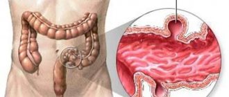
The human intestinal wall consists of several layers (mucosal, submucosal, muscular and external serous). At the initial stage, paresis of smooth muscle cells occurs. This leads to decreased peristalsis (rhythmic contractions of the intestines).
Over time, stool and fluid stagnate. This leads to increased pressure. At the next stage, signs of intoxication of the body appear due to stagnation of metabolic products.
Risk factors for the development of paresis include impaired water and electrolyte status.
How does paresis manifest?
With intestinal paresis, symptoms are nonspecific. They resemble inflammatory diseases (enteritis, colitis). The intestines of sick people contract poorly.
Local symptoms include moderate pain, flatulence (bloating), nausea, vomiting, and constipation-type bowel movements. Impaired intestinal patency leads to increased gas formation.
Patients experience heaviness in the abdomen and bursting pain.
It is diffuse and has no specific localization. Postoperative ileus palsy (PSP) often presents with nausea and vomiting. In the early stages, vomit may contain remains of semi-digested food. Subsequently, the contents acquire a fecal character. This is due to the fact that chyme is thrown into the overlying parts of the gastrointestinal tract.
Common symptoms include a mild increase in body temperature, rapid heartbeat, decreased blood pressure, shortness of breath, and shallow breathing. Respiratory disorders are caused by displacement of the diaphragm and compression of the lungs and heart. With intestinal paresis in children and adults, fever is a rare symptom.
It indicates the development of complications. In the presence of postoperative paresis with regular vomiting, there is a risk of dehydration (dehydration). This manifests itself as headache and dry mucous membranes. During physical examination, a positive Lothuissen's sign is often determined. When listening to intestinal peristalsis, only heart and respiratory sounds are detected.
Possible complications of paresis
Self-medication and untimely consultation with a doctor are the main reasons for the development of complications. Paresis can lead to the following consequences:
- dehydration;
- blood thickening and blood clot formation;
- ischemia of the intestinal wall;
- perforation (perforation);
- peritonitis;
- acute intestinal obstruction;
- bleeding;
- tissue necrosis;
- formation of diverticula.
If you carelessly use a colonoscope while examining a patient, you may accidentally damage the intestinal wall. The risk of perforation increases if the following symptoms are present:
- paresis lasts more than 6 days;
- increase in intestinal diameter;
- absence of feces for a week.
In severe cases, heart function is impaired. This is manifested by an increase in heart rate and a drop in blood pressure. In the presence of postoperative intestinal paresis, complications are less common. In the absence of proper medical care, death is possible.
Patient examination plan
For intestinal paresis, treatment is carried out after an accurate diagnosis has been established. The following studies are required:
- physical examination, including palpation, percussion and auscultation;
- Ultrasound;
- multislice computed tomography;
- radiography in 3 projections;
- irrigoscopy.
An x-ray reveals distended intestinal loops and fluid levels. The most informative are ultrasound and tomography. They allow you to identify the accumulation of gases, stretching of intestinal loops and signs of fecal stagnation. In this case, there is no obstruction. This is an important difference between paresis and mechanical obstruction.
The results of a survey and examination of the patient are of great importance in making a diagnosis. An indication of a recent operation is a reason to suspect paresis. Consultation with a surgeon and gastroenterologist is required. Differential diagnosis is carried out with parasitic diseases (ascariasis) and mechanical intestinal obstruction.
Conservative treatment methods
Various methods are used to combat postoperative intestinal paresis. In the acute period, hospitalization in the surgical department is required. Treatment includes the following aspects:
- removal of formed gases;
- temporary refusal to eat;
- elimination of the main cause of paresis;
- normalization of water and electrolyte status;
- improvement of metabolism;
- stimulation of peristalsis;
- limiting physical activity;
- use of medications (Proserina);
- infusion therapy.
Decompression is often performed using an endoscope. First aid for a patient is to get rid of excess gas, as it is harmful to the body. The most commonly used method is the insertion of a special tube into the rectum. Gases will be released through it. With the development of dehydration, massive infusion therapy is indicated.
The most commonly used drugs are Prozerin and Atropine. The latter is indicated for a decrease in heart rate. Conservative treatment of intestinal paresis involves the use of Proserin solution. The medicine is not used in cases of perforation, bradycardia and ischemia. The drug is not suitable for the treatment of pregnant women.
If there is no effect from conservative treatment, an increase in the diameter of the colon and paresis lasting more than 3 days, decompression is performed. It involves the insertion of a thick probe or colonoscope followed by drainage. According to strict indications, cecostomy is performed. This procedure involves creating a fistula in the cecum. It is performed when there is a high risk of complications.
Radical treatment and prevention
Cecostomy is a procedure that is performed quite rarely, but under certain circumstances it is not possible to do without it. In severe cases, open intestinal surgery is performed. The most effective types of surgical intervention are resection and cecostomy.
The method of colonoscopy with drainage helps most patients, so radical treatment is rarely performed. Health prognosis is determined by the following factors:
- timing of medical care;
- timeliness of the patient's visit to the doctor;
- person's age;
- presence of complications.
In case of perforation of the colon wall, death is observed in 40% of cases. Even after complete recovery, re-development of paresis is possible in the future. Relapses are observed mainly in elderly and weakened people. There is no specific prevention of paresis. To reduce the likelihood of developing this pathology, the following measures must be observed:
- move more;
- eat well;
- enrich the diet with fiber and pectin;
- promptly treat chronic and acute intestinal diseases;
- give preference to laparoscopic operations;
- prevent poisoning and injuries to the brain and spinal cord;
- treat existing pathologies of the heart and lungs.
Most often, paresis develops in the postoperative period after open, abdominal interventions. Recently, endoscopic treatment has become increasingly common. It is safer and less likely to lead to paresis.
Source: https://kiwka.ru/kishechnik/parez.html
Possible complications
- intestinal perforation;
- fecal peritonitis;
- necrosis of the intestinal mucosa;
- bleeding from the digestive system.
The prognosis for patients with intestinal paresis is determined by many factors: the severity of the disorders, the presence of concomitant diseases, and the patient’s age. In the absence of timely therapy, there is a very high probability of developing complications (primarily perforation and fecal peritonitis), which cause high mortality in this condition.
Forecast
The prognosis for this disease is controversial. It all depends on the patient’s age and associated complications. In most cases, the lethality of the disease can be caused by intestinal perforation. Relapses of the pathology often occur in patients after 60 years of age.
Intestinal paresis is a pathological condition that is characterized by a decrease in the level of peristalsis of the digestive system and disruption of the movement of the food bolus through the gastrointestinal tract. The disorder can occur against the background of many diseases and may indicate damage to the digestive organs and changes in the functional activity of other body systems.
Prevention
- timely consultation with a doctor in the presence of acute conditions;
- proper management of the postoperative period after abdominal surgery;
- maintaining a healthy lifestyle, proper nutrition (see here), reducing stress levels.
In continuation of the topic, be sure to read:
- Peritonitis: what kind of disease is it, how does it manifest and be treated?
- Causes of bloating and increased gas formation, treatment methods
- Rectal fissure: causes, symptoms and treatment of pathology
- Irritable bowel syndrome: symptoms and treatments
- Details about bowel cancer: stages, symptoms, treatment and prognosis
- More about hemorrhoids: causes, symptoms and treatment methods
- Diseases of the small intestine: symptoms and signs of the disease, treatment
- Rectal cancer: symptoms, stages, treatment and prognosis for life
- Intestinal infection: symptoms and treatment methods (diet, medications)
- Intestinal adhesions: causes, symptoms, treatment methods and prognosis
Be sure to read:
Causes of profuse (liquid) diarrhea and methods of its treatment