The stomach is one of the main organs of the digestive tract. It processes all the products we consume. This is done thanks to hydrochloric acid, which is present in the stomach. This chemical compound is secreted by special cells. The structure of the stomach is represented by several types of tissues. In addition, the cells that secrete hydrochloric acid and other biologically active substances are not located throughout the organ. Therefore, anatomically, the stomach consists of several sections. Each of them differs in functional significance.
Stomach: organ histology
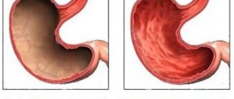
The stomach is a hollow, pouch-shaped organ. In addition to the chemical processing of chyme, it is necessary for the accumulation of food. To understand how digestion occurs, you should know what gastric histology is. This science studies the structure of organs at the tissue level. As you know, living matter consists of many cells. They, in turn, form tissues. The cells of the body differ in their structure. Therefore, the fabrics are also not the same. Each of them performs a specific function. Internal organs consist of several types of tissues. This ensures their activities.
The stomach is no exception. Histology studies the 4 layers of this organ. The first of these is the mucous membrane. It is located on the inner surface of the stomach. Next there is the submucosal layer. It is represented by adipose tissue, which contains blood and lymphatic vessels, as well as nerves. The next layer is the muscle layer. Thanks to it, the stomach can contract and relax. The last is the serous membrane. It is in contact with the abdominal cavity. Each of these layers is made up of cells that together form tissue.
Types of epithelium, their signs and properties, classification
Among epithelial tissues there are:
- single-layer flat (vascular endothelium);
- single-layer cubic (renal tubules);
- single-layer cylindrical (surface of the stomach);
- ciliated epithelium (airways);
- multilayer keratinizing (epidermis);
- multilayer non-keratinizing (oral mucosa);
- glandular epithelium (glands of external and internal secretion).
Epithelial tissues differ in their structure and functions. The following types of epithelium :
- Glandular (secretory). Epithelium, which is part of the glands. It consists of glandulocyte cells that release secretions into the blood, lymph, and glandular ducts.
- Integumentary (superficial). Lines internal organs, separates the body from the external environment, performing metabolic and barrier functions.
- Receptor (sensory). Localized in the sense organs.
Morphological classification of epithelium is based on differences in cell shape and number of layers.
The cells of the outer layer can be flat, cubic, or cylindrical. Sometimes they fit tightly together, in some cases there are narrow passages between them through which tissue fluid circulates.
Depending on the number of cell layers, the epithelium can be single-layered or multilayered.
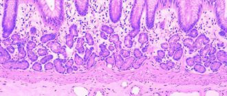
Histology of the gastric mucosa
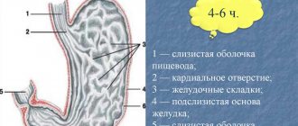
Normal histology of the gastric mucosa is represented by epithelial, glandular and lymphoid tissue. In addition, this shell contains a muscular plate consisting of smooth muscle. A feature of the mucous layer of the stomach is that there are many pits on its surface. They are located between the glands that secrete various biological substances. Next there is a layer of epithelial tissue. This is followed by the stomach gland. Together with lymphoid tissue, they form their own plate, which is part of the mucous membrane.
Glandular tissue has a certain structure. It is represented by several formations. Among them:
- Simple glands. They have a tubular structure.
- Branched glands.
The secretory department consists of several exo- and endocrinocytes. The excretory duct of the glands of the mucous membrane exits into the bottom of the fossa located on the surface of the tissue. In addition, cells in this section are also capable of secreting mucus. The spaces between the glands are filled with coarse connective fibrous tissue.
Lymphoid elements may be present in the lamina propria of the mucous membrane. They are located diffusely, but throughout the surface. Next comes the muscle plate. It contains 2 layers of circular fibers and 1 layer of longitudinal fibers. He occupies an intermediate position.
Private histology. Problem 15
15. During instrumental examination, the epithelium of the mucous membrane of the fundus of the stomach was injured. Which cells will regenerate this epithelium? Discuss the situation as you answer the following questions
1) The significance of the stomach 2) The structure of the stomach wall 3) The sources of development of the tissue systems of the stomach 4) The composition and relief of the gastric mucosa 5) The glands of the stomach, the general plan of their structure and significance. Sources of gastric epithelium regeneration
- The stomach belongs to the middle section of the digestive system. The main functions of the stomach: 1) - secretory (production of gastric juice, which includes: pepsin, chemosin, lipase, hydrochloric acid, mucus). 2) - accumulation, mixing and digestion of food by enzymes of gastric juice 3) - absorption (absorption of water, alcohol, sugars, salts) 4) - production of an antianemic factor necessary for the absorption of vitamin B12 supplied with food. 5) - excretory (release of some metabolic products through the stomach wall (ammonia, urea, etc.)) 6) - endocrine (production of gastrin, histamine, serotonin)
- The wall of the stomach in all its sections consists of four membranes: mucous membrane, submucosa, muscular and serous membranes.
- The stomach is formed in the fourth week of human embryogenesis. The single-layer prismatic epithelium of the stomach develops from the endoderm of the intestinal tube, and the connective tissue structures, blood vessels and smooth myocytes develop from the mesenchyme. Nerve structures develop from the neuroectoderm, and the mesothelium of the serous membrane from the visceral layer of the splanchnotome of the mesoderm.
- In the mucous membrane of all three sections of the stomach, three plates are distinguished. Internally, facing the lumen of the stomach - an epithelial plate, represented by a single-layer prismatic secretory epithelium (produces mucus). This is followed by the lamina propria of the mucous membrane, consisting of loose connective unformed tissue with simple tubular glands. The third plate is the muscular plate of the mucous membrane with smooth myocytes arranged in three layers: middle - longitudinal, inner and outer - circular. The relief of the mucous membrane consists of longitudinal folds (formed by the mucosa and submucosa), glandular fields - sections of the mucous membrane with glands, separated from each other by connective tissue layers, and gastric dimples - depressions of the epithelium in the lamina propria of the mucous membrane. At the bottom of the dimples, the glands of the stomach open. The deepest gastric dimples are in the pyloric region of the stomach.
- All glands of the stomach: cardiac, fundic and pyloric are located in the lamina propria of the mucous membrane and are simple, tubular, unbranched (fundic - in the body and fundus of the stomach) or branched (in the cardiac and pyloric part of the stomach with the glands of the same name). The fundic (proper) glands of the stomach are the most numerous. Each tube—gland—has a bottom and a body corresponding to the end section of the gland; neck corresponding to the excretory duct. The latter opens at the bottom of the gastric dimples. Cardiac and pyloric glands consist mainly of light mucous cells with flattened nuclei located in the basal part of the cells. The fundic glands contain chief, parietal, mucous, cervical and endocrine cells. The chief cells, the most numerous, cylindrical in shape with basophilic-stained granules in the cytoplasm, produce pepsinogen, which is converted into pepsin in an acidic environment. The cells have a well-developed granular ER and Golgi complex. Parietal (parietal) cells are large, irregular, round in shape, with oxyphilic-stained cytoplasm, in which there are many large mitochondria and intracellular secretory tubules. The cells are located outside the main cells and produce chlorides and hydrogen ions for hydrochloric acid, as well as an antianemic factor. The secretion of parietal cells is stimulated by gastrin, histamine and acetylcholine. Endocrine cells are stained with silver and chromium and produce gastrin - G-cells, histamine - Ecl-cells, serotonin - E-cells. The mucous cells of the gland are located in the body of the glands, and the cervical mucous cells are located only in the neck of the glands. In addition to secreting mucus, cervical cells are cambial for the epithelium of the glands and the integumentary epithelium of the stomach. Due to these cells, regeneration of the epithelium of the gastric mucosa is carried out.
Source
Histological structure of the gastric epithelium
The upper layer of the mucous membrane, which is in contact with food masses, is the epithelium of the stomach. The histology of this section of the gastrointestinal tract differs from the structure of the tissue in the intestine. The epithelium not only protects the surface of the organ from damage, but also has a secretory function. This tissue lines the inside of the stomach cavity. It is located over the entire surface of the mucous membrane. Gastric pits are no exception.
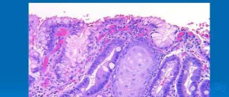
The inner surface of the organ is covered with single-layer prismatic glandular epithelium. The cells of this tissue are secretory. They are called exocrinocytes. Together with the cells of the excretory ducts of the glands, they produce secretions.
Epithelial cells in gastric contents
Epithelial cells of the gastric mucosa are found separately and in clusters together with leukocytes in patches of mucus. In an acidic environment, gastric epithelial cells look like oval (round) nuclei located nearby. In this they differ from the nuclei of epithelial cells of flat polygonal epithelium of the oral cavity and esophagus, which, due to the large size of the cells, are significantly distant from each other. In gastric contents with low acidity, epithelial cells of the gastric mucosa retain a cylindrical shape.
With hypertrophic gastritis, gastric epithelial cells are found in the form of groups, clusters, layers and casts from the gastric glands, sometimes in large numbers, in blood-stained dense patches. Sometimes they undergo mucous and fatty degeneration, as well as metaplasia, as a result of which they become rounded and flattened. A large number of epithelial cells of the gastric mucosa are detected during hypertrophic gastritis, and especially during the inflammatory process in the area of the pyloric part of the stomach.
With gastric polyps, layers of the same type of cylindrical epithelium with signs of proliferation are found: two- and three-nucleated cells with enlarged nuclei, some of them have nucleoli.
With stomach cancer, it is possible to detect atypical epithelial cells in dense shreds and mucus, sometimes located in groups and glandular formations, often with vacuolar or fatty degeneration.
With lymphogranulomatosis, Berezovsky-Sternberg cells can be found, with tuberculosis - Pirogov-Langhans giant cells, with actinomycosis - drusen actinomycetes.
Histology of the fundus of the stomach
The histology of different parts of the stomach is different. Anatomically, the organ is divided into several parts. Among them:
- Cardiac department. At this point the esophagus passes into the stomach.
- Bottom. In another way, this part is called the fundus department.
- The body is represented by the greater and lesser curvature of the stomach.
- Antrum. This part is located before the transition of the stomach into the duodenum.
- Pyloric section (pylorus). In this part there is a sphincter that connects the stomach with the duodenum. The gatekeeper occupies an intermediate position between these organs.
Lies about restoring the gastric mucosa
Hello! I have been working as a doctor for 21 years. My name is Georgy Olegovich Sapego. In this article I’ll tell you about how they fool you with all sorts of nonsense about restoring the gastric mucosa.
Again I noticed an article with lies about the stomach. People like to treat their stomach with different diets. Some people are speculating on this interest.
I will quote and immediately explain what is wrong there.
The stomach is a very sensitive organ to all irritants.
In fact, the stomach is a very resistant organ to all irritants. Acid is constantly splashing in it, which kills microbes, but we do not feel this acid.
Histology of the submucous membranes of the stomach
As in all organs, under the mucous membrane of the stomach there is a layer of fatty tissue. In its thickness there are vascular (venous and arterial) plexuses. They supply blood to the inner layers of the stomach wall. In particular, the muscular and submucosal membranes. In addition, this layer contains a network of lymphatic vessels and a nerve plexus. The muscular lining of the stomach is represented by three layers of muscle. This is a distinctive feature of this body. Longitudinal muscle fibers are located outside and inside. They have an oblique direction. Between them lies a layer of circular muscle fibers. As in the submucosa, there is a nerve plexus and a network of lymphatic vessels. The outside of the stomach is covered with a serous layer. It represents the visceral peritoneum.
Disorders of regeneration of the gastric mucosa
Disorders of regeneration of the gastric mucosa
Physiological, or reparative, regeneration of the gastric mucosa occurs in two stages. The first of them is characterized by the proliferation of germ elements represented by the epithelium of the gastric pits and the cervical part of the glands. The second stage comes down to the maturation of newly formed cells, which migrate to the corresponding sections of the glandular formations of the stomach.
The high intensity of reparative regeneration explains the fact that even relatively pronounced acute damage to the gastric mucosa is eliminated within a few days. Stomach erosions disappear within a few hours. Wounds after gastrobiopsy heal after 3-4 days. Even repeated burns of the gastric mucosa may not leave noticeable consequences.
However, the high regenerative potency inherent in the gastric parenchyma provides for the precise functioning of the regulatory mechanisms responsible for this, the constant supply of all the plastic substances necessary for the construction of new cells and, no less important, the intactness of the bulk of the cambial elements. Meanwhile, the latter are located superficially and, because of this, are easily accessible to exogenous damaging influences.
Chronic gastritis was reproduced experimentally. M. Lazovsky through repeated burns of the gastric mucosa with boiling water and other damaging effects
Brain death. Symptoms, causes and treatment Brain Death
If they are repeated, the regeneration process may be disrupted and become pathological. The basic principles of pathological regeneration of the gastric mucosa were studied by Yu. M. Lazovsky, and the main principles he established still remain in force.
Chronic gastritis was reproduced experimentally. M. Lazovsky through repeated burns of the gastric mucosa with boiling water and other damaging effects.
Pathological regeneration was primarily manifested by a violation of the process of cell differentiation. Highly specialized glandular elements (chief cells, parietal cells) were replaced by mucus-forming cells devoid of specific morphological and functional properties.
Regeneration of the gastric mucosa occurs in two stages
If the mucous membrane, which had not had time to recover, was injured again, then regeneration began to suffer at an earlier stage - the intensity of cell reproduction sharply decreased. The described experiments simulated the conditions for the development of exogenous gastritis in humans . And although, undoubtedly, such a model is simplified, the data obtained on its basis have introduced a lot of new things into the understanding of the pathogenesis of the suffering in question.
Indeed, the changes in the gastric mucosa that occur in animals turned out to be in many ways close to those established in patients with chronic gastritis.
Fracture. Symptoms, causes and treatment Fracture
Wounds after gastrobiopsy heal after 3-4 days
Damage to the glandular formations of the stomach in this disease is characterized by signs of dedifferentiation of the main and parietal cells. They undergo “mucusing” and in terms of the amount of mucoid substances they become similar to integumentary epithelial and cervical cells. Further progression of changes in the glandular apparatus of the stomach is associated with the development of its atrophy and restructuring. The expression of the latter is the appearance of elements not characteristic of the fundic mucosa: pseudopyloric glands and epithelial cells inherent in the intestine. The so-called enteralization of the gastric mucosa can be considered as a consequence of metaplasia of its epithelium.
Benign neoplasms of the stomach and intestines: histology of hemangioma
One of the benign neoplasms is hemangioma. Histology of the stomach and intestines is necessary for this disease. Indeed, despite the fact that the formation is benign, it should be differentiated from cancer. Histologically, hemangioma is represented by vascular tissue. The cells of this tumor are fully differentiated. They are no different from the elements that make up the arteries and veins of the body. Most often, gastric hemangioma forms in the submucosal layer. The typical location for this benign neoplasm is the pyloric region. The tumor can have different sizes.
In addition to the stomach, hemangiomas can be localized in the small and large intestines. These formations rarely make themselves felt. However, diagnosing hemangiomas is important. With large sizes and constant trauma (chyme, feces), serious complications can arise. The main one is profuse gastrointestinal bleeding. A benign neoplasm is difficult to suspect, since in most cases there are no clinical manifestations. An endoscopic examination reveals a dark red or bluish round spot rising above the mucous membrane. In this case, a diagnosis of hemangioma is made. The histology of the stomach and intestines is of decisive importance. In rare cases, hemangioma undergoes malignant transformation.
Gastric regeneration: histology in ulcer healing
One of the indications for histological examination is gastric ulcer. For this pathology, an endoscopic examination (FEGDS) is performed with a biopsy taken. Histology is required if an ulcer is suspected of malignancy. Depending on the stage of the disease, the tissue obtained may vary. When the ulcer heals, the stomach scar is examined. In this case, histology is needed only if there are symptoms due to which malignant degeneration of the tissue can be suspected. If there is no malignancy, then the analysis reveals cells of coarse connective tissue. When a gastric ulcer becomes malignant, the histological picture may be different. It is characterized by changes in the cellular composition of the tissue and the presence of undifferentiated elements.
Stomach under a microscope
Return to list Ask your question
Today we’ll talk about microscopying the tissues of an important human organ that functionally carries out physical and chemical processing of food. The study of the stomach under a microscope is part of the histology course. Of course, it is impossible to prepare such a microslide on your own in amateur home conditions, so for novice biologists we recommend using ready-made samples. Having studied the theoretical part, it is possible to make meaningful observations of the biomaterial.
The stomach is an integral component of the digestive tract; it is a hollow organ containing enzymes for breaking down proteins and fats. It is located between the initial part of the small intestine and the esophageal canal. The total space occupied is on average up to one and a half liters. This volume may vary depending on the content of food or water.
In addition to the named main function, the stomach performs a number of others: absorption of nutrients during the digestion of food by gastric juice (it is produced by cells of the mucous membrane), protection against parasites and bacteria (production of hydrochloric acid), production of organic compounds with high physiological activity.
Stomach tissues that can be viewed under a microscope:
The mucous membrane is a kind of protective system with bactericidal properties. Prostaglandins stimulate the formation of mucus, and they also significantly improve microcirculation;
The integumentary epithelium is a pitted epithelium, represented by stem cells capable of regeneration and renewal, which occurs within four days. The cytoplasm contains mucopolysaccharides that prevent cellular self-digestion;
Three layers of muscle and connective tissue. Smooth muscles are designed to mix (compress) incoming food and subsequently move along the digestive tract;
According to the rules of microscopy, to examine the stomach under a microscope, it is necessary to prepare a microspecimen. The biomaterial is collected in an anatomical laboratory and fixed in formaldehyde. Fixation blocks putrefaction and promotes protein folding. Next, you need to impregnate with paraffin, freeze and cut into small pieces with a microtome.
After mounting the sample on a glass slide, staining is carried out. Cellular structures are perfectly visualized by hematoxylin-eosin staining. Hematoxylin will stain the nuclei, and eosin will stain the protoplasm of the cells. After dewaxing in xylene, the section is washed with distilled water, and a hematoxylin solution is applied with a pipette for two minutes. After repeated rinsing, apply xanthene dye “Eosin” and rinse again.
The final step is to dehydrate in alcohol and add a drop of Canada balsam. Next, cover and press tightly with a glass cover slip. The prepared preparation is placed on the microscope stage and viewed under transmitted light in a bright field.
The magnification should be changed gradually. At 40 times the general outlines of the structure are already visible. You need to adjust the backlight and condenser, focus, achieving image clarity. Then we step by step increase (increase) the magnification to the maximum, changing lenses to 10x and 1000x. At a thousandfold zoom, studies are carried out in oil immersion.
For the described experiment, binocular models are suitable: Microhoney 1 var. 2-20, Biomed 3, Levenhuk 720B. If necessary, you can connect a digital eyepiece camera, for example, ToupCam 5 MP. It allows you to take photographs of what you see and measure the elements of the observed microstructure.
What is the purpose of gastric histology?
One of the organs of the digestive tract in which neoplasms often develop is the stomach. Histology should be performed if there is any change in the mucous membrane. The following diseases are considered indications for this study:
- Atrophic gastritis. This pathology is characterized by depletion of the cellular composition of the mucous membrane, inflammatory phenomena, and decreased secretion of hydrochloric acid.
- Rare forms of gastritis. These include lymphocytic, eosinophilic and granulomatous inflammation.
- Chronic peptic ulcer of the stomach and duodenum.
- Development of “small signs” according to Savitsky. These include general weakness, decreased appetite and performance, weight loss, and a feeling of abdominal discomfort.
- Detection of stomach polyps and other benign neoplasms.
- A sudden change in the clinical picture of a long-standing peptic ulcer. These include a decrease in the intensity of pain and the development of an aversion to meat food.
The listed pathologies refer to precancerous diseases. This does not mean that the patient has a malignant tumor and its location is the stomach. Histology helps determine exactly what changes are observed in the tissues of the organ. To prevent the development of malignant degeneration, it is worth conducting research as early as possible and taking action.
Functions
The epithelium of the stomach consists of villi, through which the absorption of breakdown products of various substances occurs. Due to the fact that there are a lot of villi, the surface of the mucous membrane of the small intestine becomes larger. The products of the breakdown of nutrients are absorbed into them. Absorption itself is a complex physiological process, due to the active transfer of substances through the walls of the villi. The villi perform a protective function, preventing microorganisms found in the intestines from entering the blood and lymph. They get into the intestines if we do not adhere to the rules of hygiene. Also, red blood cells, which have the shape of nuclear-free biconcave discs, carry oxygen, which contains hemoglobin, throughout the cells of the body.
Gastric histology results
The results of histological examination may vary. If the organ tissue is not changed, then microscopy reveals normal prismatic single-layer glandular epithelium. When taking a biopsy of deeper layers, you can see smooth muscle fibers and adipocytes. If the patient has a scar from a protracted ulcer, then rough fibrous connective tissue is found. For benign formations, histological results may be different. They depend on the tissue from which the tumor developed (vascular, muscle, lymphoid). The main feature of benign formations is cell maturity.
Sampling of stomach tissue for histology: methodology
To perform a histological examination of stomach tissue, it is necessary to perform a biopsy of the organ. In most cases, it is performed using endoscopy. The apparatus for performing FEGDS is placed into the lumen of the stomach and several pieces of organ tissue are cut off. It is advisable to take biopsies from several distant sites. In some cases, tissue for histological examination is taken during surgery. After this, thin sections from the biopsy are taken in the laboratory and examined under a microscope.
How long does a histological analysis of stomach tissue take?
If cancer is suspected, gastric histology is necessary. How long does this analysis take? Only the attending physician can answer this question. On average, histology takes about 2 weeks. This applies to planned studies, for example, when removing a polyp.
During surgery, urgent histological examination of the tissue may be necessary. In this case, the analysis takes no more than half an hour.