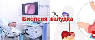
Equipment for taking biopsies
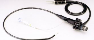
The main instrument for performing a gastric biopsy is a fibrogastroscope. It is a rigid but flexible probe. At its distal end there are light guide windows, a lens, an opening for instruments, and openings for water and air supply.
The control unit and eyepiece are located on the fiberscope handle.
Special biopsy forceps are used to take samples of the mucous membrane for examination. Sometimes a completely removed polyp is sent for a biopsy. In this case, an executory loop is used.
There should be containers in the operating room to hold the samples taken.
How to decipher the results
The biomaterial obtained as a result of the biopsy is placed in a special preservative solution and sent to the laboratory. A morphologist prepares tissue sections, stains them with special dyes and studies the histology of the gastric epithelium under a microscope. The waiting time for a conclusion is about 7 days.
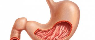
The structure of the mucous membrane is normal
The gastric mucosa is normally composed of three layers:
- Epithelial layer. It consists of a single layer of mucoid cells that synthesize mucus, which protects the walls of the organ from the action of digestive juices.
- The lamina propria of the mucous membrane. Its basis is made up of connective tissue fibers. It contains digestive glands, small blood and lymphatic vessels. Each section of the stomach has its own type of glands. In general, the main glands of the fundic and cardiac sections predominate.
- Muscular layer of the mucosa. Represented by smooth muscle cells. I have maximum muscle development in the pyloric section of the stomach.
Preparing for a biopsy
A gastric biopsy is performed during the fibrogastroscopy procedure. The patient does not even notice any difference from conventional FGDS; it is possible that the procedure will only take 5-10 minutes longer.
Special preparation for routine endoscopy is usually not required. Patients with particularly labile psyches are prescribed premedication (tranquilizer + antispasmodic + atropine).
In some cases, FGDS is performed under intravenous anesthesia (for children and patients with mental illness).
It is not recommended to eat or drink 6 hours before EGD; drink no later than 2 hours.
Sometimes preliminary gastric lavage is necessary (for example, with pyloric stenosis, the rate of evacuation of food from the stomach can slow down significantly).
Types of biopsy
The biopsy is performed:
- using endoscopy;
- surgical method.
There are several types of procedure depending on the method of performing the procedure:
- Sighting (visual). It is done using a special medical device - a gastroscope. This device looks like a long tube with a built-in lighting system. It contains an endoscope and a micro-instrument, with the help of which material is taken from the gastric mucosa.
- Search engine. Carry out with a biopsy probe. In this case, there is no visual control over the procedure.
- Open. Performed during surgery.
The most common procedure is endoscopic biopsy. Using this method, an Hp biopsy is also performed (testing for the presence of Helicobacter).
Contraindications to endoscopic manipulation of the stomach
Absolute contraindications
- Course of acute stroke.
- Acute myocardial infarction.
- Esophageal stenosis, impassable to the probe.
- An attack of bronchial asthma.
Relative contraindications
- Inflammatory processes in the pharynx.
- Feverish condition.
- Hemorrhagic diathesis.
- Epilepsy.
- Mental illnesses.
- Heart failure.
- High arterial hypertension.
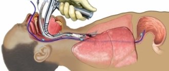
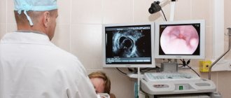
Progress of the FEGDS procedure with biopsy taking
The procedure is performed under local anesthesia - the pharynx is irrigated with a 10% lidocaine solution.
The gag reflex is suppressed (the most unpleasant thing about this procedure). After passing through the pharynx, the procedure is virtually painless. The patient lies on a special table on his left side. A mouthpiece is inserted into the mouth, and the endoscope probe is inserted through it. The doctor examines sequentially all parts of the esophagus, stomach and duodenum.
To straighten the folds and obtain a better view, air is supplied into the esophagus and stomach through a fiberscope.
If a suspicious area is detected, the doctor inserts biopsy forceps into the instrument port of the fiberscope. The material is collected by “biting out” the tissue with forceps.
Rules for collecting mucosal areas for biopsy:
- For gastritis, at least 4 sections of the mucous membrane are taken (2 fragments each from the front and back walls)
- For tumors and ulcers - an additional 5-6 fragments of mucous membrane from the center of the lesion and the periphery.
The probability of making a diagnosis when taking a biopsy from at least eight points increases to 95–99%.
Chromogastroscopy
This is an additional method of endoscopic examination.
It is used to clarify the diagnosis of diseases that are difficult to distinguish during normal endoscopic examination. Most often this concerns benign and malignant diseases, especially early forms, as well as determining the boundaries of tumor lesions and degenerative changes in the mucous membrane.
The method involves spraying the dye over the gastric mucosa. Methylene blue, Congo red, and Lugol's solution are used as dyes.
As a result, the altered areas of the mucosa appear more colored compared to the normal mucosa. A biopsy is taken from these areas.
Indications for the study
Histological examination is a specific diagnostic method, which is based on the study of tissues under the magnification of a microscope, with the help of which one can obtain information about the structure of cells, the rate of their division, and the state of the microvascular bed. This procedure allows the specialist to make the correct diagnosis and select the optimal treatment.
Indications for histology of the gastric mucosa are:
- Hyperacid gastritis (inflammation of the mucous membrane, accompanied by increased production of concentrated gastric juice).
- Atrophic (hypoacid) gastritis is a precancerous condition.
- Peptic ulcer (active and chronic forms).
- Special forms of gastric inflammation (granulomatous, lymphocytic, eosinophilic gastritis).
- Barrett's esophagus, polyposis and other gastrointestinal pathologies.
- Clinical observation of patients at risk (complicated family history, exposure to carcinogens), people who have undergone surgery on the gastrointestinal tract.
- To determine the extent of surgical intervention during planned surgery on the digestive tract.
- Evaluation of the effectiveness of treatment (radiation, chemotherapy, surgery).
A biopsy with a histological examination of the gastric mucosa should be performed for the purpose of differential diagnosis for dysphagia (impaired and difficulty swallowing), dyspepsia (abdominal pain, heartburn, belching, discomfort after eating) and other alarming symptoms (weakness, weight loss, aversion to meat foods) ).
In addition to the ability to detect the inflammatory process and pathologically altered cells, histological analysis allows us to identify some pathogens of infectious diseases, for example, Helicobacter pylori.
How is a biopsy specimen examined?
A tissue sample taken during gastroscopy is placed in a container with a preservative, labeled, numbered and sent to the histology laboratory.
The study is carried out by a pathologist. The tissue sample must be cut into thin sections suitable for examination under a microscope (that is, almost transparent). To do this, the material must be compacted and cut with a special cutting device.
Paraffin is used for compaction (for routine research) or the sample is frozen (for urgent analysis).
Next, microscopic sections are made from the frozen dense sample. A microtome is used for this.
The sections are placed on glass and stained. The finished preparations are examined under a microscope.
The pathologist, when examining the biopsy specimen, states in his conclusion:
- Thickness of the mucous membrane.
- The nature of the epithelium with clarification of the degree of secretion (atrophy, hypertrophy or normal secretion).
- The presence of dysplasia and metaplasia of the epithelium.
- The presence of inflammatory infiltration, the depth of its spread, the degree of inflammation activity. It is assessed by the number of lymphocytes, plasma cells, eosinophils infiltrating the mucosa.
- Signs of atrophy or hyperplasia.
- Presence of Helicobacter pylory and degree of contamination.
Detection of dysplasia, metaplasia, and atypia is based on visual analysis of cells. Cells belonging to a certain tissue have the same structure. If cells are detected that are not characteristic of a given tissue, altered, and not similar to neighboring ones, this is called dysplasia, metaplasia or atypia.
The main signs of malignant cell atypia:
Other cell sizes (tumor cells tend to be much larger than normal tissue cells).- Cell shape. Polymorphism is noted, the cells are completely different in shape, which is not typical for normal tissue.
- Increase in nuclear size, polymorphism, fragmentation of nuclei.
- A large number of dividing cells in smears.
- Disruption of normal communication between cells: indistinguishability of cell boundaries or, conversely, separation of cells.
- Inclusions in the cytoplasm, vacuolization of the cytoplasm.
There are reliable morphological changes that are classified as precancerous conditions, that is, in the presence of such changes, the risk of developing stomach cancer is several times higher:
- Adenomatous polyps. These are benign neoplasms originating from glandular cells. They have a very high probability of cancerous degeneration.
- Intestinal metaplasia of the gastric mucosa. This is a situation where part of the gastric epithelium is replaced by villous epithelium of the intestine.
- Chronic atrophic gastritis. With this gastritis, a sharp decrease in the number of glands is revealed in a biopsy of the mucous membrane.
- Chronic gastritis type B. This is chronic antral gastritis associated with Helicobacter pylori infection.
- Xanthomas of the stomach. These are accumulations of fat cells in the stomach lining.
- Menetrier's disease. A disease in which overdevelopment of the gastric mucosa occurs with the development of adenomas and cysts in it.
What result can you get?
The histology of the biopsy material and correct decoding make it possible to accurately confirm almost any disease of the stomach. The most common of them include:
- Erosive and ulcerative-necrotic changes in the mucous membrane and the wall of the organ being examined with a history of prolonged gastritis or gastric ulcer.
- Atrophic changes in the wall of the stomach - a decrease in the amount of glandular tissue, which leads to a decrease in the protective properties of the mucous membrane of the organ.
- Complete or incomplete metaplasia of the gastric epithelium is a precancerous condition. In this case, normal glandular tissue is replaced by epithelium, which contains the intestines, i.e. accumulation of intestinal-type epithelium is observed.
- Benign neoplasms, gastric polyps - the cellular elements of the tumor have a high degree of organization, tissue and cellular differentiation.
- Malignant neoplasms or stomach cancer - most often adenocarcinoma is confirmed by histological examination.
Today, biopsy is the gold standard test for confirming the clinical diagnosis of most diseases of the upper gastrointestinal tract.
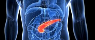
Stomach cancer
It's no secret that taking a biopsy is aimed primarily at excluding a malignant process.
Gastric cancer is one of the most common malignant tumors. The early stage of stomach cancer usually occurs without any symptoms. Therefore, it is so important to identify the tumor and begin treatment in the early stages. It is impossible to overestimate the importance of a biopsy from suspicious areas here.
According to histological type, the following forms of stomach cancer are distinguished:
- Adenocarcinoma is the most common type of cancer, originating from glandular cells, and can be differentiated or undifferentiated.
- Signet ring cell carcinoma.
- Squamous cell carcinoma.
- Adenosquamous cell carcinoma.
- Small cell cancer.
- Undifferentiated cancer.
The histological type of cancer is very important in determining prognosis and treatment tactics. Thus, poorly differentiated adenocarcinoma, undifferentiated and signet ring cell carcinoma are considered the most malignant. The cells of these tumors are poorly connected to each other and spread quite easily through the lymphatic and blood vessels.
It has been proven that contamination of the gastric mucosa with the bacterium Helicobacter pylory increases the risk of developing stomach cancer in patients with chronic gastritis. This microbe causes atrophy of the epithelium and leads to metaplasia and dysplasia.
Therefore, in recent years, the histological report has been required to indicate the presence of this bacterium in the material, as well as the degree of contamination.
Prices in the European clinic for the diagnosis of stomach cancer
- High-definition EDGS with digital recording, diagnostic - from 14,300 rubles.
- X-ray contrast examination of the esophagus and stomach - 20,700 rubles.
- Ultrasound examination of the abdominal cavity (liver, gallbladder, bile ducts, spleen, pancreas), including determination of the level of free fluid - 7,000 rubles.
- Diagnostic laparoscopy - 57,500 rubles.
But it’s not just about prolonging life. The goal of treatment in the majority of even advanced cases is to improve its quality, relieve the person from many painful symptoms, including pain, provide him with the opportunity to lead a normal lifestyle, engage in activities that are important to him, and communicate with loved ones.
Modern methods of treating stomach cancer used in the European Clinic allow treatment with minimal hospitalization and the use of minimally invasive techniques that improve not only the prognosis for life, but also the quality of life of our patients.
Additional modern research
Examination of a tissue sample under a conventional light microscope is usually sufficient. An experienced doctor is able to quickly assess the morphological picture and see cell atypia. But sometimes other methods are used to clarify:
- Electron microscopy. Examination under an electron microscope allows you to examine all cell organelles. Images can be photographed and stored in computer memory for later comparison. The disadvantage of electron microscopy is that only a few cells are visible in the field of view.
- Immunohistochemical methods. The method is based on the principle of antigen-antibody interaction. In some doubtful cases, special sera are used that contain antibodies to certain molecules that are unique to certain tumor cells.
Main conclusions
- This procedure is almost painless.
- A biopsy is necessary to establish a definitive histological diagnosis.
- The quality of the analysis largely depends on the skill of the doctor taking the biopsy and the morphologist conducting the histological examination.
- The doctor may issue a dubious conclusion, which will indicate a suspicion of malignancy of the process. In this case, a repeat biopsy will be required.
When dysplasia and metaplasia are detected in tissues, particularly careful observation and repeated examinations at certain times, as well as treatment, are necessary.
© Operation.Info
Treatment of the disease depending on the results of the examination
A biopsy is considered a diagnostic procedure, after which the treatment tactics will be correct.
It all depends on the test results. If a biopsy is performed if a tumor is suspected, then using this method it is possible to determine the type of formation and composition. The result will be final. If the conclusion is negative, then the specialist may prescribe the test again.
Surgical treatment is carried out only if the atypical cellular composition is confirmed. Treatment of the disease depends on the results shown by the biopsy.