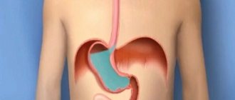
Hyperplasia refers to the abnormal activity of cells, which results in their excessive growth and deformation (metaplasia), and the formation of benign formations that can develop into malignant ones (malignization). All organs are susceptible to pathology, but gastric hyperplasia is more common. Literally translated as “overeducation.” All tissues and layers of an organ can change.
Gastric hyperplasia is a fairly common phenomenon.
The concept of pathology
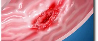
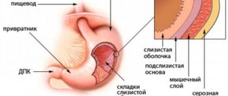
The disease is based on the natural process of cell division, which is normally necessary for the body. However, under the influence of certain factors, the process becomes excessive, which is fraught with the development of oncology. Most often, changes occur at the external level - hyperplasia of the gastric mucosa. Due to cell division, it thickens and polyps appear. Why is this phenomenon popularly called “fiery polyp”.
This is one of the most common stomach diseases. In the early stages it is easily treatable. While advanced forms can become chronic, which cannot be eliminated. Against this background, foveal hyperplasia of the gastric mucosa occurs (destruction in the endometrium). In addition, the disease can affect the antrum and cardiac regions, body and bottom of the organ.
Reasons for development
The main reason is prolonged irritation of the mucous membrane, leading to injuries and wounds. The reasons are:
- Chronic diseases (gastritis, ulcers and other inflammations) and advanced infections (intestinal, rotavirus). Excessive division is a defensive reaction to the aggressor. For example, against the background of chronic lymphoid gastritis (focal accumulation of lymphocytes in the epithelium in the form of follicles), lymphofollicular hyperplasia of the stomach of the 1st degree may develop. It is important to note that it begins to manifest itself only from stage 3, before which it can be detected by chance during FGS.
Various inflammatory processes in the stomach can lead to hyperplasia
- Predisposition at the genetic level.
- Hormonal imbalance or formation affecting hormones. For example, a pancreatic tumor provokes excess acid production in the stomach, to which the organ responds with additional cell proliferation.
- The parasite Helicobacter pylori is a bacterium that contaminates the body with its waste products, weakening its defenses and destroying the upper layer of the stomach, gradually penetrating deeper. It is fraught with the development of the most dangerous type - hyperplasia of the integumentary pitted epithelium of the stomach. Structural and secretory changes occur, and cancer may develop.
- Unhealthy diet, which is dominated by additives, preservatives, carcinogens (additives of group E), excessive addiction to alcohol.
- Long course of taking non-steroidal drugs.
- Stress, regular overexertion.
- Impaired functioning of the parasympathetic nervous system and secretory function of organs. Changes in the functioning of the duodenum provoke the release of gastrin, which irritates the mucous membrane. Against this background, lymphofollicular hyperplasia of the antrum of the stomach can develop.
Stress can also trigger this disease
Types and forms of flow
Depending on which parts of the stomach and tissue are affected, several types and forms of the disease are distinguished. All of them are reflected in the table.
| View | Description |
| Foveal hyperplasia of the stomach | Deformation of the folds of the stomach occurs (increase in length and curvature), gastric pits and their epithelium. The most common and least dangerous type. More often caused by taking non-steroidal drugs. |
| Antrum | Proliferation of tissue at the junction of the stomach and duodenum (antrum). Externally it is expressed by multiple small growths. The reason is nutritional deficiencies, since this department accounts for the bulk of the digestion work. |
| Lymphofollicular | Multiple lymphocytes accumulate in the follicles, the tissue thickens and grows. It is caused by all the previously discussed reasons, gastritis is especially dangerous. Since this combination can result in oncology. |
| Lymphoid with mucosal lesions | An increase in lymphocytes, thickening of the mucosa and its hyperplasia. Causes infections and ulcers. |
| Lymphoid hyperplasia of the antrum of the stomach | Reproduction of lymph node tissue. The consequences are similar to integumentary pitting and lymphofollicular. Caused by infection and ulcer. |
| Glandular | The glandular epithelium grows, round and oval polyps are formed. Caused by an increase in the size of the stomach. The rarest type. |
| Polypoid | The formation of multiple polyps in any part of the stomach. |
| Integument of pit epithelium | The cells responsible for the production of protective mucus grow. |
| Fine grain | Characterize the size of the lesion. |
| Coarse grain | |
| Diffuse | Overgrowth of all types of tissue over the entire surface and cavity. Often combined with a chronic course. |
| Focal hyperplasia of the gastric mucosa (“wart”) | Formation of additional tissue in one or more places. Characteristic of the first stages of the disease, the formations are benign. |
Regeneration of epithelial tissues
Read:- II. Traumatic regeneration.
- Alteration. Dystrophies. Regeneration.
- ANI SOFT TISSUE.
- Types of muscle tissue.
- Types of epithelial tissues
- Age-related changes and regeneration
- Opening abscesses and soft tissue phlegmons.
- Histogenesis and regeneration of muscle tissue
- Degeneration and regeneration of nervous tissue.
- Degeneration and regeneration of peripheral nerve fiber
Epithelial tissues have a high ability to regenerate. Regeneration can be physiological, which occurs constantly in a healthy body - the integumentary epithelium is constantly exposed to environmental influences, so the epithelial cells wear out and die relatively quickly; and reparative regeneration, which occurs as a result of damage. Regeneration is carried out due to cambial cells, the location of which is different. In the single-layer multirow ciliated epithelium of the trachea, the intercalated cells are cambial. In the mesothelium, cambial cells are located diffusely. In the single-layer prismatic glandular epithelium of the stomach, cambial cells are located at the bottom of the gastric pits. In the single-layer prismatic bordered epithelium of the small intestine, cambial cells are located at the bottom of the crypts. In all other epithelia, cambial cells are predominantly found in the basal layer.
Glands, classification, sources of development. The glands are divided into 2 groups: 1) internal secretion - endocrine - they produce hormones that enter the blood, the glands do not have excretory ducts - the pituitary gland, pineal gland, adrenal glands, parathyroid and thyroid glands. 2) external secretion - exocrine glands - produce secretions released into the external environment, have secretory or terminal sections or excretory ducts - salivary glands, gastric glands, sebaceous glands. The chemical composition of the secretion can be different: protein, mucous, protein-mucous, sebaceous. According to the method of secretion removal: Meracrine, apocrine and holocrine type. Sources of development. Adrenal gland: cortex of coelomic epithelium, medulla of neural origin. Pituitary gland: neurohypophysis of neural origin, adenohypophysis of ectodermal origin. parathyroid and thyroid glands from epithelial tissue. All exocrine glands develop from epithelium (parenchyma). The salivary glands develop from the epithelium of the oral cavity. Mammary gland from ectodermal epithelium.
Secretory cycle. Types of secretion. Secretion is a complex process, including 4 phases: 1) absorption of various organic and inorganic substances of water from the blood of lymph by glandulocytes. 2) from these products, secretions are synthesized in the EPS and enter the AG, where they accumulate and are modified. 3) Secretory granules are released from the cell. Secretion is released differently, and therefore three types of secretion are distinguished. Types of secretion: 1) Merocrine - the glandular cell completely retains its structure (salivary glands). 2) Apocrine - partial destruction of glandular cells occurs, that is, the apical part of the cytoplasm of glandular cells (mammary gland) is separated along with secretory products. 3) Holocrine – complete destruction of glandular cells (sebaceous glands). 4) Restoration of the original state of glandular cells.
Date added: 2015-05-19 | Views: 1255 | Copyright infringement
| | | 4 | | | | | | | | | | | |
Symptoms
In the first stages of development, the disease does not make itself felt, therefore the symptoms listed below refer to the moment when hyperplasia has significantly developed at the internal level. Some species are especially dangerous. For example, lymphofollicular hyperplasia of the gastric mucosa, being a harbinger of oncology, requires special attention and timely treatment.
A sign of hyperplasia may be severe pain in the stomach
Common signs include:
- Constant pain of different nature: aching, cutting, stabbing, burning, “hungry.”
- Loss of appetite, belching (in advanced stages - with blood), hiccups.
- In later stages - nausea and vomiting.
- Bloating and flatulence.
- Abnormal stool (usually diarrhea due to involuntary contraction of the muscles of the digestive organs).
- General weakness, signs of intoxication (fever, aches, headaches and dizziness).
- Pale skin due to poor circulation.
- Feeling of muscle tension or cramps, cramping.
General malaise, weakness and fatigue are common
As you can see, the symptoms are not specific; they are similar to the manifestations of gastritis, ulcers, common intestinal disorders and a number of other inflammations. At the same time, the more advanced the situation, the more external manifestations arise, and their severity intensifies. That is why great importance is given to the diagnostic stage, which allows us to determine the specific type and nature of the disease. This way, it is possible to promptly identify and prescribe effective treatment for hyperplasia of the integumentary pit epithelium of the stomach - the most common and susceptible to the influence of therapy type, but no less dangerous than others.
On the problem of proliferation of the integumentary epithelium of the stomach: specifics of the disease
Thanks to modern medical research (fibroscopy), proliferation of the integumentary pituitary epithelium of the stomach can be detected with great accuracy, because if treatment is started in advance, it has a favorable outcome.
Proliferation is the pathological growth of cells. It can be benign or malignant. A biopsy is performed to confirm the diagnosis. There are other modern methods for detecting the disease at any stage. Focal hyperplasia of the gastric mucosa is more common.
Although this disease has many varieties.
Origins of the problem
With gastric hyperplasia, cells divide and multiply in increased numbers. Moreover, in the advanced stage of the disease, changes begin in the cells themselves. They turn malignant.
What causes cells to multiply so rapidly?
There may be several reasons:
- hormonal imbalances in the body;
- advanced chronic inflammation in the digestive organ - gastritis, polyps, ulcers;
- stomach infections that have not been completely cured;
- impaired nervous regulation of the digestive organ;
- regular exposure of the stomach to all kinds of carcinogens (including from smoking and alcohol);
- a hereditary component that promotes the proliferation of epithelial cells.
If you suspect hyperplasia, you should immediately consult a doctor. And if an illness is detected, treatment must begin immediately.
What should you be wary of?
The insidiousness of the disease lies in the fact that hyperplasia is asymptomatic for a long time, so a person does not worry.
As a result, when a chronic advanced form of proliferation of the integumentary epithelium of the stomach has developed, treatment begins with a delay.
The thin layer of epithelium that lines the walls of the stomach begins to grow rapidly.
How not to be late and seek treatment on time:
- any recurring indigestion, stomach pain, heartburn, belching should be a reason to consult a doctor;
- You should regularly undergo a preventive examination and medical examination, during which a stool test for occult blood is performed;
- If someone in your family suffered from chronic gastritis, peptic ulcer, or hyperplasia of the integumentary pitted epithelium of the stomach, you should also undergo a mandatory preventive examination by a gastroenterologist with appropriate examination.
Here are the signs that begin to worry when epithelial growth is already in force:
- cramping and very noticeable pain in the abdominal area due to involuntary contractions;
- symptoms of anemia may appear;
- it is possible that indigestion may occur during or after a meal;
- Pain in the digestive organ also occurs at night on an empty stomach.
To think that this is ordinary, simple gastritis and that everything will go away on its own is naive. Self-medication is strictly forbidden! You need to go to the doctor immediately.
In different variations
Gastric hyperplasia has different manifestations.
The types of this disease differ in that they affect different parts of the stomach. Thus, we distinguish:
- follicular hyperplasia;
- lymphoid hyperplasia;
- hyperplasia of the integumentary pitted epithelium of the stomach;
- glandular hyperplasia;
- polypoid;
- antral;
- foveolar.
The exact causes of the disease have not yet been established. There are only provoking factors. But sometimes hyperplasia occurs when the mucosa is healthy.
Starts from the hearth
The focus of hyperplasia in the stomach is a polyp in its early development, which has a benign course and is located in one specific place, the focus. Hence the name.
This disease is determined by the endoscopic method and using special dyes. The lesion is immediately colored. It can be large, small, multiple or single. It is usually formed as a result of erosion of the gastric mucosa.
Erosion of the mucous membrane, as a rule, provokes quite noticeable pain. Therefore, those suffering from this disease may experience cramping pain. They recur periodically, often arising as a reaction to provoking foods.
In addition, another sign of the presence of focal hyperplasia is developing anemia. Its symptoms may appear from time to time. And this is also a reason to contact a gastroenterologist.
In the treatment of focal hyperplasia, medications are used, as well as dietary nutrition that completely excludes fats. Medicines are selected depending on the root cause of the disease. In most cases these are hormonal drugs.
Foveal, lymphoid, glandular hyperplasia
During an endoscopic examination, an asymptomatically developing disease, foveolar hyperplasia of the stomach, can be discovered completely by accident.
The foveal form of the disease is the uncontrolled proliferation of cells in the mucous membrane or tissues of the digestive organ. It leads to curvature and thickening of the folds of the mucous membranes of the stomach, lengthening its sections.
The disease does not form either a malignant or benign tumor. But it is the initial stage to the development of hyperplastic polyps.
Lymphoid hyperplasia leads to inflammation of the lymph nodes.
Glandular hyperplasia leads to the formation of growths in place of the internal glands.
Polyp-like hyperplasia occurs with the formation of polyps, which can affect not only the digestive organ, but also the intestines.
Follicular problem
Follicular hyperplasia of the stomach is also disguised as other diseases in the early stages. But this type of disease is insidious in that it can unnoticeably develop into a malignant form.
It should be noted that follicular gastritis is quite rare - one in a hundred. What it is is when white blood cells accumulated at the site of mucosal damage transform into follicles.
The cells grow gradually without causing problems. And only when there are a lot of these cells does the patient experience painful and unpleasant sensations.
The following symptoms should alert you:
- seemingly causeless increase in body temperature;
- weakness in the body;
- occasional pain in the digestive organ.
When a person has another disease, for example, gastritis, the patient will feel symptoms characteristic of it.
The presence of follicles can only be determined if you go to the clinic about disturbing problems.
Important! If follicular hyperplasia begins to bother you with frequent, severe pain, there is a possibility that the disease has turned into a malignant form.
Diagnostics
Due to its asymptomatic onset, the disease is difficult to diagnose in time; its presence is often discovered by chance during a routine examination. Therefore, it is recommended to undergo them once every six months, especially if a person is aware of his predisposition and risks of developing hyperplasia.
It is necessary to contact a gastroenterologist, and, if necessary, an oncologist.
The main diagnostic method is fibrogastroduodenoscopy
An examination in a doctor’s office begins with collecting an anamnesis (the course of the disease according to the patient, a story about his usual lifestyle and family). FGDS (fibrogastroduodenoscopy) is the main diagnostic method. Allows you to examine the stomach from the inside and evaluate the lesions, their scale, nature and specificity. It is during this procedure that focal foveal hyperplasia of the stomach becomes noticeable.
Sometimes FGDS is supplemented by a biopsy (sampling of foreign tissue), which, through histological laboratory examination, helps determine the presence of bacteria and the nature of the neoplasm (benign, malignant).
An X-ray with contrast is indicative - the patient drinks barium, after which an examination is carried out. Allows you to determine the size of polyps, their shape and contours. Since the root cause may be another disorder in the functioning of the body, to complete the picture, they take a blood test (general and chemical), feces and urine, and sometimes gastric juice. They also help identify Helicobacter, which can be diagnosed by the presence of antibodies in the blood, antigens in the stool, the bacterium itself in a biopsy, or a positive urea breath test. In addition, to establish the root cause, an ultrasound of internal organs (pancreas, liver) can be performed.
Additionally, ultrasound of internal organs is recommended
Follicular hyperplasia of the stomach develops and is asymptomatic, except for a general deterioration in health. It can only be detected during a special examination!
Functions:
- separates the internal environment of the body from the external environment;
- participates in the metabolism of the body with the environment, performing the functions of absorbing substances (absorption) and releasing metabolic products (excretion). For example, through the intestinal epithelium, products of food digestion are absorbed into the blood and lymph, which serve as a source of energy and building material for the body, and through the renal epithelium, a number of nitrogen metabolism products are released, which are waste products for the body.
- protective - protecting the underlying tissues of the body from various external influences - chemical, mechanical, infectious, etc. For example, the skin epithelium is a powerful barrier to the penetration of microorganisms and many poisons.
- the epithelium covering the internal organs located in the cavities of the body creates conditions for their mobility, for example, for contraction of the heart, excursion of the lungs, etc.
Treatment
Treatment of gastric hyperplasia depends on the results of a comprehensive study, first of all, on the identified root cause.
Almost all types of hyperplasia are characterized by the formation of polyps, which come in different types. Therefore, the treatment has its own specifics. Large polyps (more than 1 cm) are eliminated exclusively endoscopically. Polyps caused by heredity are more often malignant. As a result, removal is required: endoscopic or open. Glandular polyps have the same character and the same fate.
Polyps are removed using endoscopic surgery
Small polyps of other origin do not require removal (unless malignancy is individually detected). Often they are not touched because they do not cause harm. But in this case, it is recommended to monitor their development (inspection once every six months) and, if necessary (increase in size, transition to a malignant neoplasm), remove them immediately.
Treatment of foveal hyperplasia of the stomach begins with discontinuation of the drugs that caused it. Due to the fact that it is provoked by the loss of the ability of cells to regenerate (ulcers and erosions), the course of therapy is aimed at eliminating inflammation (irritation) of the mucous membrane and the primary disease. The course is selected individually. As a rule, these are antibiotics, enveloping and restorative drugs.
If it is a bacterium (parasite), a chronic infection, then therapy is carried out to eliminate it: antibiotics (Tetracycline), bismuths (De-nol) and inhibitors (Omeprazole). Approximate course – 1-2 weeks.
Tetracycline is prescribed to eliminate the infection.
If the biopsy reveals a precancerous stage, which is characterized not only by excessive cell proliferation, but also by structural changes, then urgent treatment of proliferation of the integumentary pit epithelium of the stomach is necessary. The malignant formation is removed, and the root cause (bacteria, ulcers, gastritis) is treated according to the classical scheme: antibiotics, gastroprotectors, acid-reducing or increasing agents. If the course is advanced, then general restorative procedures are added; if cancer develops, chemotherapy is added. In rare cases, surgical treatment is used and part of the organ is removed.
Traditional medicine is permissible strictly with the permission of a doctor, as it can have the opposite effect if used incorrectly!
Infusions and decoctions are effective: parsley, fireweed, ginger, mint, sea buckthorn. Drink a tablespoon 3 times a day. A mixture of horseradish and honey (1 tsp each) three times a day before meals. The nutritional recommendations are the same as for ulcers, gastritis and any digestive problems: balanced, split five meals a day at a temperature of about 37-38 degrees.
An infusion of ginger root is very useful for this disease.
Products that irritate the mucous membrane are prohibited: spices and salt, alcohol, solid foods, chemical additives, coffee and strong tea, fats, soda, desserts and fresh baked goods. Steamed and boiled dietary foods, cereals, low-fat dairy products, processed vegetables and fruits are welcome. The diet for gastric hyperplasia involves following medical table No. 5. Indications vary depending on the individual case.
This video shows the process of removing a focus of gastric hyperplasia:


