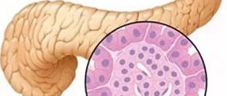
Anatomy of the pancreas
Normally, the pancreas in an adult weighs about 80 grams, has a length of 14-18 cm, a width of about 3-9 cm and a thickness of approximately 2-3 cm.
The pancreas is located in the retroperitoneal space, in the epigastric region, at the level of the first and second lumbar vertebrae, and has an oblong shape, approximately located transverse to the midline. For various pathologies determined by ultrasound, it can have a ring-shaped, spiral, split shape, or have doubled individual parts, an additional lobule.
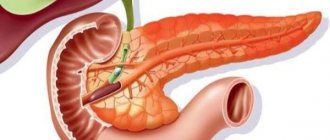
The main parts of the pancreas are the head, the body in the middle, and the tail, on the far left. The longest part of the pancreas is located to the left of the midline, and the tail near the splenic muscle is usually slightly above the head. The rather complex shape of the pancreas and its close location to nearby structures can make it difficult to recognize, but experienced ultrasound physicians are able to use surrounding structures to determine some of the boundaries of the pancreas. For example, the head and body of the pancreas are located below the liver, in front of the inferior vena cava and aorta, usually located behind the distal part of the stomach. On the far left, the tail of the pancreas is located below the spleen and, accordingly, above the left kidney.
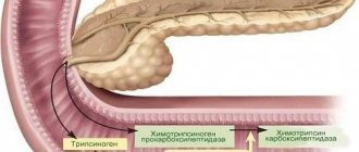
The pancreas has the form of small lobules that produce enzymes for digestion, and pancreatic islets that secrete an important hormone - insulin - into the bloodstream. It is this that allows energy to penetrate every cell of the human body and actively participates in carbohydrate metabolism. Digestive enzymes or pancreatic juice take part in the digestion process and are excreted into the duodenum.
Indications and contraindications
A biopsy is indicated for patients who have severe pain in the projection of the pancreas, jaundice, changes in biochemical blood parameters, and increased levels of pancreatic tumor markers (CA-242, CA 19-9). At the same time, during instrumental studies, space-occupying formations in a given organ should be identified, the nature of which cannot be reliably determined. Sometimes it is possible to make a diagnosis without taking a biopsy, but in some cases it is impossible to do without this procedure.
Contraindications include: blood clotting disorders, acute infectious diseases and severe general condition of the patient.
Indications for ultrasound
Ultrasound examination of the pancreas is usually included in a comprehensive examination of all abdominal organs. After all, it is closely related to the functions of other internal organs, mainly the liver. The indication for research is any pathological condition of the digestive system. Many diseases can occur with hidden or completely erased clinical signs. Therefore, once a year it is necessary to conduct an ultrasound examination of the abdominal cavity for the early detection of many diseases.
The most common conditions for which an ultrasound is recommended:
- prolonged or periodic pain, discomfort in the upper abdomen or left hypochondrium;
- tension in the anterior abdominal wall or local pain in the epigastric region, which was detected by palpation;
- frequent bloating (flatulence), nausea and vomiting that does not provide relief;
- diarrhea (stool disorders), constipation, detection of undigested parts of food in the stool;
- presence of low-grade fever for a long time;
- when observing the patient's yellowness of the skin and mucous membranes, deviations of laboratory parameters from the norm;
- with a sharp increase in blood sugar levels and an unreasonable decrease in body weight;
- after an x-ray of the abdominal organs has been performed and changes in size, shape, structure, distortion of contours have been identified, and pneumatosis of the pancreas has been detected;
- if you suspect the presence of a cyst, tumor, hematoma, stones, or abscess in the gland.
Mandatory use for abdominal injuries and elective surgeries is indicated.
Features of preparation for ultrasound in different groups of people
Age, body type, general health, expectation of pregnancy are factors that must be taken into account when preparing for an ultrasound of the pancreas. Below are important preparation points that apply to specific groups.
Fat people
Fat prevents accurate examination of the abdominal cavity, therefore, in addition to the general rules described above, obese people should do a cleansing warm enema on the eve of the examination. Also, in their case, the list of prohibited foods will be replenished with fresh fruits and vegetables. Recommended food is only porridge.
Elderly
As people age, more and more people suffer from chronic diseases, so there are some exceptions for older people. On the eve and on the day of examination of the pancreas, they are allowed to eat medications prescribed by the attending physician for the purpose of preventing chronic diseases. These include coronary heart disease, hypertension, and diabetes.
It is important to know: such “concessions” such as taking prescribed medications reduce the accuracy of ultrasound results by 30-50%.
The above rules apply to children, but you need to be very vigilant with them. It is worth making it clear to the child that soda, sparkling mineral water, fatty foods, rich pastries, ice cream, which he has time to eat 3-4 days before visiting the clinic, will negatively affect the results of the analysis.
Pregnant women
It is not advisable to undergo an ultrasound during fetal maturation. Since the frequency of monitoring of the gastrointestinal tract organ varies from 6 to 12 months, expectant mothers are advised to pick the right moment, visit a specialist in advance, undergo an examination and make sure the organ is healthy.
Compliance with the rules discussed above will allow the patient to obtain the most accurate, reliable picture of the current state of his pancreas. It is not recommended to neglect examination of the abdominal organs, since their normal functioning is an important component of a person’s well-being and longevity.
The pancreas plays an important role in the digestive system, performing a dual function: exocrine and endocrine. The first consists in the regular release of digestive juice , of which about 1 liter is released per day. The second is the production of hormones and the regulation of metabolic processes in the body. In light of the critical importance of the organ, preparation for an ultrasound scan of the pancreas in the event of such an appointment is an important element in making the correct diagnosis.
Ultrasound examination of the pancreas is included in the complex of general examination of the abdominal organs. However, there are a number of symptoms for which an ultrasound of the pancreas should be performed as an independent procedure and the organ examined separately.
Preparing for the study
Ultrasound of the pancreas can be performed both routinely and as an emergency in acute conditions. When carrying out a planned procedure, it is necessary to follow some rules and prepare for this procedure. After all, the most problematic thing during ultrasound examination is the presence of air in adjacent hollow organs. It is he who can interfere with a detailed study, distort visualization and expose the patient to an incorrect diagnosis. Doctors recommend undergoing pancreatic diagnostics, preferably in the morning. After all, it is during this half of the day, following all the rules and recommendations, that you can get the most accurate results.
During routine diagnostics, it is necessary to follow a gentle diet three days before the procedure. It is advisable to exclude foods that cause fermentation and bloating in the intestines, and do not consume foods rich in fiber and whole milk. The day before the test, it is recommended to take a laxative to cleanse the stomach and intestines. For twelve hours before the planned ultrasound examination, you must refrain from eating and drinking water, avoid taking medications, and prohibit smoking. Carbonated drinks should not be consumed as they cause excess gas formation. This can specifically affect the results and spoil the ultrasound visualization.
In case of emergency indications for an ultrasound scan of the pancreas, the patient does not need preparation. But this can reduce the information content to approximately 40%.
Features of nutrition before ultrasound
It happens that the study does not give an absolutely accurate result. The main reason for failure is the increased formation of gas inside the gastrointestinal tract (hereinafter referred to as the GIT). The organ located behind the stomach is not easy to study using ultrasound equipment, especially if the gastrointestinal tract is exposed to gases, so it is paramount to pay attention to proper nutrition.
Helpful tip: the emptyer your stomach, the more accurate the test result will be.
The maximum effect of the study can be achieved if three days before the ultrasound the patient begins to control the food entering the esophagus.
Conventionally, nutritional preparation is divided into three periods:
- 2-3 days before the examination, the following are excluded from the diet:
- legumes (peas, chickpeas, beans);
- vegetables that increase gas formation (cauliflower or white cabbage, cucumbers, asparagus, radishes, onions, broccoli);
- some fruits (melon, grapes, pear);
- protein products of animal origin (eggs and meat);
- fatty dairy products (kefir, cream, sour cream).
- 1 day before the ultrasound:
- approximately 12-14 hours before the procedure, take your last meal;
- if necessary, use drugs that reduce the amount of gas inside the gastrointestinal tract (discussed in more detail below).
- On the day of the study:
- do not eat any food in the morning;
- It is permissible to drink plain boiled water 5-6 hours before the ultrasound examination, but not more than 1 glass (200 ml).
Method of performing ultrasound of the pancreas
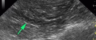
Sonography of the pancreas is a completely painless and highly informative, accessible procedure for most patients. In addition, it takes very little time - about ten minutes. It is necessary to free the abdominal area from clothing; it is on this area that the doctor will apply a special gel called mediagel. And then he will conduct an examination with an ultrasound sensor. The patient should lie quietly, first on his back, and later, with the doctor’s permission, turn on his right and left side to examine the pancreas from all sides. The ultrasound specialist examines the gland while holding the breath while inhaling to the maximum and while the patient is breathing calmly. He must also decipher the diagnostic results and give the patient a complete report and pictures of the pancreas.
During this procedure, the position of the pancreas in relation to the vessels and spinal column, the structure of the pancreatic ducts and the gland itself, shape and size are studied.
The doctor will immediately be able to see whether the gland is compacted or swollen, whether calcifications are present, whether there is an inflammatory process or not, whether pathological formations, cysts and pseudocysts are present.
If the inflammatory process has already been present for a long time, then the pancreas can significantly decrease in size, scar tissue can grow, fat deposits can increase, the capsule of the internal organ thickens, and the echogenicity of the gland increases.
Preparation
High-quality diagnostics is impossible without preliminary preparation. To do this, you need to consult with a specialist who will perform an ultrasound.
Usually the patient is prescribed a special diet that excludes fatty and stomach-heavy foods and alcohol.
It is advisable to exclude foods from your diet that can cause bloating. There should be a light dinner the day before, but not earlier than 12 hours before the examination. You can take a laxative, but only on the recommendation of a doctor. Before the examination, you should not drink or eat, or smoke.
If you are taking any medications, you must notify the specialist before the examination.
Often, the results of ultrasound are influenced by proper preparation. If you strictly adhere to your diet and follow your doctor’s recommendations, you can count on more reliable information.
Indicators of a healthy pancreas
With an ultrasound scan, the doctor will normally see a “sausage-shaped”, S-shaped pancreas. It will have clear and even edges, have a homogeneous, fine-grained or coarse-grained structure, the vascular pattern will be visible on the screen without deformation, the central duct of the gland - the Wirsung duct will not be enlarged (normally - 1.5-2.5 millimeters). It looks like a thin hypoechoic tube; it can decrease in diameter in the tail and be larger in the region of the head of the gland.
The size of this organ varies depending on the age and body weight of patients, with varying amounts of fat. The older the person, the less iron and the more echogenic it will be during scanning. A study was conducted in which 50% of people had increased echogenicity of the pancreas, and in children, on the contrary, it was decreased. An indicator of a healthy pancreas is its uniform structure.
In an adult, the size of the head of the gland can be from 18 to 30 millimeters, the body - from 10 to 22 millimeters, and the tail - from 20 to 30 millimeters. In children, everything will depend on the height, weight and age of the child: the body is from 7 to 14 mm, the head of the gland is from 12 to 21 mm, and the tail is from 11 to 25 mm.
Norms and deviations of ultrasound results
The diagnostic results of the pancreas are normal, both in women and men, with the following indicators:
- the body of the gland, located in the pancreas, can have a width from 21 to 25 mm;
- the width of the head, bordering the duodenum, protruding to the right side of the spine, should be from 32 to 35 mm;
- the length of the entire pancreas can vary from 16 to 24 cm;
- The Wirsong duct should have a thickness of one and a half to two mm.
All people may have slightly different normal ultrasound results. To identify the development of pathological changes in a given organ, the upper limits are taken into account.
It is very important that not in all cases where minor deviations of the upper limits from the norm are detected, pathology develops.
The heterogeneous structure of the pancreas and other types of its changes are of great importance in identifying a pathological disorder. An ultrasound scan of the pancreas should normally have the following characteristics:
- the edges should be smooth, the shapes should have a clear outline with easily identifiable individual parts of the parenchymal organ;
- the presence of homogeneous tissues having a structure density corresponding to the density of tissue structures in organs such as the liver and spleen;
- The Wirsung duct passing along the organ under study should not have zones with visible expansion and should be clearly visible.
Ultrasound of the pancreas for various pathologies
For pancreatitis
Pancreatitis is an inflammation of the pancreas and can be easily detected using an ultrasound scan. After all, it is the acute onset of this disease that greatly affects the structure, size, and structure of the gland tissues. The disease occurs in several stages and each stage, of course, will have its own characteristics.
Pancreatitis can be total, focal, segmental. They can be distinguished from each other by determining the echogenicity of the organ. A change in echogenicity can occur both in the entire gland and only in a certain part of it.
Initially, the pancreas will actively increase in size, the contours will be distorted, and the central duct will expand. As the gland enlarges, compression of large vessels will occur and the nutrition of neighboring organs will be disrupted, with an increase in echogenicity in them. The liver and gall bladder will also enlarge.
In the last stages of this serious disease, an experienced doctor will be able to consider when the necrotic stage will progress, organ tissue will disintegrate, and there may be the presence of pseudocysts or foci with abscesses in the area of the walls of the abdominal cavity.
What pathologies does ultrasound show?
What does an ultrasound of the liver show:
- the presence of tumors in the organ (malignant and benign) and other liver diseases caused by neoplasms;
- cirrhosis of the liver;
- hepatitis;
- changes (including necrosis) that occurred under the influence of progressive diagnoses;
- signs of parasites;
- fatty hepatosis;
- expansion of the walls of blood vessels - signs of atherosclerosis.
Diseases detected during ultrasound examination of the gallbladder and pancreas:
- gallstone cholecystitis and signs of chronic cholecystitis;
- pancreatitis;
- inflection of the gallbladder;
- enlarged spleen;
- changes in organ size as a result of pathologies;
- development of neoplasms.