Formations that arise on the walls of the esophagus are divided into benign and malignant. A man is much more susceptible to the disease than a woman. And, unfortunately, it is problematic to independently determine the disease, since the initial stage is not accompanied by symptoms.
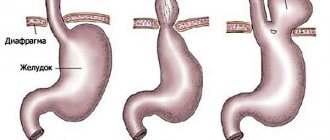
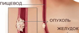
The esophagus is a tubular organ up to twenty-five centimeters long. Connects the oropharynx and stomach. It consists of three parts: cervical, thoracic and abdominal. In these three areas, physiological narrowings are formed: at the beginning and at the point of contact with the main left bronchus, and the passage through the diaphragm.
A tumor of the esophagus is the formation of benign and malignant cells from the layers of the wall of the esophagus. A lot of different types of disease have been recorded. According to ICD-10, types of oncology are coded differently, depending on the type and location. Malignant – C15, benign – D13.0.
Symptoms of the disease
The condition of patients with a benign disease does not differ from the ordinary condition of a healthy person. There may be slight fluctuations in weight due to anxiety. The infected cell grows slowly and does not appear for a long time. To determine the presence of the disease, you will need to undergo an x-ray and endoscopic procedure.
Signs of the disease
The reasons for the appearance of a tumor are alcohol abuse, frequent smoking and neglect of the rules of heat treatment of consumed food, chronic diseases of the mucous membrane, and the presence of benign formations in the body. Regardless of the location of the tumor, the symptoms are the same.
Leiomyoma of the esophagus
General features of the disease:
- Dysphagia. The process of swallowing food is difficult. The first signs of the disease occur in 90% of patients. Occurs not only due to a mechanical obstruction in the passage of food, but also due to a violation of the reflexes of the upper parts of the digestive tract. It happens that violations arise due to two of the listed factors. A mechanical obstruction manifests itself as discomfort when eating solid food passing through the esophagus. When the disease begins to progress, the patient has to drink water with each dose. So the portion size is gradually reduced and gradually reduced to zero. Moreover, there is a refusal not only from solid food, but also from liquid consistency.
- Odynophagy. The occurrence of pain in the lower part of the neck and pain in the chest. Regardless of food intake, a burning, stabbing, tearing pain is felt in the chest area, radiating to the area between the shoulder blades.
- Hypersalivation. Profuse salivation. It is a protective reflex to facilitate the passage of food. It has a thick foamy consistency.
- Bad breath. When the tissue begins to disintegrate and necrosis occurs, a purulent, unpleasant odor is felt from the mouth, which is difficult to hide.
- Regurgitation. Reverse swallowing process. Different from vomiting. Swallowed food does not have time to reach the stomach to be treated with acid, but immediately returns to the oral cavity.
- Heartburn. Increased acidity is caused by an enlarged lumen of the esophagus and improper functioning of the nervous system.
A hoarse voice, pain when eating, persistent cough while eating and varying degrees of dysphagia will help the doctor pay attention to the possible presence of a tumor of the esophagus. After conducting a series of diagnostic examinations, the doctor will make an accurate diagnosis. Cancer of the digestive tract ranks sixth among neoplasms of other organs and, unfortunately, ranks third in cases of death.
The presence of a benign tumor in the esophagus is 1% and reveals a number of varieties. These include polyp, myxoma, hemangioma, adenoma, which can be located in any part of the organ with a smooth or tuberous structure.
How to recognize cancer?
In order to promptly diagnose this difficult disease, it is important to identify the main signs of esophageal cancer:
- Dysphagia is a disorder of the process of swallowing food. Difficulty in passing food through the esophagus into the stomach. The patient may feel imaginary retention of food in the walls of the esophagus, creating a feeling of obstruction in the throat.
- Systematic regurgitation and esophageal vomiting . This symptom appears soon after eating.
- Deterioration of appetite , rapid weight loss and anemia, against the background of general malaise, weakening of the body.
- Painful sensations in the chest area, usually a burning sensation . This symptom indicates tumor growth beyond the esophageal wall. Characteristic of the late period of the disease.
Diseases and conditions characteristic of this oncology
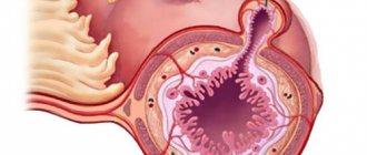
Hemangioma is a fairly rare form of benign neoplasm, which has a soft consistency and very vague boundaries. Esophageal hemangioma is a nonepithelial vascular tumor that spreads along the wall of the esophagus and adjacent tissues. Despite the benign nature of the neoplasm, hemangioma can grow to significant sizes and compress blood vessels and organs, thereby disrupting cellular nutrition processes.
Leiomyoma is one of the most common benign tumors. Leiomyoma is a submucosal formation of the esophagus. What it is has become clear, and it grows from its muscular shell. This formation has a round shape and protrudes into the esophageal lumen. Like all submucosal tumors, leimioma has a virtually asymptomatic course for a long time.
Blastoma is a general name for any tumors and neoplasms that have extremely active tissue growth. There are 2 types of blastomas: benign and malignant. The main difference between these types is that the development of a benign tumor does not invade other tissues and does not spread metastases, unlike a cancerous tumor that grows into neighboring and distant tissues, destroying the walls of blood vessels.
Modern medicine successfully copes with the treatment of non-cancerous formations, however, malignant esophageal blastoma is considered the most complex pathology, due to the implicit initial symptoms.
Esophageal hyperplasia is a precancerous condition in which the tissue of the organ grows and its natural cellular structure is replaced by a pathological one. The main prerequisite for the appearance of esophageal hyperplasia is considered to be its systematic chemical damage, which occurs as a result of the patient having a prolonged gastroesophageal reflux pathology.
In this case, the functioning of the digestive tract is disrupted and gastric acid refluxes into the esophagus, which leads to regular chemical damage to the esophagus.
Dysplasia is a pathologically abnormal development of organs and tissues of the body at the cellular level. Esophageal dysplasia is characterized by a structural disorder of the esophageal mucosa, in which the normal cells of the esophageal mucosa are replaced by cells of the intestinal mucosa. This increasing process of degeneration of the cellular structure is an absolute risk factor for the development of esophageal cancer.
There is also a type of this group of diseases known as esophageal metaplasia.
Diseases of the esophagus are often accompanied by glycogen acanthosis. Acanthosis of the esophagus is an oval white plaque that spreads along the wall of the organ. In the area where acanthosis of the esophageal mucosa forms, it thickens and becomes covered with rather large compactions filled with glycogen.
This pathology occurs in 13% of patients with any diseases of the digestive system. Glycogen acanthosis of the esophagus - what is it? Its nature has not been fully studied, but experts are inclined to believe that the pathology progresses due to chronic irritation of the mucous membrane. Irritating factors include: abuse of alcoholic beverages, smoking and spicy foods.
Neighboring organs and systems - characteristics of the lesion
When the neoplasm crosses the borders, clinical symptoms of other organs arise. It happens that the symptoms appear much brighter than those of the underlying disease. In such cases, types of esophageal tumors are distinguished.
- Laryngotracheal. Occurs in patients with a tumor in the cervical spine. Accompanied by a cough, the frequency of the voice decreases, complete loss is possible, and there are troubling hot flashes of suffocation. At short intervals, particles of undigested food are visible in the cough sputum.
- Gastric. Present in the lower third of the esophagus. The characteristic features are similar to gastritis. Discomfort in the upper abdomen, heartburn, nausea, pain.
- Neuralgic. The vagus and phrenic nerves are affected. It manifests itself in malfunctions of the heart muscle, unstable blood pressure, chest pain, leading to angina pectoris.
- Pulmonary. Manifests itself in frequent pneumonia, bronchial fistulas, and bronchitis.
In addition to the above organs, growth occurs in vessels located nearby.
Symptoms
Typically, hemangioma on the tonsils does not cause any problems or discomfort. At the initial stages of its development, there are no clearly defined symptoms. A person begins to feel the presence of a growth when it begins to grow. During this period, the patient may experience pain as the neoplasm begins to put pressure on adjacent tissues and nerve endings.
If the tumor occurs in the upper part of the larynx, the patient is overcome by a dry cough. It seems to him that there is a “lump” in his throat. Gradually, the sick person begins to wheeze when trying to speak, loses his voice for no apparent reason, and blood clots come out along with phlegm. If the vocal folds are affected, the patient's voice changes. If the tumor has formed in the lower part of the larynx or has reached a large size, the patient complains of shortness of breath and breathing problems. The danger is that a large tumor can completely block the trachea while the patient is sleeping, limiting the flow of air.
Very often, having felt a sore throat and experiencing breathing problems, patients mistake these symptoms for signs of a cold and try to get rid of unpleasant symptoms with the help of pills. However, as a rule, there is no improvement in the condition.
A hemangioma may not bother you for a long time and not let you remember it, but the presence of a neoplasm in the larynx, albeit benign, is a good reason to contact an ENT doctor to prescribe the correct treatment.
Why does a tumor occur?
Until now, the exact reasons for the appearance of cancer cells in the esophagus have not been established. But the research and experiments of scientists have led to the conclusion that the occurrence of foreign formations is influenced by external factors. An example is the consumption of alcohol and hot food. Statistics show that benign and malignant tumors of the esophagus are encountered by people living in Iran, Central Asia, and China. In these geographic locations, the consumption of a huge number of foods that provoke the formation of tumors is common.
Causes of the disease
The causes of cancer in the esophagus have been well studied, in particular, the sources lie in the following factors:
- Excess body weight provokes pressure on the abdominal walls, this leads to the occurrence of reflux with the subsequent development of various pathologies.
- Strict diets have a negative effect on the body and organs of the digestive tract.
- Abuse of fatty, salty, canned, fried foods provokes the development of tumor-like formations.
- Chemical solutions accidentally entering the body leave severe burns.
- Addiction to alcoholic beverages and tobacco products.
- Genes that are inherited mutate and become the cause of the disease.
- The presence of papillomavirus in the blood causes the development of malignant diseases.
- The use of minimal nutrients and microelements necessary for the normal functioning of the body and the structure of a strong immune system.
Symptoms
The formation, which appeared in the patient quite recently, is small in size, which means the disease does not manifest itself. As tumors grow, various symptoms are observed. Thus, in the first stages, esophageal tumors are accompanied by the following symptoms:
- loss of appetite;
- there is a sharp decrease in body weight;
- feeling weak;
- there is constant fatigue.
Symptoms, at first glance, do not indicate the occurrence of a serious change in the body and the patient does not attach any importance and is in no hurry to consult a doctor, but with the development of the tumor, it acquires other signs of the disease:
- the process of swallowing food becomes more difficult due to narrowing of the esophagus;
- nausea, vomiting, and bad breath appear;
- there is pain in the chest area, which occurs due to pinched nerve endings;
- tumors are accompanied by shortness of breath, cough, pain in the chest, the voice may become hoarse and the body temperature may rise.
Classification
Any changes in the cellular composition of the esophagus are 99% malignant. Medicine has recorded four hundred cases of benign tumors in this organ. Therefore, when it comes to tumor-like diseases in the digestive organs, they most likely mean cancer.
Classification of tumor growth:
- Endophytic - the appearance of neoplasms in the thickness of the tissue walls or from the submucosal layers in the glands.
- Exophytic – the appearance of tumor-like cells inside an organ, which, due to their location in the mucous membrane, block the lumens.
- Mixed formations - the occurrence of formations in all layers, as a result of which the tissues disintegrate.
There are two types of tumors: benign and malignant. Pathologies of non-cancer cases most often demonstrate a non-epithelial structure.
Benign – determined by the growth rate of the affected cells and the presence of a pathomorphological sign.
Benign:
- intramural - located inside the wall of the esophagus;
- polyp-shaped - located in the lumen of the organ.
Polyp-like formations affect the epithelium of the esophagus, namely:
- adenomatous polyps;
- papillomas;
- cysts.
Intramural formations are not related to epithelial origin:
- lipoma;
- fibroma;
- angioma;
- leiomyoma;
- neurofibroma.
Malignant
Among malignant formations, they are classified by such characteristics as location, structure, morphology and pathological anatomy.
Epithelial:
- keratinizing and non-keratinizing cancer cell damage can be 95% squamous cell;
- basal cell;
- transitional cell;
- anaplastic and mucoepidermoid cancer;
- less than 5% of lesions are adenocarcinoma.
Non-epithelial:
- sarcoma;
- lymphoma;
- melanoma.
Malignant tumors quickly affect the lymph nodes and other organs associated with the symptoms of the disease.
Type of malignant neoplasms:
- Cancer is a malignant formation of the epithelium: basal cell carcinoma, squamous cell carcinoma, adenocarcinoma.
- Leiomyosarcoma - affects smooth muscle tissue.
- Rhabdomyosarcoma - affects the upper part of the striated muscles.
- Lymphosarcoma - lymph nodes and lymphatic tissue in the wall of the esophageal tube.
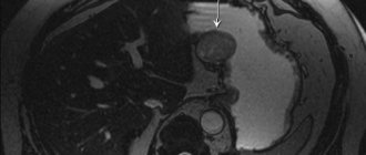
What is a submucosal formation of the esophagus?
All organs of the gastrointestinal tract, including the esophagus, are susceptible to cancer pathologies.
Oncology, or submucosal formation of the esophagus, is a rather rare phenomenon in comparison with other disorders of the digestive system, but is considered the most complex. The development of a neoplasm is influenced by external and internal factors: heredity, ecology, deterioration of a person’s well-being, food intake, age, associated pathologies.
According to doctors, this form of cancer affects men aged 55 years. In women, the disease occurs much less frequently.
Varieties
The disease is divided into benign and malignant formations, which arise from various layers of the walls of the organ, mainly in the middle third of the esophagus. Each of them has a personal histological classification.
The main types of tumors are as follows:
- Epithelial - appear from epithelial cells, skin, and organ mucosa. This type is characterized by the appearance of a benign neoplasm (adenoma, papilloma), malignant (adenocarcinoma, squamous cell carcinoma, etc.).
- Nonepithelial - are the most extensive. Tumors can be of various natures: hemangioma, lipoma, chondrosarcoma, leiomyoma and others.
- Mixed - this type damages all layers of the walls of the organ, has instant disintegration with replacement of ulcers in that place. This type implies carcinoid formation, malignant lymphoma, carcinosarcoma.
Growing benign neoplasms are intraluminal and intramural. The first include polyps, adenomas, papillomas, the second - leiomyomas, esophageal cysts. Other types are observed extremely rarely.
Symptoms
When the formation is localized inside the wall of the esophagus in the lower region, symptoms may not express themselves for a long time. Only when there is strong constriction of the walls, when the lumen of the organ is blocked, the following may become signs:
- dysphagia – difficulty swallowing food, difficulty moving food from the esophagus to the stomach, a person may feel a lump in the throat;
- loss of appetite, weight loss, anemia;
- regular regurgitation, vomiting (occurs immediately after eating);
- general malaise, weakness;
- characterized by chest pain and a burning sensation. This sign indicates the growth of the formation beyond the boundaries of the esophageal wall, which is typical for the last stage of cancer.
With a significant size of the esophageal tumor, shortness of breath, debilitating cough, rapid heartbeat, and cyanosis may occur due to compression of the mediastinal organs. Malignant transformation of benign formations occurs relatively rarely.
Diseases characteristic of oncology
The development of a tumor can lead to the following pathologies:
- Hemangioma is a very rare disease, it has a soft consistency, with rather vague boundaries. The pathology is a non-epithelial vascular neoplasm of a benign nature, located along the wall of the organ, as well as tissues close to it. Hemangioma can expand to a large diameter, compress blood vessels and organs, disrupting the nutrition of cells.
- Blastoma is characterized by rapid tissue growth. It can be benign or malignant. It differs in that the first does not cover other tissues and does not expand metastases. Current medicine effectively cures non-cancerous tumors, but the malignant form is very severe due to hidden initial signs.
- Leiomyoma is a common type of tumor. Submucosal formation appears from the muscular lining of the esophagus. It has a round shape and makes its way into the lumen of the organ. It also appears asymptomatically for a long time.
- Hyperplasia is a precancerous condition, where tissue growth is observed with the replacement of the natural cellular structure with a pathological one. The main assumption for the occurrence of hyperplasia is its chemical disorder, which occurs due to the presence of a protracted GERD disease in a person. There is a disorder of the gastrointestinal tract functions, reflux of acid from the stomach into the esophagus.
In addition, dysplasia and metaplasia of the esophagus are distinguished. Pathologies of the organ are often associated with glycogen acanthosis (an oval-shaped white plaque located along the wall of the esophagus).
Complications
If esophageal cancer is not treated in a timely manner, large neoplasms can lead to complete blockage of the lumen, as well as its obstruction. The patient will not be able to swallow food, even liquid food, and hemorrhage and thinning of the walls of the organ may occur.
A person refuses to eat, a paroxysmal cough occurs, and tracheal perforation and fistulas with subsequent expansion into blood vessels become complications. The state of health noticeably worsens when metastases spread to the liver, pancreas, lungs, and brain.
Diagnostics
To establish the nature of the tumor, the degree of damage to the esophagus, and existing metastases, gastroenterology involves the following diagnostic methods:
- blood test - biochemical examination of the esophagus to determine tumor markers;
- X-ray using barium sulfate. This contrast agent helps to identify the lesion and location of the tumor;
- CT, MRI - to determine the degree of development and recognition of metastases;
- the use of endoscopic biopsy - to identify the extent of formation, stage and nature. Endoscopy is considered the main way to study this organ.
Diagnosis of benign tumors involves a biopsy of biological material, which is done by throwing tissue particles for later study.
Treatment
There are several ways to treat an existing tumor in a person:
- surgical operation - a section of the esophagus, nearby tissues, and lymph nodes are removed;
- Radiation therapy – eliminates cancerous tissue using ionized radiation;
- hysteroscopy – visual examination of the lumen and walls of the internal cavity;
- chemotherapy – the tumor is affected through strong poisons and toxic substances;
- a combination of the above methods.
cancer therapy - surgery to remove submucosal formation of the esophagus. It is very important not to cause harm to the mucous membrane of the organ, to avoid the development of a purulent course. The operation is performed using an endoscope to effectively remove the tumor.
Forecast
If a malignant tumor is not actively treated, the subsequent prognosis will be very poor. If there is no action, the patient will not live long, a maximum of 9 months.
When the pathology is revealed at stages 1-2 of development, surgical excision is performed, the patient’s condition returns to normal, and nothing threatens him.
Stage 3-4 of the disease has an uncertain prognosis due to the expansion of metastases. Here, current medicine can slow down the course of metastasis or stop it for a while. The prognosis for stage 3 cancer is quite unfavorable, the survival rate is no more than 15%.
Therefore, it is very important to recognize the disease in time, establish an accurate diagnosis, and determine a suitable treatment regimen in combination with current medications. All this will be the key to a successful recovery.
The information on our website is provided by qualified doctors and is for informational purposes only. Don't self-medicate! Be sure to consult a specialist!
Rumyantsev V. G. Experience 34 years.
Gastroenterologist, professor, doctor of medical sciences. Prescribes diagnostics and carries out treatment. Expert of the group for the study of inflammatory diseases. Author of more than 300 scientific papers.
to the doctor We recommend: What is ligation of esophageal varicose veins?
Source: https://gastrot.ru/pishhevod/podslizistoe-obrazovanie
Diagnostics
During a visual examination, the doctor does not diagnose the presence of the disease. Before prescribing productive treatment, a diagnosis is carried out using special tools. A tumor of the esophagus can be detected by X-ray with a contrast agent (barium). The solution envelops the organ, and the image shows disturbances in the normal structure.
X-rays help visually determine the degree of narrowing of the esophagus, identify any changes in wall thickness or the presence of ulcerative wounds.
Using an endoscope, the doctor examines whether the wall has spasms or swelling. This examination technique helps to identify the stage of the oncological process, the presence or absence of metastases in the lymph nodes and nearby organs.
Using ultrasound, the exact size of the tumor and secondary formations, if any, are recorded.
To determine the presence of histology, an abdominal biopsy is performed. A laparoscope equipped with a video camera is inserted through a puncture near the navel, and biological material is collected.
To determine an accurate diagnosis, they take a general biochemical test of blood and urine and monitor the dynamics of changes in indicators. Such studies are needed during chemotherapy to monitor the level of red blood cells, platelets, and white blood cells. Diagnosis of tumor-like formations is carried out for comparison with diseases that cause dysphagia.
Types of esophageal cancer relative to the depth of tumor germination
The esophagus is a muscular tube, lined on the inside with mucous membrane, and on the outside covered with an adventitial (below the diaphragm - serous) membrane. Between the mucous membrane and the muscular base there is a submucosal layer. Tumors arise in the internal layers (mucous or submucosal), gradually spreading to the muscle layer, and then extending beyond the organ. Depending on the depth of germination and spread of the tumor, the stage of esophageal cancer is determined.
In total, there are 4 stages during the course of the disease:
- The tumor is located within the mucous and submucosal layers. The lumen of the esophagus is not changed, there are no metastases.
- The tumor affects not only the mucous and submucosal layer, but also the muscle layer, but does not extend beyond the esophagus. The lumen is somewhat narrowed. Single metastases to regional lymph nodes are possible.
- The tumor spreads beyond the muscular layer, to the outer (adventitial or serous) membrane, into the periesophageal tissue, but does not affect other organs. The lumen of the esophagus is significantly narrowed (stenosis), Multiple metastases in regional lymph nodes.
- The tumor affects all layers of the esophagus, extends beyond it, metastasizing not only to regional lymph nodes, but also to other organs.
The stages of esophageal cancer play a primary role in developing a treatment strategy. Thus, for stage 1 cancer, organ-preserving surgery with removal of the tumor within the affected tissue may be sufficient; for stage 2 or 3 cancer, total organ resection with subsequent plastic surgery, as well as radio and/or radiochemotherapy may be necessary. At the fourth, terminal stage, radical treatment is not always possible; for palliative purposes, it may be necessary to insert a tube into the stomach for esophageal cancer (elimination of stenosis) and a number of other conservative measures. It should be noted that in the treatment of esophageal cancer abroad at any stage, the most progressive and effective methods are used, which make it possible to achieve remission, sometimes in the most difficult cases.
Treatment
Treatment of tumor-like formations in the esophagus is carried out depending on the degree and size of the affected cells, as well as the stage of progression of the process. Common treatments for esophageal masses rely on combination therapy. Prescribed individually by the attending physician.
The fight against esophageal tumors involves surgery and the method of endoscopic surgery, chemotherapy and radiation therapy.
Endoscopic surgery
It is performed during the course of the disease in an uncomplicated form, by inserting an endoscope, which is equipped with appropriate devices. During the procedure, bougienage is performed. This is required to eliminate the narrowing of the esophagus and to restore the lumen.
Radiation therapy
One of the effective methods of struggle. It is performed in the absence of contraindications for diseases of the respiratory organs and cardiovascular system. As a rule, such treatment is indicated to avoid relapse of the disease and infection of the lymph nodes and adjacent organs. Radiation therapy plays an important role in reducing tumors by affecting only the affected cells, leaving healthy ones without negative effects on them.
Chemotherapy
The main method of combating malignant tumors. Based on the principle of using a cytostatic substance with toxic substances that can destroy cancer cells. When carrying out this procedure, there are a number of side effects due to the effect of the drug on both diseased and healthy cells of the body. The consequences include: hair loss, damage to the oral cavity with stomatitis, a significant decrease in the body's immunity. But all these factors return to normal immediately upon completion of treatment.
Carrying out a chemotherapy procedure
Chemotherapy is necessary for the following forms:
- small cell and granular cell;
- oncological process with a poorly differentiated form.
Treatment algorithm
- All drugs that the doctor prescribes are aimed at destroying cancer cells and the further possibility of new ones arising. Effective in the second and third stages.
- The entire treatment process is palliative in nature and, as a rule, is aimed at maintaining a person’s life and significantly slowing tumor growth.
Surgery, radiation and polychemotherapy procedures. The dosage of radiation therapy before surgery is 3000-5000 rads. The procedure will be effective when the affected cells have not invaded nearby organs. Remember that you should resort to alternative medicine treatment methods only when you have the consent of doctors.
Surgery is only possible for benign tumors and is performed using a gastroscope. When performing abdominal surgery, the entire affected organ is removed or partially removed. If the surgeon finds that the lymph nodes are also affected, they will be removed.
The operation at the last stage is not performed. It is important to diagnose the disease as early as possible. Based on a first stage diagnosis, the survival rate is high. However, the listed signs do not confirm a formation in the esophagus, and the diagnosis is made by an experienced doctor.
During the operation, the surgeon removes part of the affected organ or the entire esophagus. In some cases, the doctor may have to remove part of the stomach. It should be taken into account that surgical intervention is performed if the tumor is located in the middle or lower part of the organ. If during surgery it is necessary to remove the upper part, then an alternative to the removed organ is a part of the intestine or a gastrostomy is formed.
Unconventional treatment
This type of treatment for this disease is ineffective, and sometimes can cause colossal harm to the body. Traditional medicine, unfortunately, is powerless in the fight against cancer. Therefore, you should strictly follow the recommendations and instructions of your doctor.
On the Internet there are a lot of methods that verbally promise to get rid of the disease. But rest assured that the recommendations posted on dubious sites have not been confirmed in any way by top-category specialists in the field of oncology!
Contraindications for surgery:
- metastases in lymph nodes and organs located nearby, the number of which is much greater than normal;
- patients whose age is over 70 years;
- the presence of chronic diseases occurring in the body;
- disruptions in the functioning of the cardiovascular and respiratory systems.
Medical examinations suggest that the probability of death from esophageal cancer and surgical intervention does not cross the line of 10%. This does not mean that the patient can fully perform physical and mental labor. Productivity decreases significantly or becomes completely questionable.
With a tumor of the esophagus, the patient should pay attention to food intake. Strictly follow and adhere to the doctor’s recommendations. Rough food is excluded from the diet; the amount of food consumed per day does not exceed three kilograms, and portions should be divided into 6 or 7 times. In this case, the temperature of the food consumed should be warm and accompanied by an additional vitamin complex.
Treatment of benign tumors
Due to the fact that esophageal tumors are often complicated by ulceration, bleeding, suppuration, and malignancy, surgical tactics are indicated for them. Epithelial intraluminal tumors that have a long narrow stalk can be removed by electroexcision through an endoscope. It is more advisable to excise tumors on a wide base during open esophagotomy. Resection of the esophagus is resorted to in cases where malignancy cannot be excluded, or in cases of significant tumor size.
Intramural tumors of the esophagus require thoracotomy, enucleation of the tumor and subsequent restoration of the integrity of the esophageal wall. If there is significant destruction of the muscle wall, resection of part of the esophagus is performed with its plastic surgery using a gastric, small intestinal or colonic graft or esophagogastroanastomosis.
Complications and prognosis
Tumors that are benign in nature suggest a favorable outcome. But due to the high degree of possible relapse, lifelong observation at the dispensary is necessary.
In case of malignant neoplasms, the time at which the disease is detected and treatment is prescribed plays a role. Unfortunately, the absence of metastases does not guarantee a favorable outcome.
Until now, the specifics of preventing tumor-like formations of the esophagus have not been identified.
What does improper treatment or lack thereof lead to:
- partial obstruction of the alimentary canal or complete blockage;
- lack of passage of food;
- due to tissue breakdown, hemorrhage is possible;
- death.
It all depends on the degree of the disease. If the disease is detected in time, there is a high chance of complete recovery of the body and relapse in the future. If the disease is detected at an early stage, 90% of patients have a chance to live for five years. The subsequent stage will be favorable only for half of cancer patients. 10% of 100 expect full recovery of the body. And no matter how disappointing it may sound, patients with the fourth stage are incurable. After diagnosis, the patient dies soon after.
It happens that a benign tumor develops into a malignant stage. It proceeds slowly. If surgical intervention is no longer advisable, the average life expectancy of the patient is 5-10 months.
Benign tumors of the esophagus
Benign tumors of the esophagus are relatively rare findings in gastroenterology, accounting for 0.5 to 5% of all esophageal neoplasms. Most often, esophageal tumors develop in men; The predominant age of patients is from 25 to 60 years. The etiology of benign esophageal tumors is unknown; The exception is esophageal cysts, which are embryonic malformations. Favorite places for tumors to localize are natural narrowings and the lower third of the esophagus.
Prevention of occurrence
If we take into account the causes of the tumor, it is possible to identify preventive measures. This is medical and personal prevention of the disease.
Personal prevention consists of giving up bad habits: using tobacco products, alcoholic beverages, abstaining from spicy foods and spices, maintaining a normal temperature for the food consumed.
The diet includes the required amount of vegetables and fruits that facilitate the movement of food, foods rich in fiber. Don't forget about your oral health. Food that enters the esophagus must be chewed well so that there is no difficulty in swallowing.
Medical prevention consists of periodic examination and treatment of timely detected chronic diseases associated with the gastrointestinal tract, leading to inflammation of the esophageal mucosa.