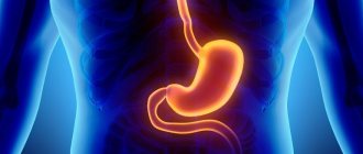
Leiomyoma of the stomach is a benign neoplasm. The disease occurs as a result of the proliferation of smooth muscles. This type of tumor is located not only in the stomach, but also in the uterus or intestines. Most often it is small in size and for this reason it may not manifest itself for a long time. The first serious symptoms begin to worry when the tumor is large. Lack of proper treatment leads to the transformation of leiomyoma into a malignant state.
Causes
Doctors have still not been able to find the exact reason why leiomyosarcoma occurs. A large number of negative factors lead to the development of this disease:
- improper or inadequate nutrition, abuse of fatty and spicy foods leads to the destruction of the walls of the stomach, which provokes the active growth of smooth muscles;
- lack of vitamins and minerals in the diet;
- exposure to radiation and ultraviolet radiation on the body, for example, people who often fly on airplanes and undergo access control through X-ray arches are more susceptible to any types of tumors;
- areas with poor ecology provoke not only pollution of the lungs, but also of the entire body;
- weak immune system;
- hormonal imbalance causes tumor growth due to significant cell proliferation;
- the influence of certain bacteria and viral diseases, for example, the bacterium Helicobacter pylori can cause not only leiomyoma, but also antral gastritis;
- inflammation of the gastric mucosa;
- nervous tension and stress;
- hereditary predisposition to stomach diseases.
Causes of stomach cancer
The exact causes of stomach cancer are unknown. DNA mutations occur in the mucous membrane of the organ, resulting in “wrong” cells that can acquire the ability to grow uncontrollably. Why this happens is not entirely clear. But risk factors are well studied - conditions that increase the risk of stomach cancer.
Heredity and stomach cancer
Some people carry a “time bomb” hidden in their genes. Sometimes not even one. This is confirmed by some facts:
- If a close relative (parents, siblings, children) of a person is diagnosed with stomach cancer, the risk is increased by about 20%.
- Men get sick more often than women. It is difficult to say what exactly this is connected with, but it can be assumed that a difference between male and female genes is involved.
- Japanese who migrated to the United States suffer from stomach cancer less often than their compatriots, but more often than “native” Americans. This suggests that the matter is not only in the nature of nutrition, but also in heredity. The main suspect is a gene called RNF43.
- Carcinoma, the most common type of stomach cancer, is more common in people with blood type A (II), which they, of course, received along with their genes.
- The risks are increased in some hereditary diseases: pernicious anemia (3-6 times), hypogammaglobulinemia, non-polyposis colon cancer.
- The incidence of stomach cancer increases after age 70, thought to be due to the accumulation of unwanted mutations in body cells as we age.
Symptoms
It is difficult to diagnose the initial stage of gastric leiomyoma, since when it is small in size it is not a concern. The main symptoms appear when the tumor begins to increase in size and bleed. Most often, leiomyosarcoma is diagnosed with the following symptoms:
- dizziness and weakness, which are typical for internal bleeding;
- a sharp decrease in body weight, which is associated with the destruction of the gastric mucosa and its inability to absorb nutrients;
- a decrease in the level of hemoglobin in the blood and, conversely, an increase in the number of leukocytes may indicate bleeding of internal organs;
- severe pain in the stomach after eating;
- dark color of stool is a sign of intestinal or gastric bleeding;
- constant fatigue;
- pale skin due to blood loss and anemia;
- Frequent heartburn occurs due to the release of gastric juice into the esophagus, as the stomach decreases in size due to the tumor.
Benign stomach tumors
A benign tumor of the stomach is a neoplasm that develops slowly and does not have manifestations characteristic of malignant tumors. The basis for a benign tumor can be epithelial cells, nervous tissue, fatty and vascular structures. Locations in the layers of the stomach: submucosal, subserosal, muscular. Sometimes a benign tumor transforms into a malignant one.
TYPES OF BENIGN TUMORS
Taking into account which cells served as the basis of the tumor, epithelial and non-epithelial tumors are distinguished.
Epithelial:
- polyps are round-shaped formations with a wide base, similar to a stalk. They can be located singly or in group clusters. They grow from glandular and epithelial cells and have a vascular network. The risk of progression to a malignant course is very low;
- adenomas are formations that look like growths of glandular tissue. They are found less frequently than polyps. They can degenerate and lead to stomach cancer.
Tumors that grow from other non-epithelial cells are rare:
- fibroids - from muscle tissue;
- neuroma - from the cells of the myelin sheath of the nerve trunk;
- fibroma - from connective tissue fibers;
- lipoma - from adipose tissue;
- lymphangioma - from the lymphatic vascular system;
- hemangioma - from cells that line the walls of blood and lymph vessels;
- tumors from mixed tissues.
More often diagnosed in women. Distinctive characteristics of nonepithelial tumors: clearly defined contour, flat surface, round shape. They can grow to large sizes. The likelihood of degeneration into a malignant tumor is very high.
CAUSES OF BENIGN TUMORS
The exact reasons that can trigger the occurrence of benign tumors of the stomach have not been determined.
There is a list of risk factors:
- chronic gastritis;
- infection with bacteria Helicobacter pylori, due to which the protective mechanisms of the gastric mucosa are reduced;
- hereditary predisposition;
- uncontrolled drinking of alcohol, smoking;
- nutritional disorders: predominance of salty, smoked, fatty foods in the diet;
- environmental prerequisites;
- a malfunction in the body's immune response.
SYMPTOMATICS
The clinical picture does not have any clear manifestations, and in the absence of tumor growth, there are no symptoms at all. Benign tumors can be suspected by mild signs or accidentally discovered during an endoscopic examination.
Signs:
- disruptions in the gastrointestinal tract, similar to gastritis: aching pain in the stomach, nausea, flatulence, abnormal stool;
- stomach bleeding, which can cause anemia;
- poor appetite, fatigue.
An increase in tumor size is manifested by severe symptoms:
- vomiting with blood streaks;
- bloody stools;
- persistent decrease in hemoglobin;
- frequent dizziness;
- rapid loss of body weight;
- general decrease in performance.
Clinical manifestations may vary depending on the type, location, size and growth of the tumor.
DIAGNOSIS OF BENIGN TUMORS
Diagnosis includes:
- Collection of a complete medical history, clarification of hereditary factors, lifestyle;
- Examination of the patient;
- Complete blood count to determine the degree of anemia;
- Analysis of stool to detect traces of blood;
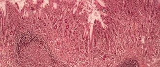
- Endoscopy is a method in which the doctor examines and evaluates the appearance of the inner surface of the stomach using a special visual device (endoscope). Also during the procedure, it is possible to immediately remove growths if they are detected. Next, the biomaterial is sent for research, which will determine what cells the tumor consists of;
- Tests to detect Helicobacter pylori. There are several methods: stool, blood and urease breath test;
- An X-ray of the abdominal organs helps determine whether there are irregularities in the stomach, which may be indirect signs of neoplasms;
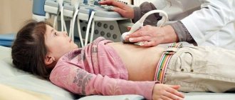
- Ultrasound examination of the abdominal organs;
- Computed and magnetic resonance imaging.
TREATMENT
The pathology can be cured exclusively by surgery - removing the tumor.
The polyp can be removed during gastroscopy or esophagogastroduodenoscopy (EGD). A special flexible hose is inserted into the stomach, a polyp is found, a metal loop is thrown over it, and diathermy current is applied, so that there is no bleeding after excision. Then they check how effectively the procedure was carried out and, if necessary, repeat it; sometimes it is necessary to cut off part of the stomach wall along with the leg. The removed tumor is sent for histological examination to confirm its benignity.
Non-epithelial tumors also require surgical removal. To do this, the following procedures are used:
- wedge-shaped or partial resection (partial removal) of the stomach;
- enucleation: the tumor is removed using a special method, without damaging nearby tissues.
If there are a large number of polyps formed from mixed cells, an operation to remove the stomach is performed - gastrectomy.
After surgery to excise a polyp or any other neoplasm, a course of medicinal and restorative therapy is required, the following are prescribed:
- gastroprotectors based on rebamipide to restore the mucous membrane of the gastrointestinal tract;
- drugs that reduce the secretion of hydrochloric acid in the stomach;
- antibiotics if the analysis confirmed the presence of Helicobacter pylori;
- therapeutic diet with a predominance of protein products. Food must be chewed thoroughly, take a break before each sip, stretching the eating time for at least half an hour.
COMPLICATIONS OF BENIGN TUMOR
- Degeneration of a tumor from benign to malignant: changes occur in the cells that make up the tumor, they become different from the cells of the organ from which they originated.
- The appearance of a through defect in the wall of the stomach at the site of the tumor, which is accompanied by the development of a life-threatening inflammatory process in the abdominal organs.
- A steady decrease in the lumen of the stomach, which occurs due to the fact that the tumor grows to a very large size and blocks the cavity.
- Areas of necrosis, ulcers on the surface of the neoplasm, which do not go away for a long time, since the regeneration process is disrupted.
- Strangulation of the polyp. Due to the fact that the stalk of the polyp can grow in length, there is a risk that the tumor will prolapse into the duodenum and be pinched by the muscle ring, which is located between the stomach and the duodenum. In this case, severe paroxysmal pain occurs.
PREVENTION AND PROGNOSIS OF MALIGNANT TUMORS
There are no special preventive measures to prevent benign tumors of the stomach. General recommendations:
- abstaining from alcohol or drinking it in moderation;
- organization of proper and balanced nutrition (excluding salty, fried, fatty, smoked foods, adding fruits and vegetables);
- timely treatment of gastritis and other gastrointestinal diseases;
- At the first complaints, you should immediately contact a therapist or gastroenterologist and undergo all the necessary examinations. If tumors have been removed, control gastroscopy must be performed every 3 months in the first six months after surgery, then at least 1-2 times a year.
The prognosis can be called favorable. To prevent unwanted complications, you need to undergo routine examinations throughout your life.
Diagnostics
To make an accurate diagnosis and detect leiomyoma, complex diagnostics is used, which includes the following methods:
- collecting anamnesis about the patient, that is, living conditions, place of work, recent health status;
- during a physical examination, palpation is performed in the stomach area and painful sensations are identified;
- it is necessary to take a blood, urine, and stool test to detect blood in it;
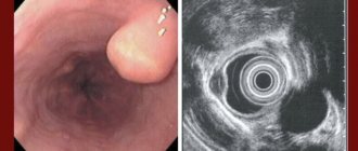
- fibrogastroendoscopy will allow you to assess the condition of the digestive tract;
- Ultrasound diagnostics (ultrasound) makes it possible to determine the size of the tumor and its location;
- Additionally, a bacterial analysis is prescribed for the presence of bacteria that cause ulcerative conditions;
- Using computed tomography, you can find out about the origin of this type of sarcoma, as a full scan of the organ is performed.
Classification of stomach cancer: what is it like?
Malignant tumors of the stomach, according to the WHO International Histological Classification, are divided into 11 types, depending on which cells they originate from. The predominant cancer is from glandular cells that line the mucous membrane and produce mucus - adenocarcinoma. It accounts for 90-95% of all cases. There are also tumors from immune (lymphoma), hormone-producing (carcinoid) cells, and from nervous tissue.
One of the oldest classifications divides malignant tumors of the stomach into 3 types:
- Intestinal. As the name suggests, in the surroundings of the tumor there is intestinal metaplasia, that is, the gastric mucosa becomes similar to the intestinal mucosa. This type of cancer is more common in older people and has a more favorable prognosis. This is a typically “Japanese” type of cancer.
- Diffuse. Tumor cells spread along the wall of the stomach, they are surrounded by normal mucous membrane.
- Mixed.
Gastric adenocarcinoma
Stages of stomach cancer
Stomach cancer is divided into early (initial) and advanced. In early cases, the tumor does not grow deeper than the mucous membrane and submucosa. Such tumors are easier to remove (including endoscopically) and have a better prognosis. The TNM classification is also used, which takes into account the size and growth into different tissues of the primary tumor (T), metastases in regional (nearby) lymph nodes (N), and distant metastases (M).
Classification depending on the condition of the primary tumor (T):
- Tx – primary tumor cannot be assessed;
- T0 – the primary tumor is not detected;
- T1 – severe dysplasia of mucosal cells, the tumor is located in the superficial layer of the mucous membrane (“cancer in situ”);
- T2 – the tumor has grown into the muscle layer of the organ wall;
- T3 – cancer has reached the serous (outer) lining of the stomach, but has not grown into it;
- T4 – the tumor has grown into the serosa (T4a) and into adjacent structures (T4b).
Treatment methods for gastric leiomyoma
When prescribing a treatment method, the size of the tumor and the general condition of the patient play an important role. If the submucosal formation is up to 3 cm, in this case gastroendoscopy is performed. Local removal of the tumor will preserve healthy gastric tissue. Abdominal surgery is used only for large tumors. In the postoperative period, anti-inflammatory medications and antibiotics are prescribed. Medicines that reduce hydrochloric acid levels are also prescribed. In this case, the submucosa recovers much faster.
Classification
Malignant tumors of the antrum of the stomach are classified according to:
- Histological structure - mucinous adenocarcinoma, medullary cancer with lymphoid stroma, undifferentiated form, etc.
- According to Lauren - intestinal, diffuse, mixed, unclassified types.
- The macroscopic criterion is early and advanced cancer.
- Japanese classification based on lymphatic drainage pathways and damage to regional lymph nodes.
- International TNM system with definition of process stage.
Other classifications are used less frequently.
Unconventional treatment
It is imperative to avoid spicy, fatty, and smoked foods.
In the postoperative period, the patient must follow a strict diet and follow the recommendations of the attending physician. Mandatory avoidance of spicy, fatty, smoked, and heavy-for-the-stomach foods; coffee and alcoholic drinks. The diet should include various types of cereals, lean meats, vegetable purees, dairy products and fish.
Folk remedies will help to effectively restore the body after surgery. Taking antitumor preparations will significantly improve the general condition and have a beneficial effect on the submucosal tissues: collecting plantain herbs, celandine, chamomile, St. John's wort, galangal, calendula, tartar, calamus root. In addition to the above-mentioned herbs, tinctures of periwinkle, calamus, mistletoe, and tartar can be used.
Treatment of stomach polyps
As stated earlier, all adenomatous and large polyps larger than 10 mm should be removed. How is endoscopic polypectomy or mucosal resection performed?
After clearly defining the boundaries of the formation, an injection of saline solution colored with indigo carmine blue dye is performed under its base into the submucosal layer in order to prevent perforation and clearly visualize the vascular structures. Then the endoscopic loop is positioned on the unchanged mucosa around the formation and is gradually tightened. Next, electrocoagulation is performed using an electrosurgical knife/block. A post-resection defect with a white scab without “plus tissue” indicates the radicality and reliability of the surgical intervention, thereby eliminating possible complications. The stages of the operation are presented in the pictures (in our Clinic we use a knife - ERBE VIO 300D).
Large exophytic (protruding) polyps on a wide base or flat formations that cannot be removed with endoloops are subjected to a technically more complex version of endoscopic removal - dissection in the submucosal layer. This type of operation is aimed at radical removal of the formation as a single block: after creating a “hydrostatic cushion”, a circular incision is made around the formation with special instruments, after which the connective tissue plexuses of the submucosal layer are dissected with complete elimination of the pathological focus.
Postoperative follow-up
The first day after polypectomy, a gentle regimen of activity and nutrition is recommended. As mentioned earlier, a post-resection defect of the mucous membrane remains at the site of the polyp, and therefore an anti-ulcer diet is prescribed - table No. 1 according to Pevzner. Its essence is to limit foods that stimulate secretion in the stomach, long-lasting and difficult to digest foods (spicy, fatty, fried foods). This diet and restriction of physical activity are followed for a month. It is also necessary to remember not to take anticoagulants for 3 days both before and after surgery, in order to prevent early and delayed bleeding.
According to modern concepts, the most rational is to monitor patients after polypectomy after 1 year. At the same time, the condition of the mucous membrane is assessed for the appearance of new formations or relapse at the site of a previously performed operation. If, after resection of the formations, the histological response confirms the presence of high-grade dysplasia or early cancer, then control gastroscopy should be carried out in the interval: 1.5 months - 3 months - 6 months. – 1 year and every year thereafter.
Prognosis and prevention
After surgery, it is necessary to get rid of bad habits.
After following doctor's orders or after surgery, gastric leiomyoma usually has a favorable prognosis for recovery. This also depends on early diagnosis and the correct method of tumor removal.
There are no specific methods for preventing this type of disease. Since leiomyoma often develops asymptomatically, the best option for prevention is regular visits to specialists. Methods for preventing gastric cancer include a healthy lifestyle, proper nutrition and giving up bad habits.
Reasons for development
The following factors lead to gastric antral cancer:
- Lifestyle - stress, weakness, bad habits, lack of routine, etc.
- The nature of the diet is an excess of table salt and carbohydrates, a lack of vitamins and microelements, rough food that injures the surface of the epithelium and promotes the colonization of the bacterium Helicobacter pylori on the mucous membranes of the duodenum and stomach.
- Infection with the bacterium Helicobacter pylori.
- Composition of consumed water. If it contains nitrates and nitrites, under the influence of microorganisms they are converted in the stomach into nitrosamines, which are powerful carcinogens.
- Endogenous causes - duodenogastric reflux, heredity, decreased local and humoral immune response, etc.
The process of carcinogenesis in the stomach is usually triggered by free radicals formed during inflammation. Therefore, cancer almost always occurs against the background of chronic pathology. Precancerous diseases include:
- adenomatous polyp-like growths (gastric adenomas);
- chronic ulcer;
- stomach operated for benign pathologies;
- chronic inflammation of various nature, including Ménétrier's disease and autoimmune gastritis type A.
Dysplasia of the mucous membrane plays a major role in the development of malignant neoplasms.
Localization and metastasis
Malignant neoplasms can be located in any part of the stomach. If its antral region is affected, the tumor may be localized closer or further from the pylorus, which will determine the clinical course and surgical tactics.
In antral cancer, metastasis occurs in several ways:
- Along the wall of the stomach with invasion into the surrounding healthy mucous membrane.
- With blood flow to distant organs - liver, lungs, bones, pancreas, etc.
- Lymphogenously to regional lymph nodes.
- With the help of implantation of cancer cells in the peritoneum - carcinomatosis.
Lymphogenous metastasis is more common. In case of antral cancer, tumor “dropouts” in the lymph nodes are found in half of the patients.
Complications and relapses
After removal of a tumor from the stomach, especially during major operations, there is a high risk of developing subsequent complications. These include:
- cancer recurrence;
- bleeding and anemia;
- purulent-septic complications, up to peritonitis;
- failure of the gastric stump or created anastomoses;
- hypoxia;
- pancreatic necrosis;
- intestinal obstruction;
- thromboembolism;
- myocardial infarction, pneumonia and other complications from the surgery and anesthesia.
After operations for gastrointestinal cancer, the risk of death remains high, especially with voluminous, technically complex interventions, for example, after complete removal of the stomach - from 11 to 25%. The level of training of the surgeon and anesthesiologist has a great influence on the incidence of complications and mortality.