Description of the procedure
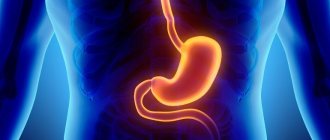
Ultrasound is an informative, harmless method of instrumental examination. This type of examination of the esophagus does not take much time: its duration is about 15 minutes. The main advantage of ultrasound examination is the resolving property of the equipment, which allows one to examine all the features of organs and their sizes. Using ultrasound of the esophagus and stomach, the following examinations are performed:
- study the walls of the stomach and intestines, tissue structure;
- detect motility disorders (movement of food in the tract);
- determine the condition of the vessels;
- lymph nodes are analyzed.
Indications
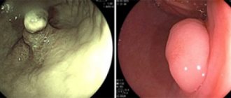
To identify some types of diseases, it is impossible to do without ultrasound. An ultrasound examination will determine the stage of the disease. Using this method, you can quickly identify the symptoms and characteristics of the disease, make a diagnosis, and, therefore, prescribe appropriate drug therapy. The organs of the digestive system are checked using an ultrasound examination if there is a suspicion of formations in the esophagus. In such cases, patients may complain of abdominal pain. This method of research helps to make a thorough analysis of inflammatory processes in the colon, to study erosions and ulcerative lesions of the mucous membrane of the gastrointestinal tract. In addition, ultrasound can reveal the location of polyps.
The specialist prescribes the procedure if there are suspicions of the following pathologies:
- neoplasm in the esophagus;
- peptic ulcer of the stomach or duodenum;
- tumor lesions in the stomach;
- hernia development.
Also, ailments that can be diagnosed using the ultrasound diagnostic method include:
- gastroduodenitis;
- polyps;
- any ulcerative lesions;
- cancerous tumors.
Ultrasound of the esophagus should also be done as a preventive diagnosis. In addition, it is sometimes possible for stomach acid to enter this organ. As a rule, this indicates the development of an infectious disease. To determine it, doctors prescribe such a study. Once the disease is diagnosed, doctors will prescribe appropriate therapy.
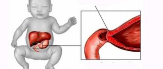
An organ ultrasound in a small child is primarily necessary to diagnose developmental anomalies . The method is widely used for the initial diagnosis of various diseases in a child, since it is safe for the child’s body.
Types of research
Depending on the anatomical features of the person, preliminary diagnosis, certain circumstances, and general condition, the type of ultrasound is chosen. There are several types of research:
- Percutaneous is the classic, first to appear, type of ultrasound. It consists in the fact that the sensor is moved over the skin of the abdomen and chest lubricated with a special gel, and a two-dimensional image of the organ being examined is reflected on a special monitor. In case of examination of the esophagus, the doctor passes the sensor from the neck to the epigastrium. In this way, all its departments are examined. The patient does not experience any discomfort during the procedure, and no complications can arise. The gel is necessary to improve the conductivity of ultrasound. During the procedure, the patient lies on his back with his head thrown back. Additionally, the study can be performed with the patient lying on his side.
- Intraesophageal - a sensor is inserted into the lumen of the esophagus. With its help, you can more accurately and efficiently examine the structure of the esophagus, detect changes in its walls and mucous membrane, and identify even minor neoplasms. This type of ultrasound is not suitable for children due to the danger of damaging the mucous membrane of the walls of the esophagus with the sensor, the lumen of which is much narrower than that of an adult.
- Endosonography is another name – endo-ultrasound. An ultrasound sensor with an optical device is inserted into the lumen of the esophagus. Reminiscent of the fibrogastroduodenoscopy method. Allows you to visualize and study those minute pathological changes in esophageal tissue that cannot be detected using conventional ultrasound. For example, the depth and degree of germination of neoplasms.
- Water-siphon - helps to assess the motility of the esophagus. During the study, the patient is asked to drink water, during which time the behavior of the walls and the patency of the organ are monitored.
Thanks to the development of such a diagnostic direction as ultrasound, it has become possible to timely detect various types of pathology of the esophagus, stomach, adjacent tissues and other organs.
Modern ultrasound diagnostic devices allow you to reproduce a 3D image in color on a monitor, which helps determine the characteristics of inflammatory processes in the gastrointestinal tract.
But the quality of the examination largely depends on the resolution of the device, the year of its manufacture, the competence of the doctor, and the thoroughness of preparing the patient for the procedure. It is unlikely that it will be possible to identify all problems of the digestive system using a first-generation device, so you need to clarify its characteristics before starting the study.
Preparation for the procedure
A couple of days before the diagnostic procedure, you should adhere to a strict dietary regimen. Before the study, you should not smoke or drink alcohol. Ultrasound is performed on an empty stomach, with the exception of half a glass of tea and a small cracker. Such preparation will allow doctors to make a more accurate diagnosis. In addition, patients are prohibited from eating the following foods before an ultrasound:
- peas;
- bread made from rye flour;
- cabbage dishes;
- carbonated drinks;
- fresh fruits and vegetables.
Why do patients trust medical networks?
The medical holding "SM-Clinic" has been successfully operating in Moscow since 2002.
Many private and corporate clients use the services of the centers. Only modern, expert-class equipment is available to patients - ultrasonic scanners of the latest generation. The staff are professionals in their field. Each of the diagnostic doctors is a qualified specialist in the application of ultrasound technologies of modern medicine. High accuracy of the examination is guaranteed, as well as a high level of service and availability of diagnostics. In addition, it is possible to perform diagnostics at home. The medical center has created comfortable conditions for every patient who needs accurate research not only in the stomach, but also in other organs and systems of the body. You need to undergo an ultrasound of the stomach in Moscow - contact SM-Clinic.
How is the procedure performed?
Before the ultrasound begins, the patient must lie down, then a special gel is applied to his stomach. The doctor moves the sensor and examines the organs on the screen, after which the person drinks a small amount of water - this will help the doctor see how it penetrates into the stomach. In this way, the esophagus section can be examined. The diagnostic procedure takes approximately half an hour. During decoding of the results, the echogram is described, pathologies are detected or not, and the person’s health status is determined.
There are several methods for ultrasound examination of an organ. For example, we are talking about the use of fluoroscopy and gastroscopy methods. The latter method involves inserting a special sensor through the pharynx into the esophagus. Thanks to such manipulations, it is possible to determine the disease at the initial stage, when the patient does not experience pain and does not have other unpleasant symptoms. The doctor records changes in the structure of tissues. An ultrasound is performed in the morning, on an empty stomach.
Kinds
Ultrasound includes a number of methods, including:
- Transdermal. During the procedure, a special sensor is located on the surface of the patient's skin on which the gel is applied. The sensor is localized in the cervical region and upper abdominal cavity. Due to this position of the device, different parts of the organ can be visualized. The technique is often used to diagnose diseases in a child.
- Intraesophageal. This method involves inserting a sensor through the lumen in the esophagus, due to which changes in the tissue structures of the organ can be determined. The technique is usually used to diagnose adult patients, since there is a risk of injury to the child. This method is also used to diagnose the heart.
- Using a water-siphon test. This technique allows you to determine the motility of organs. When performing a diagnosis, a person must drink a certain amount of liquid. It, in turn, contrasts in the stomach cavity.
- Endoultrasound. The endoscopic procedure allows you to view organs in detail. EndoUS helps to see pathological changes that are inaccessible to conventional ultrasound. EndoUS is performed using an endoscope with a special optical device and a sensor located at the tip of the device.
The doctor determines the type of diagnostic technique based on the patient’s test results and the characteristics of his anatomy. Thus, you can choose the appropriate method for each case.
Methodology
Ultrasound is a non-invasive research method that allows you to obtain a two-dimensional image of internal organs. Some devices have such high resolution that they can see areas less than 1 mm in diameter. If the doctor performing the study has the necessary level of knowledge and experience, this greatly facilitates the diagnosis of diseases.
When performing an ultrasound, you can obtain data on the condition of the stomach and esophagus, as well as information about neighboring tissue structures and general motility of the digestive tract. For children, ultrasound is considered the more preferable diagnostic technique due to its high level of safety.
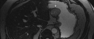
On the monitor, the image of the abdominal part of the esophagus is represented by an oval located between the aorta and the left lobe of the liver. When the sensor is placed in a transverse position, the esophageal cavity has a star-shaped shape. This is due to the fact that in the absence of incoming food, the mucous membrane collapses and acquires a certain folding. The echogram shows that the esophagus is normally divided into 3 layers:
- Internal – mucous;
- Medium – muscular;
- External – adventitia.
The thickness of the wall of the esophagus in a child normally does not exceed a few millimeters. When swallowing liquid, the lumen of the esophagus increases in size, and areas of reduced density begin to appear inside. If the patient swallows pieces of food, the diameter of the esophagus increases even more.
Article on the topic: Magnesium sulfate - a laxative for quick bowel cleansing
results
It should be noted that ultrasound cannot be a complete replacement for endoscopy or radiography. Nevertheless, this method allows doctors to obtain information regarding the condition of tissues, organ contours, and monitor the development of pathologies. What do ultrasound results show? The method helps to study the peculiarities of organ functioning and reflexivity. The results will provide information regarding the thickness of the gastric walls, the presence of inflammation, and tumors. The technique will make it possible to reliably determine pathologies, blood flow characteristics, and study tissue structures in detail. This diagnostic method will help answer many questions and identify many ailments.
Norms and pathologies
Normally, sections of the esophagus are visible as oval-shaped formations. Its walls are 6 millimeters thick. A glass of water can be evacuated in approximately 20 minutes. The walls must be uniform, the condition of the tissues surrounding the esophagus must also remain normal. In the presence of organ pathologies, reflux is sometimes diagnosed. In rare cases, cysts are found. The formation of hernias and other pathologies is possible.
In a healthy child, the thickness of the wall of the section in the esophagus is several millimeters. In sick babies, polyps, small hernias, disruption of gastric motility, obstruction in the esophagus, and other diseases are detected.
Reviews and features
Most people consider ultrasound an effective technique. At the same time, reviews from the majority of patients who underwent the procedure show that some do not fully understand the features of this diagnostic method. When undergoing an examination for the first time, people are interested in what an ultrasound should show. Those who have completed it claim that the method helps to identify many things. Experts also consider the technique to be effective, since pathological processes can be detected in the initial stages. At the same time, the method is simple and safe for the majority of subjects.
Cost of ultrasound of the stomach in Moscow in the medical network and registration for diagnostics
All details about the medical clinic in Moscow you want to contact, as well as about examination methods and diagnostic costs, can be found on our website or from the Contact Center operators. In addition, if you want to do an ultrasound of the stomach in our center, the operators will be able to schedule you for an examination at one of the network clinics of your choice. You can also use the feedback form to register online on the website. Take advantage of it at a time convenient for you and appreciate the quality of work of our specialists! We guarantee a decent level of service and availability of diagnostic procedures.
| Name of service | Price, rub.)* |
| Ultrasound of the stomach | 1,200 rub. |