Structure
The ileum and jejunum are quite similar in structure. The entire inner layer of such an organ is a mucous membrane, which is abundantly covered with villi (raise by about 1 millimeter). In turn, the surface of these elements consists of columnar epithelium. In the center there is a lymphatic sinus, as well as capillaries (blood vessels).
It should be especially noted that there are much fewer villi in the ileum than in the jejunum. However, they all participate in the process of obtaining useful and nutritious substances. Fats are absorbed through lymphatic vessels, and amino acids and monosaccharides through venous vessels. The entire mucous membrane of the ileum has a rather uneven surface. This is due to the presence of crypts, villi and circular folds. These formations significantly increase the total surface of the intestinal lining, which undoubtedly affects the process of absorption of digested food.
Small intestine
This section of the gastrointestinal tract got its name due to its small diameter, which varies from 2.5 to 6 cm. The structure of the walls differs between the mucous membrane and the submucosa, the muscular layer, and the outer serous membrane. It can be compared with the width of the lumen of the colon - from 6 to 10 cm. If the structure of the intestine is presented in good quality pictures, then the differences are better visible.
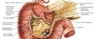
In addition to its own glands located in the wall of the department, ducts open into its lumen through which pancreatic juice and bile flow. Anatomically, the size of the duodenum is small (finger is the ancient name for a finger). However, this department is very important for changing food.
- Pancreatic juice entering the duodenum is necessary for the digestion of carbohydrates, proteins and lipids. The composition of the juice is greatly influenced by the type of food eaten. Thus, when consuming large amounts of fat, the lipase content is higher. If proteins predominate, then the concentration of enzymes that break them down is higher.
- Lipase, which breaks down fats, is activated in the presence of bile. It “breaks” fats into tiny droplets, making them more accessible to enzymes. Trypsin and chymotrypsin are involved in the decomposition of protein molecules.
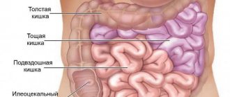
- The absorption of amino acids, simple sugars, and vitamins begins in the walls of the duodenum. The transition of molecules from food to lymph and blood continues in the jejunum. The length of this section is 0.9–2 m. The walls are relatively thick and well supplied with blood.
Features of the location of the jejunum in the abdominal cavity: located on the upper left side of the abdomen. The ileum, 2.5 to 3.5 m long, is located in the right lower abdominal cavity.
Features of the ileum
The intestinal juice of this organ begins to be released under the influence of chemical and mechanical irritation of the walls by chyme. In 24 hours its production can reach 2.4 liters. In this case, the reaction of the juice is alkaline, and its dense part consists of lumps-epithelial cells that produce and accumulate enzymes. At the right moment, the cells begin to be rejected into the intestinal lumen and then destroyed, thereby ensuring cavity digestion.
It should be noted that on the surface of each epithelial cell there is a microvilli. They are peculiar outgrowths on which enzymes are fixed. Thanks to them, another level of digestion occurs, called membrane (parietal). At this stage, food is hydrolyzed and absorbed in the ileum.
As you know, intestinal juice contains exactly 22 enzymes. The main one is called enterokinase. This enzyme is designed to activate pancreatic trypsinogen. In addition, the ileum secretes juice, which contains substances such as lipase, amylase, sucrase, peptidase and alkaline phosphatase.
The movement of chyme into other parts of the intestinal tract is carried out due to the contraction of fibers of the muscle layer. Their main types of movement can be called peristaltic and pendulum-like. The second group of contractions mixes the chyme. As for the worm-shaped (peristaltic) waves, they propel food to the distal sections.
By the way, both presented types of digestion exist in direct connection. In cavitary hydrolysis occurs of more complex substances to so-called intermediate substances. The processed foods are then broken down using membrane digestion. Next, the process of absorption of nutrients and nutrients begins. This occurs due to an increase in intraintestinal pressure, as well as the motility of muscle tissue and the movement of villi.
Ascending colon
The ascending colon is located in the right lateral region of the abdomen. Interestingly, it is located a little closer to the midline than the descending colon. The intestine originates at the point where the ileum enters the cecum, thereby being considered a continuation of the cecum. The ascending colon and the cecum are separated by two grooves. They, in turn, correspond to the frenulum of the ileocecal valve. Its main task is considered to be the passage of chyme from the ileum, that is, the last section of the small intestine, into the cecum, that is, into the first section of the large intestine. But at the same time, it should not allow contents contaminated with bacteria to pass from the large intestine into the small intestine.
The ascending colon is located in the right abdomen, then it projects to the right lateral region. Its posterior surface, which is not encumbered by the peritoneum, is located on the posterior wall of the abdomen and completely covers the extreme lateral position on the right. The colon originates just below the iliac crest, then it goes upward. As for its location, first it is ahead of the quadratus lumborum muscle, and then the right kidney. All these organs are delimited from the intestine by special fiber, as well as connective tissue membranes. The ascending colon reaches the lower surface of the right lobe of the liver, and then bends to the left and moves slightly forward or ventrally, thereby transitioning into the transverse colon. Its anterior part and sides are adjacent to the anterolateral abdominal wall. In some cases, it may be covered in some places by the greater omentum, as well as by loops of small intestines.
In an ordinary person, the length of the ascending colon is twenty centimeters, but it also happens that its position, as well as the length itself, can change, both up and down. If your cecum is higher than it should be, then the length of the ascending colon may be twelve centimeters or less. As for the diameter, it reaches approximately seven centimeters.
A free band, an omental band, and a mesenteric band were placed on the ascending colon. Accordingly, they are located on the anterior, posterolateral and posteromedial surfaces.
In the place where the ascending colon borders the cecum, the Busi sphincter is located. It is a circular bundle of smooth muscle fibers that has the shape of a notch or deep circular groove at the point where these sections of the colon connect. In some cases it looks like a crescent or petal.
The ascending colon is the initial section of the colon, but its continuation is considered to be the transverse colon. The functions of this part of the large intestine include the absorption of water and electrolytes, so that the practically liquid chyme that passes from the small intestine to the large intestine can thicken, turning into feces. It cannot be called one of the participants in the digestive process.
Disorders in diseases of the ileum
The ileum (where this organ is located is described a little above) quite often undergoes inflammatory processes. All diseases of this part of the small intestine have similar manifestations. As a rule, they are based on a violation of the digestive, excretory, absorption and motor functions. In medical practice, these deviations are usually combined under one common name - malabsorption syndrome.
General symptoms of diseases
The ileum, diseases of which can arise for various reasons, almost always makes itself felt by general signs of malaise. These include the following:
- pain syndromes;
- stool disorder;
- rumbling in the intestines;
- increased gas formation.
Quite often, patients complain to their doctors that they have diarrhea for a long time with trips to the toilet up to 4-7 times a day. In this case, undigested food remains may be found in the stool. In the first half of the day, the patient often feels rumbling in the intestines, which usually subsides only in the evening.
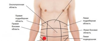
The affected ileum sometimes causes pain. They can have different localization (in the umbilical region, to the right of the midline of the abdomen and under the “spoon”) and character (bursting, pulling and aching). As a rule, the intensity of such pain decreases noticeably after the gases that have formed are released.
External symptoms of ileal diseases
Diseases of this part of the small intestine may also be accompanied by extraintestinal manifestations. They are caused by impaired absorption and breakdown of nutrients, vitamins and minerals. At the same time, patients lose weight quite quickly and cannot gain weight. Deficiency of B vitamins and iron often leads to the development of anemia, the formation of cracks in the corners of the lips and inflammation of the oral cavity. If the body begins to lack vitamin A, this can manifest itself in dry conjunctiva and night blindness. If there are hemorrhages on the patient’s body, this indicates a vitamin K deficiency.
Diseases of the small intestine
There are disruptions in the production of enzymes necessary for the complete breakdown of food. Insufficiency of digestive function - maldigestion. The condition in which absorption is impaired is called “malabsorption.” As a result, the body does not receive the substances it needs. The following processes may develop: destruction of bone tissue, splitting of nails and hair loss.
Symptoms of diseases of the small intestine:
- pain in the navel area;
- bloating, heaviness in the abdomen;
- loose stools, light-colored feces;
- "boiling" in the stomach;
- weight loss.
Inflammation of the small intestine - enteritis - can be caused by bacteria. The production of enzymes and digestion in general are disrupted. In the absence of enzymes responsible for digesting carbohydrates, intolerance to this food component develops. For example, lactase deficiency is the inability to break down the milk sugar lactose. Celiac disease is the absence of enzymes that break down gluten in cereals. Undigested substances become toxic products that poison the intestines.
To restore microflora, it is recommended to take probiotics along with prebiotics. In case of enzyme deficiency, the patient is prescribed medications that contain the missing substances. Treatment of intestinal dysbiosis is carried out with antibiotics and probiotics.
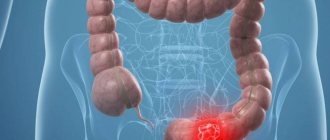
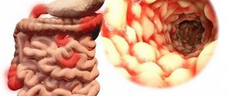
Crohn's disease
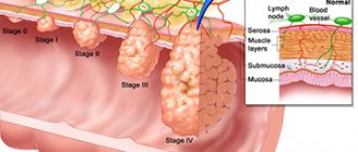
The most severe and common disease of this part of the small intestine is Crohn's disease (or so-called terminal ileitis). Typically, with this diagnosis, inflammation is localized in the last 15-20 centimeters of the ileum. Rarely, the process involves the blind, thick and duodenal sections of the gastrointestinal tract.
Inflammation of the ileum, the symptoms of which we will consider below, should be treated in time. Otherwise, after 3-4 years the patient may develop complications such as intestinal obstruction, fistulas, abscesses, peritonitis, amyloidosis, bleeding and others.
Diagnostics
To establish an accurate diagnosis, it is necessary to undergo a series of examinations that will allow you to estimate the percentage of disorders and select effective therapy.
Be sure to read: Antiulcer drugs for the stomach and duodenum Before sending for examination, the gastroenterologist must inform the patient about the preparation, which consists of following a two-week diet. This is done in order to maximally free the sections of the gastrointestinal tract from food. During this period, it is best to consume liquid porridges that have been boiled in water. In the morning and evening it is necessary to do cleansing enemas. One day before the expected diagnosis, you should completely avoid eating food, and six hours before you should not even drink water.
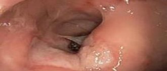
There are difficulties in diagnosing this section and the entire rectum. This is explained by the peculiarity of the location of the organ, which is almost inaccessible for detailed examinations. That is why the key role in making the correct diagnosis is given to the combination of endoscopic, immunological, histological, and radiological methods together with the general clinical picture. The most common diagnostic methods:
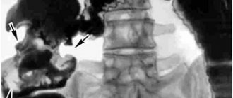
- X-ray. This method will help detect intestinal obstruction, including the ileum. In addition, the presence of inflammatory processes that change the structure of the organ is diagnosed.
- Ultrasound. The most universal diagnostic method that will allow you to diagnose the presence of foreign inclusions in the intestine. In addition, using this method you can accurately see the location of the organ and its size.
- Enteroscopy. The main advantage of this procedure is the ability to perform a biopsy for histological and cytological examination. Also, with this manipulation you can eliminate minor bleeding and polyps.
The presence of any deviation may require additional consultation with gastroenterologists, oncologists, endocrinologists and other specialists. That is why differential diagnostic methods should be carried out only in gastroenterological departments of a multidisciplinary hospital.
In continuation of the topic, be sure to read:
- Rectal cancer: symptoms, stages, treatment and prognosis for life
- Small intestine: location, structure and functions
- Duodenum: location, structure and functions
- Jejunum: location, structure and functions
- Caecum: location, structure and functions
- Sigmoid colon: location, structure, functions and diseases of the organ
- Ileocecal valve (Bauhinian valve): concept, location, structure and functions
- Colon: sections of the intestine, structure and functions of the organ
- Rectum: sections, structure, functions and diagnosis of the organ
- Large intestine: location, structure and functions
Be sure to read:
Ileocolitis (inflammation of the ileum and colon): symptoms and treatment
Symptoms of Crohn's disease
The signs of this disease vary.
- Intense pain in the right region (often reminiscent of acute appendicitis). In this case, the patient has a fever and is bothered by constant nausea and vomiting. Typically, pain occurs 3-5 hours after eating.
- Development of anemia and exhaustion.
- Scar changes in the ileum, which cause intestinal obstruction.
- Constant constipation or diarrhea, as well as rumbling in the intestines.
- Heavy bleeding or slight blood in the stool
Other diseases
Lymphoid hyperplasia of the ileum occurs against the background of an immunodeficiency state and proliferative changes in the intestinal walls. Typically, such changes are transient and often disappear without a trace on their own. The reason for the development of such a deviation may be an inadequate reaction of the intestinal lymphoid tissue, which occurs to external stimuli.
Major diseases
Chronic ileitis occurs due to improper treatment of an acute disease, concomitant dysfunction of the liver, gall bladder and intestines. The process is characterized by severe pain and diarrhea. It responds well to treatment. This includes proper nutrition and taking enzyme preparations.
Diseases of the ileum
One of the most serious chronic diseases is Crohn's disease. The inflammatory process of the intestinal walls alternates with periodic scarring. The resulting scars provoke intestinal obstruction. Ulcers and fistulas lead to bleeding. In this regard, anemia develops. Due to lack of nutrients, the patient loses weight and becomes completely exhausted.
From the respiratory system, cough, bronchospasm, hoarseness, and constant nasal discharge are observed. Neurological disorders are expressed in dizziness and pain, frequent fainting. The blood pressure is low and arrhythmia develops. Dry mouth and throat. Urticaria, dermatitis, and Quincke's edema occur on the skin.
Immunodeficiency contributes to the development of lymphoid hyperplasia. This phenomenon occurs against the background of complications of tonsillitis, polyposis, and pathologies of the large intestine. The submucosal layer of the terminal intestine is affected. Cells grow by dividing and new growths appear. Lack of blood circulation leads to tissue death. The body's metabolism is disrupted and intoxication develops. This condition is transient and often disappears forever.
Whipple's disease is more common in men. Specific fungi appear, they multiply and grow. Thickening of the mucous membrane occurs. The digestion process is disrupted, the body does not receive enough nutrients. General intoxication develops. The condition is accompanied by an increase in temperature and aching joints. Loose stools are observed periodically.
Among neoplasms, benign ones are more common. The disease has no symptoms for a long time. When the tumor reaches a large size, pain, intestinal obstruction, and bleeding occur. Colon cancer rarely develops. A predisposing factor is poor nutrition. This is the consumption of large quantities of fatty, fried and smoked foods. Ignoring the disease contributes to damage to neighboring organs.
Treatment of diseases
Typically, lymphofollicular hyperplasia affects only the terminal ileum.
This disease is a concomitant condition and therefore does not require treatment. As for Crohn's disease, cancer and other inflammatory processes, if treatment is delayed, they can affect the entire gastrointestinal tract, which subsequently leads to death. In this case, therapy consists of the use of medications, including antibacterial ones, which are prescribed only by an experienced gastroenterologist. By the way, such diseases in the later stages of development are often treated with surgical intervention. It is also worth noting that, along with medications for the treatment of diseases of the ileum of the small intestine, a strict diet is also prescribed. As a rule, it includes only light, quickly digestible and vitamin-rich foods. In addition, the patient is strictly prohibited from consuming alcoholic beverages, highly salted, spicy, fatty, fried and heavy meat, fish, and mushroom dishes. The patient's diet should include warm porridge prepared with semi-milk, day-old white wheat bread, sometimes butter, egg omelet, tea, compotes, fruit drinks, decoctions of rose hips, blueberries, and bird cherry. If you follow a diet and take all medications prescribed by your doctor, the outcome of treating an inflamed ileum will certainly be favorable.
Treatment
Ignoring and neglecting pathologies of the ileum is dangerous, the same applies to all other gastrointestinal diseases. Otherwise, the entire intestine will gradually be affected, especially if Crohn's disease, enteritis or cancer is suspected. Concomitant conditions, such as hyperplasia, go away on their own if the underlying disease is treated. Patients must be prescribed a strict diet, consisting of foods and dishes that do not irritate, do not overload the gastrointestinal tract, and are easily digestible.
At the initial stages of inflammation in the small intestine, it is treated with a conservative method. In cases of acute infection, antibacterial drugs are prescribed, if necessary, the deficiency of enzymes is compensated, and inflammation is relieved with anti-inflammatory drugs.
In advanced conditions or in cases of necrosis, intestinal obstruction, cancer, surgery is performed followed by rehabilitation or continuation of therapy.
We recommend: The most effective folk remedies for the treatment of intestinal colitis