What is undifferentiated (adenogenic) gastric cancer?
Stomach cancer is a disease that concerns many people.
Thousands of scientists around the world are developing new methods for treating oncology. In order to allow doctors to plan the necessary treatment and more likely establish the correct diagnosis, a classification of stomach cancer according to degrees of differentiation was created. The degree of differentiation is determined by microscopic examination based on what the tumor cells look like and what activity they exhibit.
Adenogenic stomach cancer: symptoms and manifestations
There are 4 types of cancer, depending on the degree of cell differentiation:
- well-differentiated gastric cancer. Such neoplasms are of low malignancy, their cells are practically no different from healthy ones, so the prognosis for them is favorable;
- moderately differentiated gastric cancer. It belongs to the average degree of malignancy and is, so to speak, a transitional form;
- low-grade gastric cancer. Cells of poorly differentiated formations have lost almost all similarities with normal tissue. They actively multiply and spread throughout the body;
- undifferentiated. The cells are completely atypical, do not resemble healthy ones in any way and cannot perform their functions. They just receive nutrients and constantly divide. Quite often, specialists cannot determine the histogenesis of the tumor. Adenogenic neoplasms are the most aggressive form of oncology, the cells of which are incapable of differentiation.
Features of undifferentiated types of gastric carcinoma:
- rapid rates of growth and metastasis;
- predominance of infiltrative growth (expansive forms are practically not found). The tumor does not have clear boundaries and spreads over a wide area;
- are more often localized in the proximal part of the stomach, or completely affect the entire organ;
- the structure of which the tumor consists has a loose growth pattern. Parenchyma predominates over stroma;
- The cell nuclei are irregular in shape and differ in size from each other.
Features of stomach tumors
Low-grade gastric cancer is quite common. In terms of the severity of the course and the danger of consequences, only undifferentiated stomach cancer can be called more dangerous. Such formations are characterized by pronounced mitotic activity. In addition, their sizes increase especially quickly.
The tissue from which the tumor is formed completely loses its properties as a result of degeneration into cancer.
The main distinguishing feature of such a tumor is that it consists mainly of units that are similar in structure to the mother cells. They are only capable of dividing and consuming substances. It is due to this that rapid growth in education is observed.
Cancer cells grow rapidly and invade new tissue
Pathology Clinic
As is already clear from the description of the pathology, adenogenic gastric cancer is the most aggressive form. The stages and symptoms of the disease change especially quickly. At the same time, it is important to emphasize that even an advanced stage may not have significant symptoms.
In most cases, the pathology develops against the background of peptic ulcer disease. Smoking, dietary errors, and so on can also be provoking factors. Accordingly, the symptoms will be similar to stomach pathologies. It is for this reason that patients are in no hurry to see doctors, which in many cases leads to late detection of cancer.
First signs
Gradually, as the pathology progresses, symptoms begin to appear. Most patients note the following changes:
- constantly present pain in the abdomen of an aching nature, not associated with food;
As cancer develops, aching pain in the stomach appears
- nausea, sometimes vomiting, possible presence of blood impurities;
- change in stool to dark, pasty-like;
- severe weakness;
- loss of interest.
Undifferentiated adenogenic cancer of gastric tissue especially often causes the appearance of common symptoms.
As a result of the fact that the degenerated cells no longer fulfill their role, the digestion process is greatly disrupted. As a result, the patient’s body no longer receives all the nutrients required.
All this leads to complaints such as increased fatigue, weakness, sleep disturbances, and weight loss.
Poorly differentiated stomach cancer is accompanied by nausea
Cancer patients are characterized by mood changes, which manifest themselves in deep depression, apathy and suicidal tendencies.
In general, at the initial stage, patients may only notice a decrease in appetite and a disturbance in taste. Particularly characteristic of damage to stomach tissue is a complete aversion to all meat products.
If the formation is located in the cardia, complaints such as problems with swallowing may occur. Sometimes spasms occur, causing vomiting.
But the most dangerous phenomenon is that if there is a formation in this section, the walls of the stomach begin to involuntarily contract.
As a result of this, cells break off from the tumor and spread with the food mass into the underlying parts of the tract, which leads to metastasis.
Patients with stomach cancer report difficulty swallowing
Late stage symptoms
At the third and final stage of the pathology, the clinic is brighter. As the size of the formation increases, the pain increases and becomes unbearable. When neighboring organs are involved, pain of a different nature may occur, for example, girdling pain or radiating to the back.
It is also possible that cancer cells spread to the tissue of the diaphragm. In such a situation, the clinic will be similar to pathologies of the heart and lungs. But when the intestines are involved, constipation appears and bloating may be a concern. At the same time, the latter symptom can also be caused by liver damage. In such a situation, yellowing of the skin and change in the color of urine also occur.
Progressive tumor growth can cause bleeding. This condition is an absolute indication for hospitalization.
Constipation occurs in advanced stages of cancer
Poorly differentiated formations are almost always detected at later stages in the presence of metastases. Patients come in especially often when they have stage 3 stomach cancer.
Accordingly, the prognosis for the pathology is unfavorable. If it is possible to identify abnormalities at an early stage, for example, during a preventive examination, then patients can only agree to surgery and subsequent chemotherapy.
Without this, it is impossible to get rid of metastases.
Thus, up to the third stage, cancer is treated with surgical methods, but in the future, palliative therapy is used. Exactly how long it is possible to live after diagnosis depends on a huge number of factors, including the psychological mood of the patient.
Diagnosis of pathology
The first question that all patients with a similar disease ask is how long they have left to live and whether it is possible to somehow alleviate the condition. The answers to these questions directly depend on how quickly the diagnosis is made. Therefore, the diagnosis of pathology is especially important. All possible methods are used. The following are especially important:
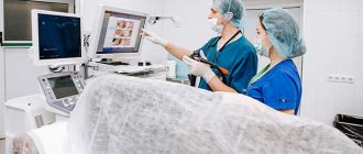
- FGDS - thanks to this method, it is possible to visually examine the stomach tissue, assess the condition and size of the formation. If necessary, tissue is taken for biopsy.
Diagnosis of stomach cancer is carried out using phagogastroduodenoscopy
- Histological examination is precisely thanks to which it is possible to accurately determine the diagnosis. In the laboratory, the obtained tissues are examined. In the presence of a poorly differentiated tumor, the cells have a characteristic shape and structure.
- Blood test - in addition to traditional studies, it is mandatory to determine markers characteristic of this particular formation.
- X-ray – a contrast agent is used during the procedure. It can be used either orally or administered intravenously, the latter option being more informative.
- Tomography is an expensive procedure, but it is precisely because of it that it is possible to assess the condition of neighboring organs and identify the presence of metastases. In addition, such a study is used in cases where surgery has already been performed and chemotherapy is being performed. Thanks to MRI, it is possible to assess the condition of the remaining nodes and their response to treatment.
MRI is performed to detect metastases
- Ultrasound is the first test ordered when cancer is suspected. The procedure is simple and accessible to everyone. In addition, the results are obtained immediately, which is especially beneficial in terms of treatment.
- Laparoscopy is performed for diagnostic purposes. Especially often during manipulation it is possible to identify nodes in the liver, peritoneum, and so on.
The final diagnosis is made only after a complete examination, and the most reliable information is provided by histological tissue analysis. Only after this can treatment begin.
If gastric cancer is suspected, an ultrasound scan is prescribed.
Features of treatment
The treatment method directly depends on the stage of the pathology. In all cases, without exception, several methods are used at once. Only the order in which they are performed changes.
The main method is to remove the tumor, with the obligatory capture of healthy tissue. Lymph nodes are also removed. In some cases, a third of the organ is removed. Only the areas necessary to perform the digestive function remain.
Types and types of undifferentiated stomach tumors
Forms of undifferentiated stomach cancer:
- solid cancer, also known as trabecular. The tumor has a dense structure. Consists of trabeculae of cancer cells that do not have a glandular structure, which are located between connective tissues;
- scirrhus (fibrous cancer) is represented by hyperchromic cells that settle between strands of coarse fibrous tissue;
- mucous cancer (ring cell, colloid), is characterized by the production of large amounts of mucus, which in volume reaches more than half of all other tissues. The structure resembles a mucous mass, in which mutation cells similar to rings are difficult to detect, which is how they got their name.
Interesting fact! In the stomach, undifferentiated variants of adenocarcinoma, small cell and large cell cancer are found.
Signs of oncology
Of course, the symptoms of different types of such malignant tumors are different. In most cases, the signs of the disease depend on the location of the compaction. But it is worth noting that poorly differentiated potato carcinoma can have identical symptoms, regardless of the type and location of the tumor. General signs of malignant formations of this type include:
- a sharp decrease in appetite;
- causeless and sudden decrease in body weight, while weight can reach critical levels;
- increasing anemia;
- general malaise and weakness;
- pathological fatigue, feeling of depression and apathy;
- general exhaustion of the body.
That is, the symptoms of the disease have a fairly serious impact on the patient’s standard of living, which is why it will be quite difficult to ignore them.
Important! As the disease progresses, characteristic symptoms may intensify. Moreover, it is worth understanding that the symptoms will be especially acute in the area where the tumor is located.
Signs of this type of carcinoma must be responded to immediately, since the disease has a very aggressive and rapid course. It is important to understand that the prognosis for poorly differentiated carcinoma depends on the stage of the disease at which the patient seeks help.
Have additional questions
Causes of undifferentiated gastric cancer
The causes of adenogenic stomach cancer are related to the standard of living of the population. During research on this matter, it was noted that this disease appears more often in poor people and less often in wealthy people. The decisive role in this is played by the nature of nutrition and lifestyle.
The risk of developing cancer increases with excessive consumption of carbohydrates, salty foods, fish, as well as due to a lack of vitamins and microelements. The quality of water and food that a person eats is of no small importance. High levels of nitrites and nitrates, which contribute to cancerous transformations, are bad for the stomach. Scientists also point out the harm of smoking and alcohol.
Interesting fact! It has been proven that the diffuse type of carcinoma can be inherited.
Internal risk factors include:
- the presence of duodenogastric or gastroesophageal reflux in a person;
- impaired absorption of nutrients;
- genetic abnormalities.
When the mucous membrane is damaged by one or more of the above factors, the likelihood of infection with the bacterium Helicobacter pilory increases. The presence of this microorganism in a person increases the risk of carcinoma by 10-12 times! Helicobacter pylory causes inflammation against which mutations and genomic rearrangements can occur in the cells of the mucous membrane. It should be noted that these processes take 30-40 years.
Also, ailments such as gastritis and ulcers, which are precancerous conditions, are closely associated with poor diet and the H. Pilory bacterium. Chronic gastritis turns into cancer in 75-80% of cases if left untreated for a long time.
Undifferentiated breast cancer prognosis
Today we will talk in an article about undifferentiated cancer. This is a fairly serious disease. In the article we will also look at the signs of this disease, methods for diagnosing it, as well as all possible methods of treating the disease. First, let us note that cancer is a general name that implies a disease associated with the mutation of cells and their spread in the body.
The disease can affect different human organs. The disease is also diagnosed at different stages. Where this disease is located is called its differentiation. It is usually denoted by the letter G.
If there are infected cells in the human body, in which the degree of modification and difference from healthy cells is high, then they are called undifferentiated and designated as G3. There are also highly differentiated cells. They are almost identical to healthy ones.
They are usually designated as G1. Highly differentiated cancer has a benign course.
Tumors that belong to this type of disease have the same name as the tissue on which they have spread. For example, adenocarcinoma, non-keratinizing squamous cell carcinoma and others.
Undifferentiated cancer is named after the shape of the infected cells. For example, undifferentiated squamous cell carcinoma, signet ring cell carcinoma and others. This disease has rapid progression and is characterized by frequent metastases.
This disease is malignant and can affect various human organs.
Undifferentiated small cell cancer has its own peculiarity - mutating cells are not able to differentiate. In other words, it has no capacity for development. That is, it does not develop to such an extent as to perform its intended function.
We can say that she does not mature, but remains at a certain stage of formation.
A cancerous tumor of this type of cancer consists of undifferentiated cells that cannot perform their intended functions to ensure the normal functioning of a particular organ.
Let's consider the types of disease. The most common types of this pathology are:
- Adenogenic breast cancer.
- Undifferentiated thyroid cancer.
- Adenogenic gastric cancer.
- Undifferentiated lung cancer.
- Adenogenic cancer of the nasopharynx.
The symptoms of a patient whose body is infected with an undifferentiated cancer may vary depending on which organ is affected by the malignant tumor. If a person has a disease such as undifferentiated stomach cancer, then he will have the following symptoms:
- Presence of heaviness in the stomach after eating.
- Unpleasant sensations (burning, dull or sharp pain) in the stomach.
- The person is constantly sick.
- There is vomiting.
- Rejection of certain foods (for example, meat, poultry and others).
- Partial or complete lack of appetite.
- A person only needs a small amount of food to feel satisfied.
- There is severe weight loss.
It is a well-known fact that the earlier a disease is diagnosed, the more likely a person is to restore his body. Undifferentiated (adenogenic) cancer is diagnosed using modern research methods.
- Endoscopy. To identify cancerous formations of internal organs, endoscopy methods such as fibrogastroscopy, bronchoscopy, and colonoscopy are used.
- Laparoscopy is a surgical intervention in the human body to detect cancer cells.
- Ultrasound (ultrasound examination of the body). Despite the fact that this research method is quite simple, it allows you to determine the presence of tumors on organs such as the liver, pancreas, uterus, ovaries and lymph nodes.
- X-ray. This diagnostic method allows you to detect the presence of undifferentiated cancer cells. They carry out such types of studies as irrigography, hysterography, computed tomography of the head and heart. This diagnostic method allows you to see the affected areas of infected cells and determine their structure.
- Biopsy. In some cases, it is necessary to take this test from a person. A biopsy is a study of the affected organ material. This procedure allows you to determine what type of tumor the tumor has. What stage is undifferentiated cancer at? A prognosis for the development of the disease can also be made through a biopsy.
It should be said that for the treatment of undifferentiated cancer it is better to use the most modern methods. It is also desirable that the approach be comprehensive.
Thus, a person has a better chance of stopping the process of proliferation of cancer cells and setting up his body for regression of the disease. Full recovery of the body is possible.
As mentioned above, it is better if the disease is diagnosed at an early stage.
Therefore, it is recommended for a person to periodically examine the body. You need to see a doctor on time and take the necessary tests. If any deviations from the norm are detected, additional studies should be carried out in order to exclude the presence of cancer cells in the body.
What therapy is recommended if a person has undifferentiated cancer? Treatment is carried out in several ways.
- Treatment using radiation therapy. This method is carried out by emitting ionizing energy. Radiation therapy is prescribed after surgery to remove remaining cancer cells. Radiation can be carried out remotely or internally. Both types of radiation therapy may also be prescribed.
Unfortunately, if a person consults a doctor at a late stage of the disease, then he can no longer undergo surgery. And for this type of cancer, the surgical method is the most effective. Therefore, the advanced degree of undifferentiated disease has a poor prognosis.
But if the disease is diagnosed at an early stage, then it can be cured. It is necessary to remove the tumor through surgery. But after the tumor is removed, the patient should undergo a course of chemotherapy and radiation.
But a person should know that even if complex treatment to remove cancer cells was successful, relapse is possible. That is, their reappearance in the body. Especially during the first three years after therapy.
There are statistics that relapse after treatment for stomach cancer occurs in 90% of cases. If it happens, then the prognosis will be disappointing, namely, on average, a person lives 3 months.
An interesting fact is that the causes of cancer cells in the human body have not yet been established. But the causes of cancer are classified into 3 large groups.
- Physical factors. This group includes ultraviolet and radiation.
- Chemical factors. Namely, carcinogenic substances.
- Biological factors. For example, viruses.
Initially, under the influence of some factors, the structure of DNA changes. As a result, the cell does not die, but changes and begins to multiply.
In addition to the above external factors, there are internal factors that disrupt the DNA structure. Namely heredity. But when making a diagnosis, it is difficult to determine what exactly became the basis for this failure.
Since the exact causes of cancer are unknown, treatment of this disease comes down to removing the infected cells. However, most scientists agree that the main cause of cancer is a violation of the DNA structure. And it is destroyed by carcinogens.
With age, the body's resistance decreases, so it is necessary to reduce the intake of carcinogens into the body. It is recommended to avoid exposure to ultraviolet radiation, infection with viruses, and be careful when taking hormonal medications.
You should also stop smoking, as this habit leads to lung cancer.
It should be said that there are various centers in the world that treat cancer tumors. If possible, you should read the reviews and results of such clinics.
It may make sense to treat cancer in a special clinic where there is an integrated approach.
Some centers offer 24-hour patient monitoring and use the latest treatment methods using modern medical advances.
Undifferentiated cancer is treatable; the main thing is to take all necessary measures to restore the body and have a positive attitude. Therefore, do not lose hope for recovery.
source
In low-grade oncology, tumor cells are not as similar in structure to healthy cells as possible. This is a sign of malignancy. Poorly differentiated breast cancer is aggressive.
It spreads very quickly throughout the gland itself, metastasizes to other organs and is difficult to treat. In this case, timely diagnosis is very important. This type of tumor is the second most deadly among cancer diseases.
However, there are a number of factors that increase the likelihood of encountering cancer:
Hormonal disorders, especially during menopause;
Lack of childbirth and lactation;
Abuse of nicotine and alcohol;
Unfavorable environmental conditions.
Exposure to radiation on the body, late first birth, breast trauma and other factors also increase the risk of cancer.
It can be common and specific to breast cancer.
Change in color of the breast skin;
Common cancer signs include weakness, constant fatigue, headaches, loss of appetite and weight loss, including cachexia. They are due to the fact that tumor cells secrete toxic substances that cause signs of intoxication.
The course of the disease is aggressive. Cancer cells appear through the degeneration of the body's own cells under the influence of negative factors.
A genetic mutation occurs, the cell becomes cancerous and begins to rapidly divide. For poorly differentiated cancer, this rate is especially high.
While the tumor is small, it is localized only in the tissues of the mammary gland, and then affects the lymph nodes and spreads through the vessels to other organs.
Source: //sklad-kotlov.ru/rak-molochnoy-zhelezy/nedifferentsirovannyy-rak-molochnoy-zhelezy-prognoz/
Symptoms of adenogenic stomach cancer
Most cases of gastrointestinal cancer are detected at late stages, which makes it impossible to carry out radical therapy, thereby reducing the patient’s chances of recovery.
Symptoms are divided into two groups:
- local;
- are common.
Local signs include:
- pressing pain in the stomach;
- heartburn;
- belching air with an unpleasant odor;
- dysphagia (depending on the location of the tumor);
- lack of appetite;
- change in taste preferences, aversion to previously favorite foods;
- satiation with a small amount of food;
- nausea and vomiting (sometimes bloody).
Common symptoms of undifferentiated stomach cancer include:
- sudden weight loss;
- anemia;
- apathy and weakness;
- temperature increase;
- nervousness, absent-mindedness.
Poorly differentiated cancer: what is it, highly differentiated cancer, difference
Malignant tumors look different under a microscope. In some cases they resemble normal tissue, while in others they are very different from it. Depending on this, pathologists and doctors conditionally divide malignant neoplasms into two types:
- Well-differentiated tumors retain many features of normal cells and tissues. They grow into neighboring organs and metastasize quite slowly.
- Poorly differentiated tumors contain cells and tissues that are very different from normal ones. They behave much more aggressively and spread throughout the body faster.
The degree of differentiation of cancer determines how quickly it can spread throughout the body. And this, in turn, directly affects the prognosis for the patient. In addition, poorly differentiated tumors respond less well to radiation therapy and chemotherapy. The oncologist takes this into account when drawing up a treatment program.
The terms “highly differentiated” and “lowly differentiated” themselves are quite general and say little. For a more accurate assessment, oncologists distinguish 4 degrees of differentiation.
What are the degrees of differentiation of cancer?
The indicator of the degree of tumor differentiation in oncology is usually denoted by the Latin letter G. There are five options:
- Gx - the degree of differentiation cannot be determined.
- G1 - highly differentiated malignant neoplasms.
- G2—medium degree of differentiation.
- G3 - poorly differentiated tumors.
- G4 - undifferentiated cancer. Tumor cells have lost all the external characteristics and functions of normal cells. They resemble each other like two peas in a pod; their only activities are growth and reproduction.
This is what the general scheme looks like, but for different types of cancer the degree of differentiation is determined differently. For example, when examining tumor tissue from the mammary gland under a microscope, the presence of milk tubules, the shape and size of cell nuclei, and the activity of cell division are taken into account. Each of the three characteristics is scored, then their total number is calculated.
For prostate cancer, a special Gleason system is used. It provides 5 gradations, each of which is assigned a certain number of points, in accordance with the numbering, from 1 to 5:
- The tumor consists of homogeneous glands, the cell nuclei are changed to a minimal extent.
- The tumor consists of clusters of glands that are separated by connective tissue partitions, but are located closer to each other than normal.
- The tumor consists of glands of different structures and sizes; the tumor tissue grows into connective tissue partitions and into neighboring structures.
- The tumor contains cells that are very different from normal ones. The tumor grows into neighboring tissues.
- The tumor consists of undifferentiated cells.
Typically, prostate cancer has a heterogeneous structure, so the doctor must select from the list two gradations that are found in most of the tumor. The points are added up and the degree of differentiation is determined from the resulting number.
Are the degree of differentiation and the stage of cancer the same thing?
Different indicators are used to describe a malignant tumor. Division depending on the degree of differentiation is only one of the possible classifications.
The stage of cancer is determined depending on three parameters, which are designated by the abbreviation TNM: how much the tumor has grown into neighboring tissues, whether it has managed to spread to the lymph nodes and give metastases.
Currently, the classification of tumors depending on their “molecular portrait”, especially in later stages, is becoming increasingly important.
How does the treatment of well-differentiated cancer differ from low-grade cancer?
It is impossible to answer this question unequivocally, since cancer can be very different. For some malignant tumors (for example, breast cancer, prostate cancer), the degree of differentiation is especially important; in cases with other oncological diseases, this indicator fades into the background.
In general, we can say that poorly differentiated tumors are more aggressive and require more aggressive treatment.
When drawing up a treatment plan, the oncologist focuses on many indicators: the stage of cancer, the degree of differentiation, the type and location of the tumor, its “molecular portrait”, the patient’s health status and concomitant diseases. Early diagnosis greatly increases the chances of successful treatment, but even in advanced cases it is always possible to help and significantly prolong the patient’s life.
Make an appointment for a consultation around the clock +78 800 100 14 98
Source: //www.euroonco.ru/terms-from-az/vysokodifferentsirovannaya-nizkodifferentsirovannaya-opuhol
Diagnosis and treatment of adenogenic cancer
There are no particular differences in diagnostic methods between various forms of gastric carcinoma. Initially, standard studies are carried out to identify symptoms of the disease: complaints and anamnesis are collected, external examination, palpation, percussion, etc. are carried out.
Patients with gastric cancer usually complain of one or another gastric disorder, as well as abdominal pain, loss of appetite, weight loss, general weakness and malaise. Depending on the stage of the disease, the general appearance of the patient changes: the skin turns pale, becomes dry, facial features become haggard, eyes become dull.
During palpation, pain is observed in the epigastric region. It is possible to palpate a stomach tumor in rare cases, but metastases in the lymph nodes, liver and lower abdomen can be detected.
The symptoms of adenogenic stomach cancer are very similar to gastritis or ulcers, so further examination is required to establish the correct diagnosis.
Doctors usually prescribe a number of tests:
- blood test. It will show the presence of anemia, proteinemia, leukocytosis, blood clotting disorders, changes in the level of protein, albumin and other substances. Although in the initial stages of the disease there may not be any significant changes.
- fluoroscopic examination. With its help, you can identify the presence of a tumor or signs indicating it, namely: irregular relief of the mucous membrane, thickening of its folds, erosion, defect in the filling of the stomach, retention of the contrast agent, narrowing of the lumen or outlet. The use of the latest radiographic techniques makes it possible to detect cancer in 80% of cases;
- endoscopic studies (FGES). This is an advanced method for diagnosing stomach cancer, which allows for a differential diagnosis and detection of a tumor at an early stage. Also, using FGES, it is possible to perform a biopsy with further histological and cytological examination, which is mandatory for verifying the tumor. After a biopsy, it will be possible to confirm that the tumor is undifferentiated.
If oncology is detected in the stomach, laparoscopic examination, CT and ultrasound of the abdominal cavity, liver scanning, lymphography and angiography are performed. All these methods are used to find out how far the tumor has grown and determine further treatment.
Poorly differentiated cancer: types and differences of the disease
Oncological diseases have their own classification, where low-grade cancer occurs, which is a pathology in which cancer cells have significant differences and a characteristic location within one tumor. In this case, the abnormal cells do not have a clear structure, which is inherent in healthy tissues.
The concept of differentiation of oncology should be understood as the degree of development of pathological cells.
If a benign neoplasm acts as a highly differentiated cancer, since its cells resemble healthy tissue in structure, then the poorly differentiated structures are changed in such a way that it seems impossible to recognize which tissue has been transformed in this way.
Characteristics of the problem
Poorly differentiated cancer is an oncological pathology that is characterized by the rapid division of cancer cells.
In appearance, they resemble stem cells that go through several stages of development in the future.
They have irregularly shaped nuclei, so they cannot perform the functions of healthy tissue, but they consume nutrients and energy, unlike high-grade cancerous tumors.
This type of cancer has a high degree of malignancy; the tumor grows rapidly, affecting new areas of the organ (spreading metastases). It can be formed in different organs of the human body.
Note! Poorly differentiated tumors are practically insensitive to chemotherapy, therefore they are the most dangerous compared to all oncological diseases.
The most common low-grade neoplasms are squamous cell and adenogenic low-grade cancer.
Types of poorly differentiated and undifferentiated cancer
Cancerous tumors that have low differentiation can affect various organs:
- Undifferentiated stomach cancer occurs due to addictions, as well as consumption of salty, spicy and canned foods in large quantities. Sometimes the appearance of the disease is provoked by a person’s existing stomach ulcer. Most often, adenogenic stomach cancer develops, which manifests itself in the form of pain in the abdomen, nausea, and intolerance to certain food components. As the malignant tumor grows, weight loss and pale skin are observed. The appearance of stomach bleeding. To confirm the diagnosis of “undifferentiated gastric cancer” and determine the degree of its malignancy, a biopsy is performed.
- Breast cancer is an aggressive form of pathology that spreads metastases throughout the body. Symptoms of the disease appear in the early stages of cancer.
- Poorly differentiated adenocarcinoma of the cervix is the most common variant of the pathology. It is diagnosed using a biopsy and laboratory tests.
- Undifferentiated lung carcinoma is characterized by the spread of metastases to the lymph nodes, liver, adrenal glands and brain. Signs of the disease manifest themselves in the form of cough, shortness of breath, pain in the chest area.
- Poorly differentiated bladder cancer is caused by painful urination, difficulty, and pain in the lower abdomen.
- Poorly differentiated colon cancer is formed from its epithelium and is characterized by a large amount of mucus production and its accumulations in the form of clots.
- Thyroid cancer of low differentiation is caused by the formation of a node in the structure of the organ, a rapid increase in its size, which provokes an enlargement of the thyroid gland itself.
Diagnostic measures
Diagnosis of low-grade cancer is carried out using several methods:
- examination and study of the patient's medical history;
- MRI of internal organs;
- CT scan of internal organs and systems;
- Ultrasound and radiography;
- blood test for cancer markers;
- puncture and biopsy of organ tissue;
- endoscopy and irrigoscopy;
- stool analysis, cytology smear, curettage.
After passing the examination, the oncologist makes an accurate diagnosis. He then prescribes the appropriate treatment, which is carried out in the clinic.
Note! In oncology, moderately differentiated cancerous neoplasms and undifferentiated tumors are also distinguished. They can all exhibit different symptoms.
Oncology treatment
Since low-grade cancer exhibits symptoms with great intensity, treatment should be carried out immediately. To do this, the doctor may prescribe the following therapy methods:
- Surgical intervention.
- Several courses of chemotherapy for cancer of the ovaries, liver, skin or other organs and tissues.
- Radiation and immunotherapy.
- Use of enzymes and hormones.
- Androgen blockade in prostate pathology.
Auxiliary treatment methods can also be used in the form of herbal medicine, painkillers, and so on. During and after treatment, in some cases it is necessary to follow a diet. The diet should be balanced, including only natural products that do not contain carcinogens.
Prognosis and prevention
The prognosis of low-grade cancer depends on the stage of the disease and the malignancy of the pathology. At the initial stage of development, survival rate is up to 80% of cases, at the second stage – 50%, at the third stage – 20%, and at the last stage of oncology development, survival is observed in 5% of cases.
Prevention of pathology consists, first of all, in avoiding the influence of unfavorable factors. It is recommended to eliminate bad habits, lead a healthy lifestyle, promptly treat various diseases, and eat right. Doctors recommend regular examinations for early detection of cancer.
Note! Low-grade cancer is a dangerous pathology that is rapidly developing. Therefore, it is important to identify it at an early stage of development, when the chances of survival are high.
(1 5,00 of 5) Loading...
Source: //OncoVed.ru/common/nizkodifferentsirovannyj-rak-vidy-i-otlichiya-zabolevaniya
Treatment of adenogenic stomach cancer
Treatment for malignant stomach tumors may include:
- An operation whose purpose is to remove the part of the organ affected by the tumor and the surrounding lymph nodes, since they may contain metastases. This is called subtotal resection. In advanced cases, the stomach may be completely removed (this is called a gastrectomy). If there are metastases in surrounding organs, then the operation is complemented by their removal.
- Chemotherapy. It involves the introduction of cytostatic drugs that inhibit the growth of a malignant neoplasm.
- Radiation therapy (exposing the tumor to radiation, which destroys its cells).
The best treatment for undifferentiated gastric cancer is considered to be complex therapy with mandatory surgical intervention. Indications for resection are determined based on the size, location and shape of tumor growth, as well as its degree of spread to surrounding tissues and structures.
One of the important problems in the treatment of stomach tumors is the inoperability of the patient. Late diagnosis is considered the main reason for inoperability. Often, a tumor is found when it has already grown beyond the affected organ and the process of metastasis has begun. Then it is not possible to completely remove the tumor.
With adenogenic types of cancer, an additional difficulty arises associated with infiltrative aggressive growth. Even an experienced specialist will not be able to clearly determine the boundaries between affected and healthy tissues. Therefore, more than half of all diagnosed patients are classified as inoperable.
In addition, radical treatment is contraindicated for people with severe exhaustion or obesity, concomitant pathologies (diabetes mellitus, renal failure, etc.). This is due to the complexity of surgical interventions on the stomach. The operation sometimes causes complications and even death in patients.
How is inoperable cancer treated?
In such situations, palliative treatment is prescribed in the form of operations to remove part of the tumor, install a bypass anastomosis between the stomach and intestines, or apply a gastrostomy. This eliminates complications that are often observed in such patients: dysphagia, pyloric stenosis, bleeding and tumor disintegration.
Also, if radical surgery for stomach cancer is not possible, palliative chemo-radiation therapy is used. This approach only slightly increases life expectancy, but can have a beneficial effect and improve the general condition of the patient. Sometimes it is even possible to achieve the tumor becoming resectable.
Although adenogenic cancer is poorly susceptible to chemotherapy, it is often used in the postoperative or preoperative period to reduce the likelihood of disease relapse and increase the effectiveness of gastric resection.
The following were recognized as the most effective:
- 5-Fluorouracil;
- platinum preparations;
- Etoposide;
- Adriamycin;
- Mitomycin.
They are combined in various schemes. Chemotherapy courses for stomach cancer are repeated several times before and after surgery.
Fact! More than half of adenogenic neoplasms are considered inoperable.
Radiation therapy for stomach cancer is prescribed 2 weeks before surgery in the absence of associated complications. The doctor selects the radiation dose according to individual indications. As a rule, irradiation is carried out 5 times a week, 2-4 Gy at a time. The total number of sessions is from 10 to 20, and the total radiation dose is 30-40 Gy. Radiation therapy may be continued after surgery.
Poorly differentiated carcinoma and its varieties are difficult to treat forms of oncology because they respond poorly to chemotherapy and radiation. Therefore, new methods are being developed to administer chemotherapy and radioactive substances directly to the cancer site. An example of this is intra-arterial polychemotherapy - the introduction of cytostatic drugs into the arteries feeding the tumor.
To increase the sensitivity of undifferentiated neoplasms to chemistry and radiation, modifying factors were invented: hyperthermia (heating of the tumor), immunotherapy, the action of magnetic fields, artificial hyperglycemia. Their use allows you to destroy a larger number of cancer cells.
Treatment
Success in the treatment of undifferentiated cancer directly depends on the stage, that is, on the size of the malignant formation and the prevalence of secondary foci (metastases). As a rule, an integrated approach is most effective when several techniques are used at once.
The greatest problem is the late detection of pathology; often the tumor turns out to be inoperable and manages to give numerous metastases
Medicines
If the tumor cannot be removed surgically, palliative treatment is prescribed. Its main goal is to improve your well-being as much as possible and try to preserve the functions of the affected organs.
Treatment is prescribed symptomatically, that is, drugs are selected that relieve pain and facilitate digestion.
Chemotherapy
Methods such as chemotherapy and radiation are used as auxiliary treatment for the adenogenic type of gastric pathology. Their use before surgery is necessary to reduce the size of the affected area.
Such treatment sometimes helps to preserve the stomach, since during surgery it will be possible to remove not the whole organ, but part of it. The use of radiation therapy and cytostatics after surgery helps reduce the risk of relapse.
Surgery
If pathology is detected in a timely manner, surgical treatment is resorted to. This is the main method of combating pathology. An operation is performed to remove tumor foci.
With this type of cancer, in most cases, it is necessary to completely remove the digestive organ, as well as adjacent tissues into which mutated cells could penetrate. Only if the disease can be diagnosed in the initial stages, it becomes possible to remove only part of the stomach.
Since damaged cells penetrate the lymph nodes first, they are removed simultaneously with the affected organ. After surgery, additional treatment is carried out using chemotherapy.
This is necessary to destroy the affected cells that have entered the bloodstream or lymphatic system. If they are not destroyed, they can become the germ of a new tumor.
Diet
Successful recovery after treatment is facilitated by a properly formulated diet. All types of food that are difficult to digest are removed from the diet. Only light foods with minimal fat content are allowed. Products are well boiled or stewed.
At the same time, the food should be nutritious enough and contain the substances necessary for the body to increase the body's endurance.
Metastases and relapse of adenogenic gastric cancer
A poorly differentiated tumor is formed from the epithelium of the gastric mucosa, penetrating deeply into its layers and forming loose clusters. The neoplasm quickly spreads from the stomach tissue to the surrounding tissues and neighboring organs. This process is called implantation metastasis.
Interesting fact! 50% of patients with this type of oncology experience a relapse of the disease, and metastases in gastric cancer are detected already in the first stages of the disease in 75% of patients.
In addition, tumor cells can enter lymphatic or blood vessels, spreading through them to different parts of the body. In gastric cancer, lymphatic metastasis occurs very quickly. More often than others, secondary lesions form in the liver, regional lymph nodes, spleen, pancreas, and intestines. Also, most patients have peritoneal carcinomatosis. The undifferentiated type is characterized by early relapse (within the first three years after surgery) and was diagnosed in 90% of all cases.
When cancer develops in the gastric stump, it is subjected to extirpation, and then esophagojejunostomy is established. Treatment may also be supplemented with radiation and chemotherapy.
Metastases in distant organs are removed only in the case of well-demarcated tumors and in good health of the patient, which is very rare in adenogenic cancer.
Signs of poorly differentiated adenocarcinoma
The disease can be asymptomatic for a long time, which makes it difficult to detect in the early stages. Common symptoms can be identified:
- The appearance of anemia;
- The occurrence of cachexia (exhaustion);
- General ailments of the body;
- Poor appetite.
Symptoms can be considered depending on the location of the cancer cells.
Poorly differentiated gastric adenocarcinoma has the following symptoms:
- There is no desire to eat;
- The digestive functions of the stomach are impaired;
- There is a rejection of eating meat products;
- There are asthenias;
- Weight is greatly reduced;
- There is rapid satiety (with a small amount of food);
- Painful sensations of various types occur;
- Vomiting, bleeding, change in stool color, constipation (in later stages).
This type of adenogenic carcinoma is characterized by the formation of metastases in certain organs - axillary lymph nodes (Virchow's gland), periumbilical space, ovaries (Krukenberg's cancer), pelvic floor. The location of these metastases is determined by the anatomy of the lymphatic system.
Poorly differentiated adenocarcinoma is the most malignant form of signet ring cell tumors.
The causes of such a tumor are considered:
- Foods high in nitrates;
- Presence of infections (for example, Helicobacter pylori);
- Abuse of spicy, fried, smoked foods;
- Insufficient amount of vitamin C;
- The release of duodenal contents of the duodenum, which first leads to reflux gastritis, and then to the occurrence of adenocarcinoma.
The development of such a tumor can also be divided into stages:
- The initial stage, when the tumor is located on the gastric mucosa;
- Stage I, cancer penetrates the epithelium and lymph nodes located near the stomach;
- Stage II is characterized by tumor growth to the lining of the stomach and into the lymph nodes surrounding the stomach;
- Stage III, which is characterized by tumor growth through the entire thickness of the gastric wall and spread to the lymph nodes;
- IV, during which cancer cells penetrate other organs and metastases occur.
When a low-grade colon tumor occurs, the signs are as follows:
- The appearance of mucus and blood in the stool;
- Disturbances in the process of bowel movement;
- The occurrence of intestinal bleeding;
- There is bloating;
- There may be abdominal pain.
With a poorly differentiated tumor of the cecum, the following symptoms occur, some of which, such as impaired bowel movements and mucus and blood in the stool, are also characteristic of colon adenocarcinoma. But there are also specific signs:
- Aching pain occurs in the right side of the abdomen, at the bottom;
- Dizziness, tachycardia and general weakness may occur;
- Stool color changes;
- Bleeding from the anus may occur.
Symptoms of a tumor of the sigmoid colon are as follows:
- Bloating and pain in the right side of the abdomen;
- Belching and nausea are possible;
- Intestinal obstruction;
- Involuntary tension of the abdominal muscles.
With low-grade adenocarcinoma of the rectum, in addition to blood and pus in the stool, and disturbances in the process of defecation, which is also typical for other tumor locations, the following specific symptoms may be observed:
- Change in the shape of feces;
- The occurrence of pain during bowel movements;
- Bloating;
- Incontinence of feces and gases;
- Feeling of a foreign body in the rectum.
Possible causes of this disease are:
- Inactivity;
- Eating large amounts of red meat (and small amounts of fruits, vegetables, cereals);
- Chronic intestinal diseases of a sluggish nature.
There are two types of malignant tumors of the cervix:
- On flat epithelial cells (85%);
- In cells involved in the synthesis of mucus (15%), they are classified as poorly differentiated adenocarcinoma.
When low-grade adenocarcinoma occurs in the body (endometrium) and cervix, the following symptoms occur:
- Aching pain in the lumbar region;
- Prolonged bleeding during menstruation;
- Uterine bleeding in the middle of the cycle (during postmenopause);
- Pain in the abdomen (lower part) of an atypical nature;
- Purulent vaginal discharge with an unpleasant odor.
Adenocarcinoma on the cervix grows in two ways:
- Exophytic form - when it grows towards the vagina;
- Endophytic form - increases towards the cervical canal (towards the body of the uterus).
Malignant tumors are more common in women between 40 and 60 years of age. The causes of this tumor are:
- Exposure to chemical carcinogens;
- Early onset of sexual activity - before 16 years of age (and promiscuity);
- Early pregnancy and childbirth;
- History of abortion;
- Immunity disorders;
- Presence of human papillomavirus.
Since such tumors are possible mainly during the postmenopausal period, they are often diagnosed at later stages of development.
Metastases in this type of adenocarcinoma often occur in the lungs, brain, and bones.
Symptoms of a poorly differentiated ovarian tumor are:
- Discomfort and pain in the abdominal cavity;
- Quick satiety when eating;
- The appearance of irregular menstruation;
- Irregular urination rhythm;
- Pain during sexual intercourse;
- There may be enlargement of the axillary, supraclavicular and inguinal lymph nodes.
This type of oncology belongs to epithelial cancer, a cancerous neoplasm without clear boundaries, and the cells from which it was formed are greatly modified in a pathogenic direction. The most serious type of tumor here is considered to be serous ovarian adenocarcinoma.
Life prognosis for adenogenic stomach cancer
The prognosis for undifferentiated forms of cancer is considered unfavorable due to its aggressive course. It responds well to treatment only at an early stage, therefore, in order to increase the chance of recovery, timely detection of the tumor is necessary, and this requires the patient to have a certain self-organization and a responsible attitude towards health.
The average 5-year survival rate after radical treatment does not exceed 20%. Life expectancy after relapse is extremely low and averages three months.
The prognosis for stage 4 undifferentiated gastric cancer is very poor. Only a few live up to 5 years, and life expectancy often does not exceed 1 year.
Invasive ductal breast cancer G3: symptoms
Poorly differentiated ductal breast cancer forms inside the wall of the breast duct. As the tumor develops, it begins to grow into neighboring tissues and affects the breast tissue; metastases affect the lymph nodes and spread throughout the body. Invasive ductal breast cancer is characterized by the appearance of a dense neoplasm adherent to adjacent tissues. The tumor causes the nipple to be pulled inward along with the isola. Poorly differentiated ductal carcinoma is characterized by a high degree of aggressiveness, a high rate of cell division and spread. Rapid onset of symptoms is common in aggressive G3 invasive breast cancers. The response of tumors to treatment depends on the degree of differentiation and stage of development of the tumor. Treatment of cancer at an early stage of development is most effective.
Oncologists at the Yusupov Hospital use their experience to diagnose the early stages of cancer, determine the type of tumor and prescribe effective treatment. Depending on the state of health, the woman receives treatment using innovative methods that are used by progressive oncology clinics around the world. In the hospital, the patient is under the supervision of various specialists - a mammologist, a gynecologist, an oncologist. The Yusupov Hospital includes a 24-hour hospital, a rehabilitation clinic, and a diagnostic center. If symptoms of trouble or discomfort appear, you should seek advice from a mammologist or hospital oncologist.