The stomach is one of the main organs of the digestive tract. It processes all the products we consume. This is done thanks to hydrochloric acid, which is present in the stomach. This chemical compound is secreted by special cells. The structure of the stomach is represented by several types of tissues. In addition, the cells that secrete hydrochloric acid and other biologically active substances are not located throughout the organ. Therefore, anatomically, the stomach consists of several sections. Each of them differs in functional significance.
Stomach: organ histology
The stomach is a hollow, pouch-shaped organ. In addition to the chemical processing of chyme, it is necessary for the accumulation of food. To understand how digestion occurs, you should know what gastric histology is. This science studies the structure of organs at the tissue level. As you know, living matter consists of many cells. They, in turn, form tissues. The cells of the body differ in their structure. Therefore, the fabrics are also not the same. Each of them performs a specific function. Internal organs consist of several types of tissues. This ensures their activities.
The stomach is no exception. Histology studies the 4 layers of this organ. The first of these is the mucous membrane. It is located on the inner surface of the stomach. Next there is the submucosal layer. It is represented by adipose tissue, which contains blood and lymphatic vessels, as well as nerves. The next layer is the muscle layer. Thanks to it, the stomach can contract and relax. The last is the serous membrane. It is in contact with the abdominal cavity. Each of these layers is made up of cells that together form tissue.
What kind of formations are these?
The cells of the stomach form 3 layers: the mucous lining, the muscular layer and the serosa. The glands lie on the inner surface of the folds. They are evenly distributed in the mucosa so that enzymes and hydrochloric acid reach all parts of the food bolus equally. The secretion is released from the glandular formations due to contractions of the muscular plate of the stomach wall. This process is stimulated by the vagus nerve. Each secretory structure performs its inherent function. The accessory cells of the stomach glands produce mucus, while the parietal cells produce hydrochloric acid.
Very important! Savina G.: “I can recommend only one remedy for the quick treatment of ulcers and gastritis” read more.
Histology of the gastric mucosa
Normal histology of the gastric mucosa is represented by epithelial, glandular and lymphoid tissue. In addition, this shell contains a muscular plate consisting of smooth muscle. A feature of the mucous layer of the stomach is that there are many pits on its surface. They are located between the glands that secrete various biological substances. Next there is a layer of epithelial tissue. This is followed by the stomach gland. Together with lymphoid tissue, they form their own plate, which is part of the mucous membrane.
Glandular tissue has a certain structure. It is represented by several formations. Among them:
- Simple glands. They have a tubular structure.
- Branched glands.
The secretory department consists of several exo- and endocrinocytes. The excretory duct of the glands of the mucous membrane exits into the bottom of the fossa located on the surface of the tissue. In addition, cells in this section are also capable of secreting mucus. The spaces between the glands are filled with coarse connective fibrous tissue.
Lymphoid elements may be present in the lamina propria of the mucous membrane. They are located diffusely, but throughout the surface. Next comes the muscle plate. It contains 2 layers of circular fibers and 1 layer of longitudinal fibers. He occupies an intermediate position.
Muscular plate of the gastric mucosa
Material taken from the site www.hystology.ru
The stomach performs secretory, mechanical and endocrine functions. There are single-chamber and multi-chamber stomachs.
Unichamber stomach . Its wall is built of mucous, muscular and serous membranes (Fig. 265).
The mucous membrane consists of an epithelial layer, a basic lamina, a muscular lamina and a submucosa.
Rice. 265. A – diagram of the microscopic structure of the fundus of the stomach:
1 – single-layer cylindrical glandular epithelium; 2 – gastric dimple; 3 – own fundic glands of the stomach; 4 – lamina propria of the mucous membrane; 5 – muscular plate of the mucous membrane; 6 – submucosa (a – blood vessel, b – fat cell); 7 – muscular layer; 8 – intermuscular nerve plexus; 9 – serous membrane; 10 – mucous membrane; 11 – oblique layers of the muscular layer; 12 – circular layer and 13 – longitudinal layer of the muscular layer. B – diagram of the electron microscopic structure of the mucous cells of the superficial epithelial layer of the stomach (according to Ito): 1 – microvilli; 2 – granules of mucous secretion; 3 – mitochondria; 4 – Golgi complex; 5 – granular endoplasmic reticulum; 6 – basement membrane.
Rice. 266. Glands of the fundus of the stomach:
I – neck of the gland; II – body of the gland; III – bottom of the gland; 1 – single-layer glandular epithelium; 2 – gastric pit; 3 – lamina propria; 4 – additional cells; 5 – parietal cell; 6 – main cells; 7 – muscular plate.
Its surface is in the form of an uneven contour, which is facilitated by the loose connection of the mucous and muscular membranes.
The mucous membrane forms folds, fields and pits. All layers of the mucosa take part in the formation of folds, and the formation of fields involves the epithelial layer and the main lamina, in which the glands are located in groups delimited by connective tissue. Gastric dimples are formed as a result of the immersion of the epithelium into the thickness of the main lamina (A – 2).
The epithelial layer is represented by a single-layer columnar glandular epithelium. Its cells are characterized by pronounced polar differentiation: the basal pole contains an oval nucleus and numerous mitochondria; Above the nucleus is the Golgi complex. The apical pole contains secretory granules and drops of mucoid secretion (B). The surface epithelial layer produces mucus, which protects the mucosal tissue from mechanical damage from the rough part of the feed and the negative effects of gastric juice. The lamina propria is built from loose connective and reticular tissue. Here, adjacent to each other, lie simple tubular unbranched (branched) glands. Their excretory ducts open into the gastric pits. The structure and function of the glands of different zones of the stomach wall vary, and therefore they are divided into fundic, pyloric and cardiac. On this basis, parts of the stomach are usually called fundic, pyloric, cardiac.
Simple, tubular fundic glands have an unbranched or weakly branched terminal section and a short excretory duct that opens into a relatively shallow gastric fossa. The gland is divided into a neck, body and bottom (Fig. 266). The cervix is the excretory duct, the body and bottom are the secretory section.
Histological structure of the gastric epithelium
The upper layer of the mucous membrane, which is in contact with food masses, is the epithelium of the stomach. The histology of this section of the gastrointestinal tract differs from the structure of the tissue in the intestine. The epithelium not only protects the surface of the organ from damage, but also has a secretory function. This tissue lines the inside of the stomach cavity. It is located over the entire surface of the mucous membrane. Gastric pits are no exception.
The inner surface of the organ is covered with single-layer prismatic glandular epithelium. The cells of this tissue are secretory. They are called exocrinocytes. Together with the cells of the excretory ducts of the glands, they produce secretions.
Functions of the stomach[edit | edit code]
The stomach serves as a reservoir for swallowed food, which mixes in it and begins to be digested under the influence of gastric juice, which contains pepsin, chymosin, lipase, hydrochloric acid, mucus and other active compounds. In addition to the chemical processing of food, the stomach also performs an endocrine function by secreting a number of biologically active substances - histamine, motilin, gastrin, substance P, serotonin, etc., as well as an absorption function - it absorbs sugars, alcohol, water, salt. An anti-anemic factor is formed in the gastric mucosa, which promotes the absorption of vitamin B12 supplied with food.
- accumulation of food mass, its mechanical processing and promotion into the intestines;
- secretion of the antianemic Castle factor (in the mid-20th century it was noted that anemia occurs after gastric resection), which promotes the absorption of vitamin B12 from food;
- excretory (increased with renal failure);
- protective (bactericidal) - due to hydrochloric acid;
Histology of the fundus of the stomach
The histology of different parts of the stomach is different. Anatomically, the organ is divided into several parts. Among them:
- Cardiac department. At this point the esophagus passes into the stomach.
- Bottom. In another way, this part is called the fundus department.
- The body is represented by the greater and lesser curvature of the stomach.
- Antrum. This part is located before the transition of the stomach into the duodenum.
- Pyloric section (pylorus). In this part there is a sphincter that connects the stomach with the duodenum. The gatekeeper occupies an intermediate position between these organs.
The fundus of the stomach is of great physiological importance. The histology of this area is complex. The fundus contains the stomach's own glands. Their number is about 35 million. The depth of the pits between the fundic glands occupies 25% of the mucous membrane. The main function of this department is the production of hydrochloric acid. Under the influence of this substance, biologically active substances (pepsin) are activated, food is digested, and the body is protected from bacterial and viral particles. Proprietary (fundic) glands consist of 2 types of cells - exo- and endocrinocytes.
Work of the stomach
The stomach is a complex reservoir for temporary storage of food before being delivered to the small intestine. The organ undergoes careful preparation of the food bolus for further movement through the gastrointestinal tract. The stomach releases some components that immediately enter the blood and lymph. Lumps of food are ground, partially broken down and enveloped in bicarbonate mucus for the unhindered, safe passage of food chyme into the intestines. Consequently, partial mechanical and chemical processing of food occurs in this part of the digestive system.
The muscle layer of the stomach is responsible for mechanical splitting. Chemical preparation is carried out by gastric juice, which consists of enzymes and hydrochloric acid. These digestive components are secreted by the parietal glands of the stomach. The composition of the juice is aggressive, so it can dissolve even small cloves in a week. But without the special protective mucus produced by other glandular centers, the acid would corrode the stomach. Special protective mechanisms always work, and their strengthening occurs with a sharp jump in acidity, provoked by rough, heavy or unhealthy foods, alcohol or other factors. Failure of at least one mechanism leads to serious disorders in the mucous membrane, which will affect not only the stomach itself, but also the entire gastrointestinal tract.
The glandular centers of the stomach are responsible for special protective mechanisms, which form:
- insoluble mucus, which contains the inner part of the gastric walls to create a barrier against the penetration of digestive juice into the tissues of the organ;
- mucous-alkaline layer, localized in the submucosal layer, with the alkali concentration equal to the acid content in the gastric juice;
- secret with special protective substances responsible for reducing the synthesis of hydrochloric acid, stimulating mucus production, optimizing blood flow, and accelerating cellular renewal.
Other defense mechanisms are:
- cellular regeneration every 3-6 days;
- intensive blood circulation;
- an antroduodenal brake that blocks the passage of food chyme into the DCP during a jump in acidity until the pH stabilizes.
It is extremely important to maintain optimal acidity in the stomach, since it is hydrochloric acid that provides the antimicrobial effect, the breakdown of food proteins, and regulates the activity of the organ. During the day, the parietal glands in the stomach secrete about 2.5 liters of hydrogen chloride. The acidity level between meals is 1.6-2.0, after - 1.2-1.8. But if the balance of the protective and acid-forming functions is disturbed, the lining of the stomach becomes ulcerated.
Histology of the submucous membranes of the stomach
As in all organs, under the mucous membrane of the stomach there is a layer of fatty tissue. In its thickness there are vascular (venous and arterial) plexuses. They supply blood to the inner layers of the stomach wall. In particular, the muscular and submucosal membranes. In addition, this layer contains a network of lymphatic vessels and a nerve plexus. The muscular lining of the stomach is represented by three layers of muscle. This is a distinctive feature of this body. Longitudinal muscle fibers are located outside and inside. They have an oblique direction. Between them lies a layer of circular muscle fibers. As in the submucosa, there is a nerve plexus and a network of lymphatic vessels. The outside of the stomach is covered with a serous layer. It represents the visceral peritoneum.
Types and functions
The secretion of hydrochloric acid and enzymes refers to the processes that regulate the breakdown of complex food components into simple molecules. Most of the glandular formations that produce these substances are located in the lamina propria of the inner lining of the stomach. There are the following types of gastric glands:
- Own. The glands are also called fundic glands due to their location. They predominate quantitatively and are located in the body and fundus of the stomach. They are represented by simple tubular formations, grouped in groups of several in the gastric pits. The glands produce mucus, pepsinogen and chymosin.
- Cardiac glands of the stomach. They are located in the same section of the gastric wall and secrete mucus.
- Pyloric. Located in the gastric section of the same name, in close proximity to the small intestine. They belong to mucus-forming glandular conglomerates.
Return to contents
Own glands
These are components of the gastric mucosa. They include several types of cellular communities:
- Main cells. They form food secretions: pepsinogen (precursor of pepsin) and chymosin.
- Parietal cells. They are also called lining. These cellular structures produce chlorine and hydrogen ions. When these 2 components combine, a hydrochloride is formed. Parietal cells act under the influence of histamine, gastrin and acetylcholine.
- Accessory glands of the stomach. They are called cervical mucocytes. They all produce mucus. Accessory cells predominate quantitatively among all subtypes of gastric glands.
- Endocrinocytes. These cells produce biologically active substances that affect digestion and human biorhythms, his mood and circulatory system.
Return to contents
Cardiac structures
PAY ATTENTION! Do not prolong gastritis or an ulcer until stomach cancer, it is better to be on the safe side, but this will be necessary. read the story of Galina Savina >>
Their cells function at the border between the esophagus and the stomach. They produce bicarbonates and chlorides of potassium and sodium. The cardiac glands have a tubular structure and branching terminal sections. Substances produced by the cells of these glandular conglomerates produce mucus to protect the inner lining of the gastrointestinal system.
Pyloric centers
These are the stomach's own glands, located at the point where it passes into the duodenum. They have a tubular structure and highly branched ends. The cells of these glandular conglomerates form an alkaline secretion that protects the walls from ulceration. The glands also produce small amounts of biologically active and hormonal substances.
Benign neoplasms of the stomach and intestines: histology of hemangioma
One of the benign neoplasms is hemangioma. Histology of the stomach and intestines is necessary for this disease. Indeed, despite the fact that the formation is benign, it should be differentiated from cancer. Histologically, hemangioma is represented by vascular tissue. The cells of this tumor are fully differentiated. They are no different from the elements that make up the arteries and veins of the body. Most often, gastric hemangioma forms in the submucosal layer. The typical location for this benign neoplasm is the pyloric region. The tumor can have different sizes.
In addition to the stomach, hemangiomas can be localized in the small and large intestines. These formations rarely make themselves felt. However, diagnosing hemangiomas is important. With large sizes and constant trauma (chyme, feces), serious complications can arise. The main one is profuse gastrointestinal bleeding. A benign neoplasm is difficult to suspect, since in most cases there are no clinical manifestations. An endoscopic examination reveals a dark red or bluish round spot rising above the mucous membrane. In this case, a diagnosis of hemangioma is made. The histology of the stomach and intestines is of decisive importance. In rare cases, hemangioma undergoes malignant transformation.
Gastric glands cellular composition
LITERATURE
1. Histology, cytology and embryology,
(edited by Yu. I. Afanasyev). – M.: Medicine, 1999.
2. Bykov V.L. –Cytology and general histology. – S.-P.: Sotis, 2000.
3. Bykov V.L. – Particular human histology. – S.-P.: Sotis, 2000.
4. Histology (edited by E. G. Ulumbekov and Yu. A. Chelyshev) - M.: GOETAR, 1997.
5. Histology in questions and answers (ed. Sluka B.A.) - Mozyr: White Wind, 2000.
6. Myadelets O. D. – Course of lectures on private histology – Vitebsk, 1996.
7. Chentsov Yu.S. General cytology. 3rd ed. – M., Moscow State University Publishing House, 1994.
8. Fawcett DW A Textbook of Histology. 12th ed. – NY – L., Chapman & Hall, 1994.
9. Junqueira LC, Carneiro J., Kelley RO Basic Histology. – Norwalk, Connecticut, Appleton & Lange, 1992.
10. Ross MN, Reith EJ, Romrell LJ Histology: Atext and Atlas. – Baltimore, Williams & Wilkins, 1989.
MATERIAL SUPPORT
1. Multimedia presentation.
CALCULATION OF STUDY TIME
| № | List of educational questions | Duration |
| Features of the structure of the esophagus | 10 min | |
| Stomach. General plan of the histological structure. | 10 min | |
| Cellular composition of the gastric glands | 10 min | |
| Small intestine. General plan of the histological structure. | 15 minutes | |
| Cellular composition of the epithelium of intestinal crypts | 10 min | |
| ARID system | 10 min | |
| Histophysiology of the small intestine. | 15 minutes | |
| Features of the histological structure and function of the large intestine | 10 min | |
| TOTAL | 90 min |
Question 1. ESOPHAGUS is a part of the gastrointestinal tract that transports food to the stomach. Let us recall the general plan of the tract structure: 4 shells. The esophagus also consists of the same 4 membranes, but they are all modified in accordance with the functions they perform. The epithelium of the mucous membrane, as in the oral cavity, is multilayered squamous, non-keratinizing and primarily performs a protective function. At the same time, it is constantly renewed due to the deep cambial layer of cells. Of particular interest is its complex embryogenesis. Its epithelium is formed from the prechordal plate, and it is of ectodermal origin. The lining of the esophagus is first single-layered, then double-layered, then turns into multi-row ciliated epithelium (as in the respiratory tract) and only by 6 months acquires a definitive appearance, retaining a tendency to keratinization in case of pathology.
The lamina propria contains simple tubular glands that secrete mucus. They are called cardiac glands of the esophagus. It is near these glands that tumors, cysts and ulcers of the esophagus most often occur.
A thick muscular plate appears in the esophageal mucosa. Its smooth myocytes, when the wall is irritated, reduce their tone so that “the lump does not get stuck in the throat.”
In the submucosa there are groups of small, also mucous glands - glands of the esophagus.
Muscular membrane. You remember that in the pharynx and oral cavity there is striated muscle tissue. And it passes into the esophagus to the depth of the upper third. In addition to it, smooth muscle cells also appear in the middle part. In the lower third, the membrane becomes completely smooth muscle. Usually the striated muscles are under our voluntary control (somatic innervation), but in the pharynx and esophagus it is innervated by parasympathetic fibers of the vagus nerve. In the oral cavity, the muscles are under our control, so we begin swallowing voluntarily, and then the act of swallowing is already a vegetative reflex to irritation of the receptors in the back wall of the pharynx.
The outer layer is adventitia, since the esophagus is not covered by peritoneum. This is a pustule that connects the esophagus with neighboring organs.
Question 2. Stomach. General plan of the histological structure . STOMACH is the contractible part of the digestive tract. Its main functions are to add hydrochloric acid to the digested food, using muscular efforts, like a mixer, mix it into chyme and continue digesting proteins with the help of a special enzyme - pepsin.
There are 3 histological areas in the stomach: 1 – cardiac part
2 – body and fundal part 3 – pyloric part.
The gastric mucosa includes the covering epithelium, which is dotted with many small rounded invaginations. These are the gastric pits. The epithelium covering the stomach and lining the pits is the same: single-layer columnar epithelium. And all its cells secrete mucus.
The mucous secretion they form creates a thick layer that protects the cells from the action of hydrochloric acid. Some substances, such as aspirin, can destroy this layer and lead to ulcers.
The lamina propria is literally clogged with simple or branched tubular glands, the ducts of which open at the bottom of the gastric pits. Each region of the stomach has its own glands. The mixture of their secretions forms gastric juice.
Cardiac glands are small, with a slightly convoluted end section and a large lumen. Composed of mucous cells that also produce lysozyme. Between them there are occasionally parietal cells that secrete HCl.
Fundic glands (or the stomach's own glands) are located in the mucous membrane of the fundus and body of the stomach.
Question 3. Cellular composition of the gastric glands
At the bottom of each gastric pit there are 5-7 such glands. They have a complex cellular composition. They contain:
1) undifferentiated cells;
2) mucous cervical cells;
3) parietal cells;
4) zymogen;
5) and enteroendocrine cells.
The glands are divided into: bottom, body (middle part) and neck (excretory duct).
Parietal (parietal) cells. Present in the body and neck of the gland. They seem to be interspersed between the mucous cells and lie at the base, as if moving away from the lumen. These are large cells with intense pink eosinophilic cytoplasm. Under an electron microscope, deep branched invaginations of the apical plasmalemma are visible in the cells. They form intracellular tubules, most developed in actively secreting cells. These cells produce hydrochloric acid in gastric juice. In diseases of the stomach, the number of parietal cells is correlated with the acid levels of the juice. With atrophic gastritis, when the number of parietal cells decreases, gastric juice has very low acidity. There are no secretory granules in these cells. It has been proven that these cells have the enzyme carbonic anhydrase, which causes the formation of H2CO3 from CO2 and water, and H2CO3 dissociates to form H+. The cation is actively transferred through the membrane into the lumen of the gland, and the Cl+ ion is also transferred, which transits through the parietal cell from the blood plasma into the lumen of the gland. And already outside the cell, hydrochloric acid is formed in the lumen of the tubules.
Gastric regeneration: histology in ulcer healing
One of the indications for histological examination is gastric ulcer. For this pathology, an endoscopic examination (FEGDS) is performed with a biopsy taken. Histology is required if an ulcer is suspected of malignancy. Depending on the stage of the disease, the tissue obtained may vary. When the ulcer heals, the stomach scar is examined. In this case, histology is needed only if there are symptoms due to which malignant degeneration of the tissue can be suspected. If there is no malignancy, then the analysis reveals cells of coarse connective tissue. When a gastric ulcer becomes malignant, the histological picture may be different. It is characterized by changes in the cellular composition of the tissue and the presence of undifferentiated elements.
What is the purpose of gastric histology?
One of the organs of the digestive tract in which neoplasms often develop is the stomach. Histology should be performed if there is any change in the mucous membrane. The following diseases are considered indications for this study:
- Atrophic gastritis. This pathology is characterized by depletion of the cellular composition of the mucous membrane, inflammatory phenomena, and decreased secretion of hydrochloric acid.
- Rare forms of gastritis. These include lymphocytic, eosinophilic and granulomatous inflammation.
- Chronic peptic ulcer of the stomach and duodenum.
- Development of “small signs” according to Savitsky. These include general weakness, decreased appetite and performance, weight loss, and a feeling of abdominal discomfort.
- Detection of stomach polyps and other benign neoplasms.
- A sudden change in the clinical picture of a long-standing peptic ulcer. These include a decrease in the intensity of pain and the development of an aversion to meat food.
The listed pathologies refer to precancerous diseases. This does not mean that the patient has a malignant tumor and its location is the stomach. Histology helps determine exactly what changes are observed in the tissues of the organ. To prevent the development of malignant degeneration, it is worth conducting research as early as possible and taking action.
Gastric histology results
The results of histological examination may vary. If the organ tissue is not changed, then microscopy reveals normal prismatic single-layer glandular epithelium. When taking a biopsy of deeper layers, you can see smooth muscle fibers and adipocytes. If the patient has a scar from a protracted ulcer, then rough fibrous connective tissue is found. For benign formations, histological results may be different. They depend on the tissue from which the tumor developed (vascular, muscle, lymphoid). The main feature of benign formations is cell maturity.
Sampling of stomach tissue for histology: methodology
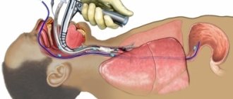
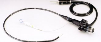
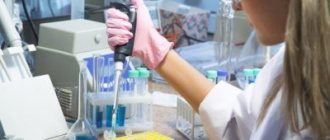
To perform a histological examination of stomach tissue, it is necessary to perform a biopsy of the organ. In most cases, it is performed using endoscopy. The apparatus for performing FEGDS is placed into the lumen of the stomach and several pieces of organ tissue are cut off. It is advisable to take biopsies from several distant sites. In some cases, tissue for histological examination is taken during surgery. After this, thin sections from the biopsy are taken in the laboratory and examined under a microscope.
How long does a histological analysis of stomach tissue take?
If cancer is suspected, gastric histology is necessary. How long does this analysis take? Only the attending physician can answer this question. On average, histology takes about 2 weeks. This applies to planned studies, for example, when removing a polyp.
During surgery, urgent histological examination of the tissue may be necessary. In this case, the analysis takes no more than half an hour.


