Ligation of the esophageal veins is a minimally invasive endoscopic procedure, which is an important component of the complex treatment of portal hypertension - increased pressure in the portal vein, through which blood flows from the intestines to the liver. Ligation of the esophageal veins helps prevent bleeding, which can be fatal for the patient.
Our expert in this field:
Ryabov Konstantin Yurievich
Chief surgeon, oncologist, endoscopist
Call the doctor
Causes of pathology
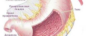
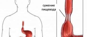
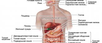
Varicose veins of the esophagus are formed as a result of disruption of blood flow to the liver through the portal vein, usually as a result of scar changes in the tissue of the organ. When the outflow of blood through the portal vein is disrupted and the pressure in it increases, the blood begins to “look” for workarounds. One of them - portacaval anastomosis (communication between the cava and portal vein) - is located in the veins of the esophagus. The pressure also increases in them and they expand. Under certain conditions they may begin to bleed.
The most common causes of increased pressure in the portal vein, which lead to esophageal varices:
- Cirrhosis is a condition in which normal liver tissue is replaced by fibrous connective tissue. It develops as a result of viral hepatitis, alcoholic liver disease, fatty hepatosis, impaired bile outflow (primary biliary cirrhosis) and other pathological processes.
- Chronic hepatitis.
- Thrombosis is a blood clot in the portal vein or splenic vein.
- Parasitic infections such as schistosomiasis.
- Compression of the portal vein by pathological formations from the outside.
- Liver cancer is hepatocellular carcinoma.
- Pathologies of the tricuspid heart valve.
According to prospective studies, 90% of patients with liver cirrhosis sooner or later develop dilatation of the esophageal veins, and 30% develop bleeding. Once a patient is diagnosed with cirrhosis, the risk of developing esophageal varices is 5% annually. The risk of bleeding is 10–15% per year. At the time of diagnosis of cirrhosis, dilatation of the venous vessels of the esophagus is detected in approximately 30% of patients.
The risk of bleeding depends on several factors:
- Portal vein pressure. The higher it is, the more likely bleeding will develop.
- The degree of dilation of the veins of the esophagus.
- Presence of “Red Markers”: If red streaks or spots are found in the area of esophageal varices during endoscopic examination, the risk of bleeding is higher.
- Degree of liver dysfunction. It is assessed using a special Child-Pugh scale. The more severe the liver failure, the higher the likelihood of bleeding.
- Alcohol consumption. If a person already has esophageal varices but continues to drink alcohol, they are more likely to develop bleeding.
- If the patient has already had bleeding, then there is a risk of it happening again in the future.
Bleeding from varicose veins of the esophagus threatens serious blood loss, the development of shock and death of the patient. Ligation helps prevent this dangerous complication.
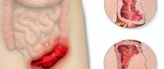
The degree of esophageal varices is assessed during an endoscopic examination. Classification of the World Gastroenterological Organization:
- Grade 1 (small varicose size): less than 0.5 cm, small dilated veins that are visible above the surface of the mucous membrane.
- Grade 2 (medium-sized node): tortuous dilated veins occupy less than a third of the lumen of the esophagus.
- Grade 3 (large varicose nodes): more than 0.5 cm, covering more than a third of the lumen of the organ.
Our doctors will help you
Leave your phone number
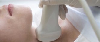
Diagnostic methods
It is quite difficult to suspect the presence of a disease based on subjective signs. Often, esophageal varices are detected only at the stage of massive bleeding. However, pathology can be assumed in patients who are part of a group of people with factors predisposing to the disease. To clarify the diagnosis, laboratory and instrumental examination methods are prescribed.
- A general blood test is necessary to assess the condition of the body as a whole, as well as the degree of blood loss, if any.
- Biochemical analysis - to determine liver function.
- Ultrasound of the abdominal organs is necessary to identify the condition of the liver, as well as other anatomical areas. Using this method, you can evaluate the blood flow of the portal system and determine the presence of free fluid.
- Plain radiography of the abdominal cavity - complements ultrasound.
- FGDS is the only objective method for assessing the condition of the mucous membrane. Allows you to visualize problem areas with varicose veins of the esophagus in real time and clarify the source of hemorrhage.
The latter method requires extreme caution, since a foreign body in the form of a fiberscope can damage the vascular wall (this can lead to a number of undesirable effects, one of which is bleeding).
Preparation for esophageal vein ligation
During the preliminary consultation, the patient must tell the doctor what chronic diseases he suffers from, what medications he takes regularly, and whether he has previously had allergic reactions to medications. If a patient is taking anti-clotting medications, they will likely need to be stopped for a while due to the risk of bleeding. It is also necessary to report if the patient suffers from diabetes and is taking medications to lower blood glucose levels. To prevent infectious complications, a course of antibiotics is prescribed.
The procedure is carried out on an empty stomach - you cannot eat anything for several hours.
Disease prevention
Prevention of varicose veins is aimed at maintaining the normal condition of the veins. First of all, it is necessary to carefully monitor the condition of the liver and follow all recommendations for the treatment of diseases. If a patient experiences a persistent increase in blood pressure, it is recommended to take measures to regulate it.
General preventive recommendations relate to maintaining a healthy lifestyle. It is necessary to strictly follow the diet and give up bad habits. It is also worth performing light exercises to strengthen the body, periodically attending a massage, and also taking vitamins according to the course.
Carrying out the procedure
During endoscopic ligation of the esophageal veins, the patient is placed on his left side, and a special plastic mouthpiece is inserted between the teeth to prevent injury to the teeth due to strong compression and damage to the endoscope. At the Medicine 24/7 clinic, the intervention is performed in a state of sedation (medicated sleep). To do this, a peripheral catheter is inserted into a vein in the arm and appropriate medications are administered through it. This helps the patient undergo the procedure as comfortably as possible.
The doctor inserts an endoscope into the esophagus, examines its mucous membrane, finds varicose veins and places latex ligatures on them. As a result, blood flow through the varicose veins stops and the risk of bleeding disappears. A different number of ligatures may be used, depending on the degree of varicose veins. For example, in grade III, up to 20 ligatures are used.
When all the ligatures are applied, the endoscope is removed, after which the intervention is completed.
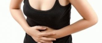
Esophageal varicose veins: symptoms
The onset of the disease does not manifest itself in any way. Therefore, patients seek medical help when the disease has already “gained momentum.” It is impossible to recognize the disease on your own, because the initial manifestations of the disease are similar to the symptoms of a digestive system disorder. So, if there is dilatation of the veins of the esophagus, the symptoms will be as follows:
- the appearance of shortness of breath ;
- the presence of heartburn (does not depend on food intake);
- pain in the area of the xiphoid process;
- swallowing disorder.
If there are varicose veins of the esophagus and stomach, its main sign is a vascular pattern on the skin of the anterior abdominal wall in the form of a “jellyfish head”. It is formed by pathologically altered veins. This symptom appears later in the course of the disease. Later, when bleeding occurs, the patient has:
- bloody vomiting;
- blood in stool;
- tachycardia;
- hypotension (sharp decrease in blood pressure);
- shock.
This is an extremely serious condition of the patient, requiring urgent hospitalization, since delay leads to death.
Recovery after esophageal vein ligation
Within 7–10 days, the ligatures leave the body naturally, and blood flow in the esophageal varices stops. For several days, the patient is in the hospital under the supervision of doctors; he is prescribed a gentle diet with liquid, cool food, and medications to reduce gastric secretion.
After 1 month, a control endoscopic examination of the esophagus is performed. If varicose veins are detected, they are re-ligated. The procedure can be repeated monthly until the dilated veins disappear completely. In the future, control endoscopic examinations can be repeated every 6–12 months.
Diagnosis and treatment
The patient's varicose veins of the stomach and esophagus are determined by such methods as:
- Ultrasound examination of the esophagus, abdominal organs;
- general blood analysis ;
- radiography using contrast;
- esophagoscopic examination of the esophagus.
When making a diagnosis, take into account that bleeding can be caused by an ulcerative lesion of the esophagus, a disintegrating tumor, or Mallory-Weiss syndrome. This is a longitudinal tear in the lining of the upper stomach or distal esophagus caused by frequent vomiting.
When the diagnosis of varicose veins of the esophagus is confirmed, treatment is prescribed according to the degree of damage to the vessels of the esophageal tube. If the disease is detected when bleeding occurs, then treatment is aimed at stopping it:
- blood transfusion;
- vasoconstrictor drugs;
- hemostatic drugs;
- electrocoagulation of the affected vessel;
- squeezing the affected vessels with a special probe.
After this, therapeutic measures are carried out aimed at preventing a relapse (repetition) of the attack. If varicose veins of the esophagus are observed with cirrhosis of the liver, then treatment of the underlying disease is carried out. It is aimed at restoring the function of liver tissue. The patient is also advised to give up bad habits and avoid strenuous physical activity.
- Antacids are prescribed to prevent unnecessary irritation of the esophageal mucosa.
- Astringents that relieve inflammation.
- Vitamin therapy.
If esophageal varicose veins are diagnosed, medication treatment can be supplemented with surgical intervention to prevent relapses.
Bandage – small rubber discs are installed over varicose veins. Sclerotation is the introduction of a hemostatic solution into the affected vein. The procedure must be carried out at least four times a year. Devascularization - all damaged veins that cannot be restored are completely removed.
With cirrhosis of the liver, the patient cannot undergo classical surgery. In this case, endoscopic alloying of the esophageal vessels is performed. The procedure involves tying the vessel with nylon loops or elastic rings.
Portosystemic shunt, in which the portal and hepatic veins are connected. This makes it possible to normalize the pressure in the vessels.
Diet for varicose veins of the esophagus
Nutrition adjustment is one of the important points in the treatment of varicose veins. The patient will have to change his lifestyle so that sticking to the diet is comfortable, since he will have to follow the diet for the rest of his life.
If a pathology is detected, the patient, first of all, needs to establish a diet: during the day you should eat 4 to 6 times in small portions. The most nutritious meal should be at breakfast, and the lowest calorie meal should be at dinner.
In order for food to be digested better, the patient should prefer cooking methods such as boiling, steaming, and pureeing foods. Definitely, you need to give up fried foods.
Reference! The temperature of the food should be such as not to cause discomfort to the person while eating. Therefore, you should avoid hot and too cold foods.
The following types of products should be avoided:
- from solid foods that can damage the gastrointestinal mucosa: crackers, nuts, hard fruits, raw vegetables;
- from products that irritate the gastrointestinal tract: pickles, marinades, concentrated juices, spices;
- from fatty foods;
- from alcoholic, carbonated drinks;
- from coffee, chocolate.
And, on the contrary, it is worth adding to the diet:
- lean varieties of meat, fish, poultry;
- dishes that envelop the gastrointestinal tract: porridge and jelly;
- low-fat dairy and fermented milk products.
Diagnostics
Diagnosis of varicose veins of the esophagus consists of several stages:
- Anamnesis collection. It is mandatory to analyze not only the patient’s complaints, but also a list of chronic diseases and external signs (for example, yellowness of the skin, swelling). The abdominal wall is palpated and tapped.
- Lab tests. The results of the CBC and blood biochemistry determine the condition of the internal organs, the number of platelets and the level of hemoglobin. In some cases, additional liver diagnostics and advanced clotting tests may be needed.
- Instrumental and hardware research. X-ray, ultrasound and esophagoscopy are aimed at a thorough examination of the esophagus and determining the root cause of varicose veins, which, along with complications of the disease, is necessarily indicated in the conclusion.
The x-ray image shows the jagged contours of the esophagus, the specifically tortuous shape of the folds of the mucous membrane. If these signs are detected, the patient undergoes the most informative endoscopic examination - fibroesophagoscopy (examination of the esophagus from the inside). This method allows you to fully examine the causes of bleeding, the degree of varicose veins, the condition of the vascular walls, and even the likelihood of rupture of the nodule in the future.
The difficulty is that it is difficult to accurately determine the source of the hemorrhagic manifestation: after a rupture, the vein loses its tone, and the site of damage becomes invisible. At the same time, at stages 3–4 of the disease, it is necessary to examine the esophagus extremely carefully so as not to touch the thinned and tense walls of the vessels.
Signs of varicose veins of the esophagus
Grade 1 esophageal varicose veins, which can develop over years, do not have obvious symptoms. Sometimes the patient may feel heartburn, heaviness in the chest, and belching. Periodically, the patient complains of discomfort when swallowing food. Sometimes VRVP is accompanied by a venous pattern on the anterior wall of the abdominal cavity.
Reference! The first symptoms, as a rule, appear several days before the onset of bleeding. It consists of significant heaviness in the chest area and a deterioration in the general condition of the patient. Ascites is sometimes observed.
Immediately after a vein rupture, the patient experiences the following symptoms of esophageal varicose veins:
- A sharp decrease in blood pressure.
- Vomiting with blood.
- Bloody stool.
- Tachycardia.
- Light bleeding is accompanied by weakness and symptoms of anemia.
Complications
The most serious complication is bleeding.
In this case, bleeding can occur even from small efforts such as overeating, since the walls of the veins become very thin. Bleeding may be small or severe and may be life-threatening. Bleeding leads to vomiting and chronic anemia. Varicose veins can also be accompanied by esophagitis, an inflammatory process of the esophageal mucosa.
Esophageal varices have a high mortality rate, mainly due to complications and concomitant diseases, such as progressive cirrhosis, causing bleeding. Despite the fact that bleeding stops on its own in approximately 79% of cases, the mortality rate from varicose veins reaches 50%. For patients who bleed and survive, the risk of recurrence in the next two years ranges from 55-75%.
You can reduce the risk of recurrent bleeding by following your doctor's instructions. However, this does not guarantee long-term survival due to advanced liver disease. Note that varicose veins of the esophagus often occur with cirrhosis of the liver.
It is important to remember, in order to prevent complications, at the first sign of any type of varicose veins (legs, uterus, pelvis, labia, varicocele, etc.), you should immediately consult a doctor. Be healthy!
Why are varicose veins of the esophagus dangerous?
Minor but regular bleeding can cause anemia. Significant - without medical intervention, they lead to the death of the patient. Before the onset of blood loss, the patient feels a salty taste and a slight tickling in the throat. After this, vomiting occurs with impurities of blood of various colors and consistencies.
The following factors can cause bleeding:
- lifting weights;
- increased blood pressure;
- binge eating;
- straining;
- ulcers;
- vomit.
Varicose veins of the esophagus: causes
The main factor causing expansion is excess pressure in the cavity of the portal vein. It transports blood from the stomach, pancreas, spleen to the liver. This phenomenon is called “portal hypertension.”
Excessive blood pressure is a consequence of such diseases:
- chronic hepatitis;
- cirrhosis;
- tuberculosis;
- neoplasms;
- amyloidosis;
- sclerosis;
- thrombosis;
- Budd-Chiari syndrome;
- cardiovascular failure.