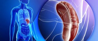
Lymphoma of the stomach has a slow course and symptoms similar to cancer of this organ, only with a more favorable prognosis. With any form of lymphoma, damage occurs to the lymphatic tissue located in the stomach and its mucous membrane.
Despite the fact that the total number of people with gastrointestinal lymphoma increases every year, malignant formation of this type can actually be found very rarely (up to five percent of all gastric neoplasias). The main task of doctors is to detect this pathology and make a diagnosis in the early stages, when the spread of cancer cells throughout the body has not yet begun. Methods of diagnosis and treatment are determined by an oncologist.
general information
The diagnosis of malt lymphoma of the stomach was introduced into practice relatively recently. Previously, this pathology was considered “pseudolymphoma,” until in 1983 scientists proved the origin of the entire tumor from one malignant cell. The term MALT stands for mucosal associated marginal zone lymphoma. They can appear in any extranodal (primary focus outside the lymph nodes) organs and structures of the body, most often affecting the digestive system (30-40%). The stomach accounts for 60-75%.
This pathology belongs to non-Hodgkin lymphoma. Women get sick somewhat more often than men. In most cases, the disease develops after 50-60 years. Usually the process occurs locally (one focus) in the antrum of the stomach (the body or cardia is less commonly affected). More often the disease is diagnosed in the initial stages (about 70%).
Symptoms
In the early stages of development, MALT lymphoma of the stomach shows few symptoms, or they are similar to the manifestations of peptic ulcer disease or gastritis.
The patient may exhibit the following symptoms:
- rare pain in the stomach (aching);
- belching, heartburn, vomiting;
- stomach bleeding;
- rapid onset of satiety when eating;
- increased body temperature, sweating in the evening and at night;
- psychophysiological exhaustion;
- weight loss;
- an increase in the size of the lymph nodes in the armpits, neck, and groin.
Enlarged lymph nodes initially retain mobility and elasticity, and the nodes merge into a conglomerate. MALT lymphoma of the gastric mucosa may be similar in its manifestations to gastric cancer.
Symptoms indicating a tumor neoplasm are quite rare.
Causes of malt lymphoma of the stomach
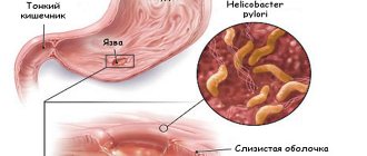
Malt lymphoma appears as a result of long-term chronic inflammation occurring in the stomach. In response to the constant presence of infectious agents, extranodal lymphatic tissue located in the mucous membranes of the digestive tract proliferates. The production of B lymphocytes increases, which then undergo multiple mutations. As a result of gene damage, cells acquire atypical properties, and tumor growth begins.
More than 90% of all malt lymphomas occur due to the negative influence of Helicobacter pylori. In other cases, the disease develops due to the systematic action of other infectious or other agents on the gastric mucosa.
Lymphomas: symptoms
The symptoms of lymphomas are very different, since they depend on where exactly the tumor began to develop, its prevalence and histological type. It is manifested by three syndromes, lymphadenopathy, intoxication of the body, its febrile states and organ damage. The first sign is an enlargement of the lymph nodes, which initially retain elasticity and mobility, but as the disease develops, they unite into large conglomerates. Other clinical manifestations:
- Unreasonable increase in body temperature;
- Intense sweating at night;
- Weight loss with the same diet;
- Constant feeling of fatigue.
Among the affected organs may be the gastrointestinal tract, brain, mammary glands, bones, and lung parenchyma. Large neoplasms put pressure on internal organs, which creates conditions that pose a threat to the patient’s life. Affected nodes:
- mediastinum - compresses the esophagus and trachea, causing superior vena cava syndrome;
- located in the peritoneum and behind it - put pressure on the intestines, leading to obstruction and accumulation of lymphatic fluid, urination disorders;
- stomach or intestines - provoke bleeding and even peritonitis.
Patients are very susceptible to infections and get them regularly, which further aggravates their condition.
Symptoms
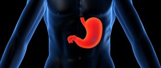
At the initial stage, the clinical picture of gastric lymphomas is absent or corresponds to that of chronic gastritis or peptic ulcer. Mostly, dyspeptic disorders are observed (heartburn, belching, nausea, vomiting, sometimes with streaks of blood) and periodic painful sensations in the epigastrium, aching in nature, independent of food intake.
Against the background of concomitant pathology, there may be complaints of bloating, poor bowel function, constipation, loss of appetite, etc. If lymphoma progresses, symptoms of a “tumor nature” appear (asthenic syndrome, severe pain).
Diagnostic methods
If the development of maltoma is suspected, the doctor conducts an examination and studies the patient’s medical history. In order to establish an accurate diagnosis and determine the course of therapy, instrumental and laboratory research methods are prescribed.
A blood test allows you to get a complete picture of the pathology. The study is prescribed to determine changes in the composition and presence of tumor markers.
The patient is also prescribed MRI, CT, and ultrasound. The techniques allow us to establish the localization and extent of spread of the pathological process.
A biopsy is also used to obtain images of changed tissue. The biopath is sent for cytological examination, which makes it possible to determine the presence of cancer cells.
Classification
Among all non-Hodgkin primary lymphomas that arise in the stomach, malt lymphomas are the most frequently reported. Their classification was developed by a special group of researchers and doctors. The following stages of the disease are distinguished:
- Stage I - pathology is limited to the stomach: 1 - tumor within the mucous membrane or with penetration into the submucosal layer;
- 2 - lymphoma affects the muscular, subserous and/or outer layer;
- 1 - paragastric lymph nodes are affected;
The last two stages were combined because they imply the same prognosis. Malt lymphomas also vary in their degree of malignancy and their aggressiveness.
Forecast
An unambiguous prognosis for malt lymphoma is impossible. The result of therapy depends on the stage of development of the formation, the individual characteristics of the patient’s body and the correct choice of course of therapy.
When treatment is started in the first stages of the disease, the prognosis is favorable in most cases. Drug treatment, radiation and chemotherapy show more than 90% survival rate in the five-year period after therapy. Delayed initiation of treatment reduces the chances of a positive effect and remission.
After completing the course of therapy, it is necessary to regularly undergo endoscopic examination of the stomach with the collection of biomaterial for research. The first such examination should be 2-3 months after the end of therapeutic measures. Subsequently, endoscopic examinations should be performed twice a year for several years.
Diagnostics
Diagnostic measures begin with a standard interview and examination of the patient. When diagnosing malt lymphoma, endoscopic examination with the collection of histological material from different parts of the stomach is of primary importance. The resulting biopsy is studied using the following methods:
- morphological assessment;
- immunohistochemical analysis;
- molecular genetic research using FISH or PCR.
The presence of an active Helicobacter pylori infection must be confirmed using immunohistochemical, serological, antigen stool or urease breath tests.
In addition, the complex of examinations for gastric lymphoma includes:
- blood test with determination of general and biochemical parameters (leukocyte formula, b2-microglobulin, lactadehydrogenase, etc.)
- fluoroscopy of the gastrointestinal tract;
- endoscopic ultrasound of the stomach and nearby lymph nodes;
- CT scan of the abdomen, pelvis and lungs;
- collection of aspiration material from the bone marrow.
The differential diagnosis of malt-lymphomas of the stomach with other lymphomas is particularly difficult for the doctor. It is also sometimes difficult to distinguish this malignant tumor from chronic diseases of the organ, since the neoplasm often occurs against the background of a peptic ulcer or gastritis.
Often, during gastroscopy, lymphomas look like mucosal ulcers or areas of hyperplastic tissue. Therefore, it is so important to always collect histological material from several points (at least 6-8) to correctly diagnose the patient.
Lymphomas (lymphogranulomatosis and non-Hodgkin's lymphomas)
HIV
Gastritis
Ulcer
Hepatitis
14198 November 23
IMPORTANT!
The information in this section cannot be used for self-diagnosis and self-treatment.
In case of pain or other exacerbation of the disease, diagnostic tests should be prescribed only by the attending physician. To make a diagnosis and properly prescribe treatment, you should contact your doctor. Lymphomas: causes, symptoms, diagnosis and treatment methods.
Definition
Lymphomas are a group of malignant tumor diseases of the lymphatic tissue, which under normal conditions is responsible for immunity. One of the first clinical symptoms of lymphoma is enlargement of lymph nodes in different locations.
Lymphomas, like other malignant tumors, have a tendency to metastasize, that is, the migration of tumor cells through the blood and lymph, as well as through contact into intact (healthy) tissues with subsequent reproduction in them.
Almost all types of lymphogranulomatosis, or Hodgkin's lymphoma, and about 65% of non-Hodgkin's lymphomas manifest as painless enlargement of the lymph nodes - 2 cm or more. However, the diagnosis of a particular type of lymphoma is established solely on the basis of a morphological examination of biopsy material taken from the tumor.
The incidence of Hodgkin's lymphoma in Russia is about 2 cases per 100 thousand population per year, and non-Hodgkin lymphoma is 5–7 cases per 100 thousand.
Causes of lymphomas
There are several possible causes for the development of lymphomas. It is important to understand that the basis for the development of any malignant disease, including lymphomas, is a dysfunction of cells.
The ability for uncontrolled tumor growth occurs when the natural process of cell division is disrupted: normally, the cell divides a limited number of times, matures, performs a certain function, and then ends its life.
Malignant cells are characterized by disruption of the processes of maturation and differentiation (the process during which the cell acquires specific characteristics and the ability to perform certain functions). Cells acquire new functions - they begin to produce proteins and toxins, and the ability to “escape immune surveillance” appears, as a result of which the process of natural cell death (apoptosis) changes.
Some viruses can directly affect the DNA of lymphocytes and contribute to their transformation into malignant cells.
For example, infection with Epstein–Barr virus (EBV) is an important risk factor for the development of lymphomas.
Lymphocytes are divided into two subtypes: T and B cells (T and B lymphocytes). Infection with human T-cell lymphotropic virus (HTLV-1) increases a patient's risk of developing certain types of T-cell lymphoma. The virus is transmitted sexually and through blood, as well as from mother to child through breast milk.
Viruses that weaken immunosurveillance are indirectly responsible for the development of certain types of lymphomas.
Thus, the human immunodeficiency virus (HIV) reduces the population of lymphocytes, while the control of mutant cells and herpes viruses type 8 and Epstein-Barr decreases with the formation of lymphomas.
Some infections can cause the development of lymphoma due to the constant activation of the immune system. Chronic infections produce a huge number of lymphocytes, which increases the likelihood of mutations during cell division and leads to the risk of lymphoma. Similar infections include Helicobacter pylori
(often found in chronic gastritis and peptic ulcers of the stomach and duodenum),
Chlamydophila psittaci
(risk of developing lymphoma in the tissues of the eye), hepatitis B and C viruses with prolonged infection.
Non-infectious risk factors for the development of lymphomas include:
- Exposure to chemicals. Numerous studies have shown that exposure to herbicides, insecticides, and benzene is associated with an increased risk of developing lymphomas.
- The presence of autoimmune diseases (rheumatoid arthritis, systemic lupus erythematosus, etc.) is a significant risk factor for the development of lymphomas.
- Radiation exposure has a great influence on the rate of cell mutations and their malignancy.
- Genetic factors. Having a close relative with lymphoma greatly increases your risk.
- Congenital immune deficiency in certain diseases.
- Age. Some types of lymphoma develop in children and adolescents, while others, on the contrary, develop in people over 60 years of age.
- Breast implants can rarely lead to the development of a type of large cell lymphoma of the breast.
Classification of lymphomas
There are two main types of lymphomas: Hodgkin's lymphoma, or lymphogranulomatosis, and non-Hodgkin's lymphomas.
Hodgkin lymphoma is characterized by the presence of specific Reed-Berezovsky-Sternberg cells in the affected lymph nodes, spleen, liver or bone marrow.
Enlarged cervical lymph nodes in a patient with Hodgkin lymphoma
The group of non-Hodgkin's lymphomas includes:
- follicular lymphoma;
- marginal zone lymphoma;
- diffuse large B-cell lymphoma;
- mantle cell lymphoma;
- Burkitt's lymphoma;
- primary lymphoma of the central nervous system;
- nodal T-cell lymphomas;
- primary cutaneous lymphomas;
- non-Hodgkin's lymphomas in patients infected with HIV;
- non-Hodgkin's lymphoma in patients infected with hepatitis B and C viruses.
Symptoms of lymphomas
The earliest symptom is enlarged lymph nodes against the background of complete well-being. Enlarged lymph nodes are painless.
Various clinical manifestations depend on the location of the tumor: if the tumor grows in the lymph nodes of the chest, compression of the airways and the appearance of hoarseness, difficulty swallowing, difficulty breathing, and obsessive cough are possible.
When lymphoma is located in the mediastinum, compression syndrome of the superior vena cava appears: headaches, swelling and dilation of the veins of the face and neck occur.
In some cases, only general symptoms are present: loss of appetite, weight loss, increased body temperature, night sweats, weakness, and sometimes itchy skin and pain in enlarged lymph nodes.
Against the background of a decrease in immunity, infectious diseases can occur - the chickenpox virus is often activated, manifested by herpes zoster.
Cases of damage to the abdominal organs (manifested by an increase in the volume of the abdomen due to tumor growth and fluid accumulation, the appearance of pain, jaundice, nausea and vomiting) and bone marrow (pain in the bones, back, frequent fractures, pallor, increased bleeding) have been described.
Diagnosis of lymphomas
The diagnosis of a particular type of lymphoma is established solely on the basis of a morphological examination of biopsy material (a piece or a whole lymph node obtained during a minor operation) taken from the tumor (histological, cytogenetic and molecular genetic research methods). The doctor will prescribe additional tests to diagnose the stage of the disease and its complications. The list of studies depends on the presumed location of the tumor and may be changed.
- Clinical blood test.
Treatment of malt lymphoma of the stomach
Since gastric marginal zone lymphoma is approximately 90% associated with Helicobacter pylori infection, treatment begins with antibacterial therapy aimed at eliminating this pathogen. Even in those patients whose disease was caused by other infections, sometimes there is a positive effect from taking drugs against Helicobacter.
Many clinical studies conducted for gastric lymphomas have proven the effectiveness of eradication treatment in 80% of patients with the first stage and confirmed infection with Helicobacter pylori. In the second stage of the disease, antibacterial therapy was successful in 40% of patients. Thus, this method is the first-line treatment for gastric marginal zone lymphoma.
After a full course of eradication therapy, after 1 year, a comprehensive examination of the patient is carried out to assess the effectiveness of the treatment. An endoscopic examination of the stomach with a biopsy is required. If resistance (resistance) of microorganisms to the treatment is confirmed, a second line of therapy is prescribed—irradiation.
Marginal zone lymphoma is very sensitive to radiation. To achieve a therapeutic effect, a total total dose of 30 to 35 Gy is prescribed. An important point is that ionizing radiation should only affect the stomach and paragastric lymph nodes. In order to protect neighboring organs (liver, pancreas, kidneys), high-precision irradiation is used, or the total load is reduced to 25 Gy.
If a patient with gastric marginal region lymphoma has not responded to antibacterial treatment and radiation therapy and for disseminated processes, chemotherapy is prescribed. The medications used include:
- chimeric monoclonal antibody to CD-20 B-lymphocyte receptors (rituximab);
- anthracycline-containing regimens;
- purine analogues of nucleosides (cladribine, fludarabine);
- regimens with mitoxantrone and prednisolone;
- alkylating agents used for non-Hodgkin's lymphomas (chlorambucil, cyclophosphamide).
For each patient with gastric lymphoma, a different treatment regimen is selected. According to indications, surgical intervention is possible, which is the method of choice for the treatment of this pathology. Gastric resection is often performed.
Just a few years ago, surgery was considered a priority for eliminating gastric marginal zone lymphomas. However, the high probability of postoperative complications and death limit the possibility of surgical interventions.
Lymphomas: treatment
The development of lymphoma treatment tactics by CELT specialists is based on the diagnostic results obtained and the patient’s individual indications. It may include surgery, radiation and chemical therapy:
| Treatment method | What is? |
| Radiation therapy | Indicated independently exclusively for localized forms of pathology and low degree of malignancy of the neoplasm. It can be used in cases where other methods of treatment are not available for one reason or another. |
| Chemotherapy | Most often it begins with the use of several cytostatic drugs of different groups. It can be carried out independently or in combination with radiation therapy, which allows achieving longer remissions. As a rule, it requires two to three courses in order to achieve complete remission. |
| Operations | Indicated for isolated lesions of internal organs, most often the gastrointestinal tract. They are aimed at complete or partial removal and require chemotherapy. |
| Additional |
|
At CELT, consultations are conducted by hematologists of the highest category, candidates and professors of medical sciences with many years of experience in practical and scientific work. You can schedule a consultation with them online or by phone. No less qualified specialists work in the urology department of our clinic. You can contact them and solve any delicate problem that reduces the quality of your life. They can undergo a diagnostic procedure such as cystoscopy of the bladder.
At CELT you can consult a hematologist.
- Initial consultation – 3,200
- Repeated consultation – 2,000
Make an appointment
By making an appointment with a hematologist, you can get a comprehensive consultation. The doctor is competent to treat various blood diseases, most of which can be identified in the early stages and prescribe timely treatment to cope with the disease quickly and easily.
Metastasis
Gastric marginal zone lymphoma rarely gives distant metastases. Metastatic seedings to regional lymph nodes are most often observed. Much less often the process spreads to distant lymph nodes.
When a tumor extends beyond the serosa of the stomach, nearby tissues and organs can be affected. Most often, lymphoma metastasizes to the liver, pancreas and kidneys. Often, patients with stage 4 of the process experience bone marrow damage.
Causes
The exact causes of maltoma have not been established. It is believed that the main cause of the development of the pathological process in the stomach area is bacteria of the Helicobacter pylori type.
Experts have also identified a number of unfavorable factors that influence the occurrence of the disease. These include exposure to chemicals and toxic substances, smoking, drinking alcohol, and infectious diseases. Maltoma can be caused by stomach diseases such as ulcers and gastritis.
Genetic predisposition is of particular importance. Many patients had close relatives who also suffered from similar diseases.
Complications and relapse
After treatment for gastric lymphoma, the risk of complications and relapse will depend on which methods were used. After long-term use of antibiotics, drug resistance, allergic reactions, intestinal dysbiosis, intoxication of the whole body or individual organs may appear. When using chemotherapy drugs, the toxic damage will be several times stronger, taking into account the regimens and doses used. There may be a decrease in the number of blood cells, suppressed immunity, damage to peripheral nerves, etc.
If during irradiation of the stomach neighboring organs and tissues are exposed to rays, typical post-radiation complications will be observed. After surgery, early and late undesirable consequences may occur. In the postoperative period the following cannot be excluded:
- bleeding;
- suture infection;
- failure of the gastric stump;
- state of shock;
- peritonitis;
- thromboembolism;
- pneumonia;
- myocardial infarction and other complications associated with surgery for lymphoma or anesthesia.
When visiting a specialized clinic, where modern equipment is used and qualified doctors work, the risk of complications and recurrence of gastric lymphoma is significantly reduced.
Treatment
Therapy for lymphoma depends on the stage and type of the tumor, as well as on the general condition of the sick person. Treatment of gastric lymphoma can be carried out separately or in parallel in several ways: with the help of surgery in gastroenterology, chemotherapy and radiation therapy, as well as with the use of medications. After diagnosis, the patient must undergo a course of antibacterial drugs that will kill Helicobacter. If this is lymphoma of the first stage of development, then surgical intervention follows antibiotics. The doctor excises the primary tumor focus and also examines peripheral lymph nodes and nearby organs. After the operation, chemical medications are prescribed that completely remove the cancer cells.
For lymphoma that has reached the second or third stage, treatment is first carried out with chemotherapy and radiation, which helps reduce the size of the tumor, and only then surgery. Not the entire body is irradiated, but only the abdominal cavity, in particular the stomach. During the operation, the doctor may decide to perform a resection, during which about seventy percent of the organ is removed. At the fourth stage, as well as in cases where the tumor has grown into large vessels and multiple metastases have occurred, or if there are possible contraindications to surgery, then treatment is reduced to palliative. This helps reduce the severity of symptoms, alleviate the patient’s condition and prolong his life, but does not eliminate lymphoma.
We recommend reading Follicular lymphoma - types, signs and methods of treatment
Prevention
Prevention of the appearance and growth of malignant tumors, including gastric lymphomas, is a priority for doctors of all specialties. If a person leads a healthy lifestyle, does not come into contact with carcinogenic factors and does not allow stressful situations, the risk of the appearance of atypical cells in the body is significantly reduced. Since gastric lymphoma is associated with Helicobacter pylori infection, the basis of prevention will be early detection and disposal of this pathogen.
Doctors at the European Clinic have extensive experience in treating lymphomas both in the initial stages and in advanced stages. For this, the clinic has everything necessary. In almost every case, we manage to achieve an improvement in the patient's condition.
Book a consultation 24 hours a day
+7+7+78
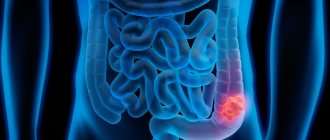
Stages of the disease
Gastric lymphoma, like other types of this disease, has four stages. Each of them differs in severity and clinical picture. The first stage is the easiest; if the disease is detected at it, then the prognosis for treatment is almost one hundred percent positive. The situation is worst at the fourth stage of the pathology.
Below are the main signs for each stage of the disease:
- Stage 1: the pathological process is localized in the stomach. At stage 1a it is limited to the mucous membrane, at stage 1b it moves to deeper layers (muscular, serous).
- Stage 2: the lymph nodes lying nearby, as well as neighboring organs, are affected.
- Stage 3: distant lymph nodes are affected, and the tumor actively penetrates into neighboring organs. Lymph nodes are affected on both sides of the diaphragm.
- Stage 4: the lymph nodes located above the diaphragm are affected, as well as the lymph nodes located in the pelvis along the aorta.
At the third and fourth stages of the disease, the pathological process affects the liver, bone marrow, spleen, and other internal organs.