Is the malignancy visible during endoscopy?
The importance of fibrogastroduodenoscopy for diagnosing gastric cancer cannot be overestimated. It is this study that allows us to identify or confirm the diagnosis of malignant neoplasm , as well as determine the type of process. This became possible thanks to a biopsy of suspicious areas of the gastric mucosa. The collected tissues are sent for cytological examination, which within 5-10 days gives a conclusion about the type of tissue.
Important International recommendations recommend performing FGDS in all patients over 45 years of age in whom the doctor, after questioning and examination, suspects the presence of stomach cancer.
This makes it possible to identify the disease at an early stage, when the overall prognosis is more positive and the average life expectancy is 10 years or more.
Symptoms
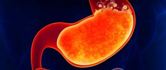
Gastric cancer is a malignant neoplasm that develops from the epithelium of the mucous membrane of the organ. According to official statistics, this disease ranks second in frequency of detection among cancer pathologies in Russia . It has been established that stomach cancer is more common in men over 45 years of age.
The problem with this disease is that it is diagnosed late. Only in 7% of cases (often by chance) can stomach cancer be detected at the first or second stage, when there are no distant metastases yet, and more than half of the patients completely recover.
In other patients, the oncological process manages to metastasize to the lymph nodes, abdominal organs, bones or brain. Then the effectiveness of even the most modern treatment is much lower, and only 5-22% of patients live another 5 years after diagnosis.
4 stages of stomach cancer
The clinical picture of stomach cancer is often subtle (especially in the first stages). The following symptoms come to the fore:
- decreased appetite;
- feeling of heaviness in the upper abdomen;
- periodic aching stomach pain;
- nausea after eating;
- belching;
- one-time vomiting;
- weight loss;
- weakness, decreased performance.
Often the disease is discovered only when surgical complications occur (eg, perforation of the stomach wall, gastric bleeding, hepatic vein thrombosis).
Sometimes situations occur when distant metastases are first detected (for example, in the liver during ultrasound). Then additional examinations are performed (FGDS, computed tomography or magnetic resonance imaging) to detect the primary tumor.
Diagnostic features
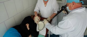
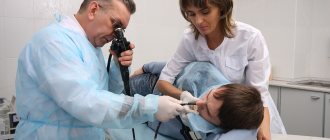
Fibrogastroduodenoscopy is performed in the endoscopy room on the patient’s “empty” stomach . Most often, local anesthesia is given to the posterior wall of the oropharynx, or a drug is administered for sedation (medicated sleep). FGDS is performed under anesthesia or sedation in a situation where emergency surgery is planned immediately after completion of the procedure.
The probe is inserted through the oral cavity, passes through the esophagus and enters the lumen of the stomach. If an oncological pathology is suspected, the doctor conducts a thorough examination of the mucous membrane. Normally, it should be pink, without defects, signs of inflammation or growth.
How is gastroscopy done?
To avoid complications after the FGDS procedure, it is important not only to prepare for it, but also to follow a diet after its completion. This article will help you cope with the insertion of the probe more easily.
Classification of the disease
Modern recommendations distinguish three types of early gastric cancer, which have different growth patterns:
- Polypous. Upon examination, a small polyp (proliferation of the mucous membrane) is discovered. It is impossible to visually determine its malignancy.
- Superficial cancer. Characterized by differences in the color of the mucous membrane, slight swelling or a reactive inflammatory reaction is possible.
- Ulcerative type. With FGDS, the neoplasm resembles an erosion or ulcer (defect of the mucous membrane). Differential diagnosis with peptic ulcer is necessary.
Visual picture
If the research is carried out at a later stage, then according to Borrman’s classification, 4 options for the visual picture are possible:
- Polypoid neoplasm. The endoscopist sees the growth of the mucous membrane on a wide stalk. Found in a third of cases.
- Polypoid cancer with ulceration. The neoplasm has a trough-shaped shape. From the edge one can see the proliferation of epithelium with erosion or a zone of necrosis, which are located in the center of the tumor. Occurs in approximately 30% of patients.
- Infiltrative-ulcerative variant. The growth of the oncological process occurs mainly in the thickness of the stomach wall. On the surface you can observe an ulcer (sometimes up to 2-3 cm in diameter), a local inflammatory process and swelling without clear boundaries.
- Diffuse-infiltrative. Immediate signs may not be detected during FGDS, since the tumor is almost entirely located in the wall of the stomach. However, the presence of compaction of the mucous membrane, rigidity and unnaturalness of the sweet tooth, and impaired motor skills are noteworthy. Detection rate – 30%. Characterized by early metastasis and poor prognosis.
A separate clinical variant of gastric malignancy is lymphoma . As you know, the stomach wall has a large amount of lymphatic tissue (MALT system).
Help Chronic Helicobacter pylori infection plays an important role in the development of the disease, so a rapid test for its presence is almost always positive.
It is necessary to highlight the following characteristics:
- the most common location is the antrum of the stomach;
- the mucous membrane may be little changed, but local redness, surface unevenness and smoothness of folds can be detected;
- proliferation of the mucous membrane;
- swelling without a clear boundary of the stomach wall.
When describing a neoplasm, the endoscopist must determine the nature of tumor growth, the presence of a defect in the mucous membrane, the consistency of the altered tissue, the location and size of the neoplasm.
This article will help you decipher the doctor’s conclusion after FGDS for common pathologies and find out what the protocol normally looks like.
How is a biopsy done?
During FGDS, if areas that resemble a malignant process are detected, a diagnostic biopsy is necessarily performed .
In this case, tissue sampling is done several times. For example, if there is an ulcer, then 3-4 samples are taken from its marginal walls and bottom. During the infiltration process, the deep-seated altered tissues are carefully collected.
After collection, tissue samples are sent for cytological examination, which should determine their type. A biopsy makes it possible to make a final diagnosis of a malignant tumor of the stomach, or to refute it. However, it alone is not enough for a complete diagnosis.
In 85% of cases of gastric cancer, metastases are found in regional lymph nodes or distant organs. Therefore, all kinds of contrast-enhanced imaging methods are used to detect them (CT, MRI, PET).
Forms of oncology
According to WHO recommendations, several histological (by origin and tissue type) forms of gastric cancer can be distinguished :
- papillary adenocarcinoma (from glandular tissue);
- tubular adenocarcinoma (from glandular tissue);
- squamous cell carcinoma (from the epithelium);
- carcinosarcoma (from connective tissue);
- mucinous adenocarcinoma (from glandular tissue, characterized by active mucus production);
- choriocarcinoma (sprouting of embryonic tissue);
- undifferentiated cancer (due to degradation of the tumor cell structure, it is impossible to determine its type);
- poorly differentiated adenocarcinoma.
A common problem in domestic laboratories is the inaccuracy of diagnostics due to the low level of qualifications of personnel (especially in small provincial hospitals) or problems with equipment.
Please note: Sometimes false positive or false negative results occur. To avoid this, it is advisable to conduct a cytological examination in a specialized oncology clinic.
The results of fibrogastroduodenoscopy are delivered to the patient within a few minutes after completion of the examination. They indicate the nature of the mucous membrane, acidity indicators and detected deviations. The results of the cytological examination are ready in 5-10 days.
Forms of oncology
According to WHO recommendations, several histological (by origin and tissue type) forms of stomach cancer can be distinguished:
- papillary adenocarcinoma (from glandular tissue);
- tubular adenocarcinoma (from glandular tissue);
- squamous cell carcinoma (from the epithelium);
- carcinosarcoma (from connective tissue);
- mucinous adenocarcinoma (from glandular tissue, characterized by active mucus production);
- choriocarcinoma (sprouting of embryonic tissue);
- undifferentiated cancer (due to degradation of the tumor cell structure, it is impossible to determine its type);
- poorly differentiated adenocarcinoma.
A common problem in domestic laboratories is the inaccuracy of diagnostics due to the low level of qualifications of personnel (especially in small provincial hospitals) or problems with equipment.
note
Sometimes false positive or false negative results occur. To avoid this, it is advisable to conduct a cytological examination in a specialized oncology clinic.
The results of fibrogastroduodenoscopy are delivered to the patient within a few minutes after completion of the examination. They indicate the nature of the mucous membrane, acidity indicators and detected deviations. The results of the cytological examination are ready in 5-10 days.
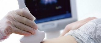
Alternative methods for examining the stomach
In addition to FGDS, there are a number of other diagnostic methods that help detect gastric malignancy. Their comparative characteristics are presented in the following table:
| FGDS | Ultrasound | CT | MRI | |
| Description of the procedure | Endoscopic diagnosis with visual examination of the mucosa | Non-invasive technique using ultrasound | X-ray technique with computer processing of the obtained images | Based on the use of magnetic resonance of atomic nuclei |
| Information content for stomach cancer | High (allows you to make a diagnosis and determine the type of cancer) | Low (only large tumors are visualized) | High (detects metastases to lymph nodes and distant organs) | High (detects metastases to lymph nodes and distant organs) |
| Using Contrast | Not required | Not required | Increases the information content of the technique | Increases the information content of the technique |
| Carrying out a biopsy | Held | Not carried out | Not carried out | Not carried out |
| Time spending | 30-40 minutes | 15-20 minutes | 10-15 minutes | 30-50 minutes |
| Side effects | · Traumatization of the gastric mucosa Bleeding Entry of stomach contents into the respiratory tract | None | Frequent use increases the risk of developing tumors. | None |
| Contraindications | · Severe condition of the patient · Blood clotting disorders Acute stage of myocardial infarction or stroke | None | · Pregnancy · Severe obesity | · Presence of metal implants · Severe obesity |
| Price for research in Moscow | 1200-9700 rub. | 700-4800 rub. | 4000-13500 rub. | 7600-21000 rub. |
Early diagnosis in the case of oncology gives a better chance of long-term remission. Want to know if stomach cancer is visible on an ultrasound? Read this article.
What to do if cancer is discovered?
If fibrogastroduodenoscopy was performed in a regular hospital or clinic, then after receiving the results the patient is referred for consultation to an oncologist .
He examines the patient and issues a referral for treatment to a public oncology hospital.
If the patient wishes, then with the results of FGDS and a referral from an oncologist (sometimes with their translation), he can also contact private foreign oncology clinics that have representative offices in Russia. They usually require a repeat endoscopic examination of the stomach with a biopsy.
When a patient enters a specialized medical institution, a full range of diagnostics is required :
- examination by a gynecologist for women and a proctologist for men;
- general blood and urine analysis;
- blood chemistry;
- specific tumor markers (CA 72-4, CEA, Ca 19-9);
- X-ray of the lungs;
- electrocardiogram;
- ultrasound examination or CT scan with contrast of the abdominal organs;
- Ultrasound of the lymph nodes of the supraclavicular region;
- colonoscopy;
- PET-CT (if possible).
If distant metastases are detected and accessible, it is necessary to perform a biopsy under ultrasound guidance.
What does the endoscopic method reveal?
Fibrogastroduodenoscopy rightfully refers to highly informative methods for diagnosing the organs of the digestive tract that make up its upper section, since during its implementation the specialist not only receives a visual picture of the internal state, but can also pay close attention to:
- anatomical features of the structure of organs;
- quality of mucous membranes and folds;
- the appearance of erosions and ulcerations;
- the presence of tumor-like or polypous formations;
- condition of blood vessels;
- the presence of hidden bleeding.
The procedure is characterized by a significant volume of manipulations, since it allows you to examine all organs related to the upper part of the gastrointestinal tract.
Esophagus
The organ is located at the beginning of the gastrointestinal tract and is a muscular tube approximately 25-30 cm long, hollow inside and slightly compressed on the sides. In accordance with the areas in which it is located, it is divided into three parts - the cervical region, the thoracic region, and the abdominal region. Its main purpose is to allow food to pass into the lower segments of the intestinal tract.
During the examination, in normal condition the following should be revealed:
- three physiological narrowings (at the beginning, end, and in the area of bifurcation of the trachea);
- normal light pink color of the mucous membrane and smooth surface without pathological changes.
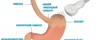
Stomach
It is a hollow muscular organ, which is an important segment of the gastrointestinal tract. In the upper part it connects with the esophagus, in the lower part - with the duodenum through two sphincters - the cardiac and the pylorus. If the closure of the latter is disrupted, the contents are thrown back into the stomach, causing damage to the mucous membrane and the development of inflammation. The inner shell has three layers, the first of which has many folds, the second contains blood vessels and lymph, and the third contains muscle fibers.
Normally, according to the results of the study, the epithelial layer should have a slightly pinkish color and a uniform surface.
Inside the stomach there is a “mucus lake” where digestive juice is produced. Depending on the color, you can accurately determine or refute the likely development of pathologies:
- transparent structure is the norm;
- greenish or yellow tint – bile impurities and the development of duodenogastric reflux;
- reddish color - blood impurities.
Duodenum
A horseshoe-shaped tubular organ, hollow inside, connecting at the beginning with the pylorus of the stomach, and at the end smoothly passing into the small intestine. It received its specific name because of its length, which in ancient times was measured by 12 transverse dimensions of the big toe. The actual length is 25-30 cm. A special feature of the organ is the presence of pancreatic and liver ducts. The examination takes into account the condition of the mucous epithelium, which should have a light pink tint, a smooth velvety surface and a uniform structure.
Choice of treatment tactics
According to national recommendations of oncologists, the choice of treatment tactics for stomach cancer depends on the extent of the tumor . If the disease is detected early, endoscopic resection of the mucosa with or without submucosal layer is performed.
Open abdominal surgery is also possible. It is carried out if there are no distant metastases, and the malignant process does not spread beyond the regional lymph nodes.
Radical tumor removal is recommended in the absence of distant metastases. Plastic surgery of the remaining part of the stomach is required.
In the terminal (last) stage of cancer, surgical intervention is necessary only if complications arise that can threaten the patient’s life (bleeding, narrowing of the lumen, disruption of the integrity of the stomach with the development of peritonitis). Radical removal of the organ then does not improve the prognosis, since distant metastases exist.
Please note National recommendations advise prescribing chemotherapy to all patients after (and in certain situations before) radical or endoscopic intervention, in the last stage of the disease.
The most commonly prescribed drugs are epirubicin, cisplatin and oxaliplatin. Only in the presence of serious concomitant diseases is chemotherapy not carried out, but symptomatic treatment is prescribed. Usually 6-8 courses are carried out until clinical remission is achieved.
Treatment of stomach cancer
Only surgical treatment is effective. It involves several different operation options:
- Removal of part of the stomach, so-called resection.
- Gastrectomy - complete removal of the stomach.
- Combined advanced operations. Resection of parts of nearby organs is also provided.
- Extending the gastrostomy tube onto the anterior abdominal wall. It is practiced in inoperable situations to maximally prolong the patient’s life.
- Formation of a bypass connection between the stomach and intestinal loops. It is performed in inoperable situations to maximally prolong the patient’s life.
In addition to surgery, antitumor treatment is carried out, which includes:
- Chemotherapy.
- Radiation therapy when it is not possible to completely remove the tumor.
Patient survival reaches 80-90% at stage I of tumor growth. At all other stages the prognosis is doubtful.
Essential drugs
There are contraindications. Specialist consultation is required.
- Etoposide (antitumor drug). Dosage regimen: IV, at a dose of 120 mg/m2 on days 1–3. The interval between courses is 3 weeks.
- Cisplatin (antitumor drug). Dosage regimen: IV, at a dose of 80 mg/m2 on the 1st day. The interval between courses is 3 weeks.