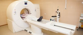
Pathology of the digestive tract requires a differential approach to establish the correct diagnosis. For this purpose, a number of invasive and non-invasive studies are used. In difficult cases, a CT scan of the stomach is prescribed, which shows changes simultaneously in the organ itself and in other parts of the abdominal cavity. Unlike barium radiography, preparation for a layer-by-layer examination is easier for the patient. If necessary, a CT scan of the stomach with contrast is performed, which shows the best result.
Do they do a CT scan of the stomach?
Examination for diseases of the gastrointestinal system includes clinical, histological and endoscopic techniques. However, in some situations, traditional directions do not provide complete information. Therefore, to clarify the pathological condition, a CT scan of the stomach and other parts of the digestive tract is done. This is a more acceptable diagnostic method for the patient, given that it is performed without swallowing a light bulb (endoscopy). The study allows us to determine the type, location and extent of the pathological process.
Indications
The diagnostic procedure with layer-by-layer sections is a high-precision technique. Specialists most often resort to such an examination when a large process is suspected in a patient. Computed tomography shows: the thickness of the walls of organs, their structure, location, changes in surrounding tissues. Research is used in the following situations:
- Clarification of the stage of the malignant process, the degree of tumor infiltration in the wall of the organ and beyond.
- To detect cancer recurrence.
- To monitor the result of the therapy.
- Determining the cause of displacement of the esophagus, stomach, duodenum or other parts of the digestive tract.
- Detailing the type of volumetric formation when alternative methods do not provide sufficient data.
- Differential diagnosis of cysts, ulcerative defects, infiltrative malignant pathology.
- Determination of probable metastatic foci in parenchymal organs, lymph nodes, bone tissue.
- To reduce the number of unnecessary surgical interventions for inoperable tumors.
Stomach cancer
Gastritis
1306 02 September
IMPORTANT!
The information in this section cannot be used for self-diagnosis and self-treatment.
In case of pain or other exacerbation of the disease, diagnostic tests should be prescribed only by the attending physician. To make a diagnosis and properly prescribe treatment, you should contact your doctor. Stomach cancer: causes, symptoms, diagnosis and treatment methods.
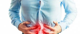
Malignant neoplasms of the stomach occupy one of the leading places in terms of prevalence among cancer diseases. Atypical cells develop from the epithelium of the gastric mucosa, subsequently forming a malignant tumor. Nonspecific (characteristic of other diseases) symptoms of stomach cancer lead to late diagnosis. Therefore, the presence of dyspeptic disorders (nausea, vomiting, etc.), discomfort in the stomach, decreased appetite, anemia and unintentional weight loss is a reason to visit a gastroenterologist. Very often, stomach cancer develops against the background of atrophic gastritis and gastric ulcer.
The bacteria Helicobacter pylori play a significant role in its occurrence.
Causes of stomach cancer
Impaired division of epithelial cells in the gastric mucosa leads to the appearance of atypical cells that contribute to the development of a tumor. Factors whose influence leads to the development of malignant tumors are called carcinogenic.
External carcinogenic factors include nitro compounds, which are formed from nitrites and nitrates found in food products. Even in small quantities, these substances provoke a tumor process. Vitamin C prevents the formation of nitro compounds in the body. Also, the formation of nitro compounds from nitrates slows down with increased stomach acidity. In contrast, low acidity is accompanied by an increase in the number of nitrite-producing bacteria and an increase in the level of carcinogenic nitro compounds.
A decrease in the acidity of gastric juice and atrophy of mucosal cells is caused by the bacterium Helicobacter pylori, the presence of which leads to the development of a malignant process in the stomach.
External provoking factors also include the composition of the diet. Frequent consumption of too hot food, carbonated drinks, salty, smoked and canned foods, as well as foods containing dietary nitrates and nitrose-containing components, increases the risk of developing stomach cancer.
Patients with diagnoses such as chronic atrophic gastritis, gastric ulcer, Ménétrier's disease (changes in the gastric mucosa with subsequent development of cysts and adenomas) should be regularly observed by a gastroenterologist and carefully examined for the appearance of tumors in the stomach.
Classification of stomach cancer
Depending on the clinical objectives, several classifications of gastric cancer are used. First of all, the tumor is classified according to its location - in the fundus, body of the stomach, in the vestibule of the pylorus or in the pylorus itself, on the lesser or greater curvature of the stomach. In addition, the histological type of the tumor is specified, which must be taken into account when prescribing therapy, since each type has biological and structural features. Some types of tumors are considered more aggressive and difficult to treat. Thus, to determine treatment tactics, it is necessary to establish the type of tumor, which is possible after a biopsy.
Depending on the biological activity and the primary affected cells, intestinal, diffuse and mixed types of gastric cancer are distinguished. The intestinal type is similar to the structure of an intestinal tumor. The specificity of the diffuse type is its rapid growth and the ability to affect nearby organs. This tumor is aggressive and has a high tendency to metastasize. The mixed type combines the characteristics of the previous two.
Cancer is also classified according to the TNM system (from the English tumor, nodus and metastasis - stages of tumor development), where the size of the tumor, damage to the lymph nodes and the presence of metastases are assessed.
Symptoms of stomach cancer
In the early stages, malignant tumors do not have characteristic symptoms.
However, doctors identify minor oncological signs of stomach cancer, and if they appear, it is recommended to consult a doctor.
These include decreased appetite and the appearance of dyspeptic disorders (feelings of heaviness, fullness in the stomach, belching, nausea). The progression of the disease leads to asthenia (severe weakness), anemia, which causes pallor, shortness of breath, and weight loss. If the tumor is localized in the cardiac (upper) part of the stomach, dysphagia (impaired swallowing) may occur. Cancer of the pyloric part of the stomach is accompanied by a violation of the movement of food into the duodenum and can cause vomiting of undigested contents (or food eaten the day before). If blood vessels are involved in the cancer process, the vomit and feces will be black. This type of vomiting is called “coffee grounds,” and loose, tarry stools are called melena. Pain and the inability to digest food lead to sudden weight loss and weakness of the patient.
Pain in the stomach appears already in the later stages of the disease. In this case, girdle pain (from the back) may indicate metastases to the pancreas, and pain in the heart area may indicate growth into the diaphragm. If metastases have spread to the intestines, symptoms may include bloating, rumbling and constipation. The disintegration of the tumor is accompanied by vomiting “coffee grounds” or blood.
Diagnosis of stomach cancer
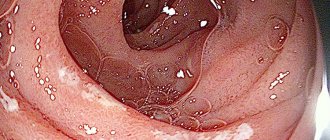
When dyspeptic disorders or discomfort in the stomach appear, patients are referred primarily to gastroscopy, which allows one to see areas of altered gastric mucosa and perform a biopsy (taking biomaterial for histological examination).
What is a CT scan of the stomach
Computed tomography is the process of obtaining information about an organ through computer processing of X-ray radiation. Refers to complex diagnostic methods. As a result of irradiation of tissues of different densities, sections with images of organs appear on digital media.
CT scan of the stomach is a study that requires special preparation. This is due to anatomical and functional features, given the hollow nature of this section of the digestive system. The examination is performed using a spiral tomograph. The MSCT procedure takes from 20 to 60 minutes.
Types of CT diagnostics
There are 3 main types of CT diagnostics, which differ from each other in technical characteristics:
- Step-by-step CT. The very first type of CT diagnostics. For one revolution of the X-ray tube, one section of the organ occurs. To make the next cut, you need to move the table a certain distance. It takes about 20 minutes. The quality of information is not inferior to spiral computed tomography.
- Spiral CT (SCT). In this tomograph, the X-ray emitter moves in a spiral around its axis with the simultaneous movement of the conveyor table with the patient, which provides information in several planes. Using SCT, it is possible to obtain up to 64 sections of an organ. High resolution makes it possible to see tumors with a diameter of 1 mm. The simultaneous movement of the table and the emitter allows you to reduce the procedure time, but sometimes the moving table causes interference in the images.
- Multislice CT (MSCT). It is a type of spiral CT. The main difference is the high rotation speed of the X-ray emitter and a larger number of special sensors that receive information. Using MSCT, you can obtain up to 320 sections of an organ, which increases the information content and accuracy of the study. It is the shortest study in terms of time, which significantly reduces the radiation load on the body.
Be sure to read: More about colon cancer: stages, symptoms, treatment and prognosis
What does a CT scan of the stomach with contrast show?
To achieve good visualization, the technique is combined with the administration of special drugs intravenously and orally (by mouth). A CT scan of the stomach with contrast shows the organ more clearly and reveals minimal changes in its structure. The patient undergoes preliminary preparation, which is agreed upon by a radiologist. The optimal route of administration and type of medicinal substance are determined based on the data obtained about the patient and the expected pathology.
Computed tomography is an expensive examination. Therefore, the equipment is installed in large treatment centers or private structures. The price of diagnostics depends on the number of sections, the type and route of contrast administration, and the quality of the equipment. The cost may also vary in different regions and ranges from $45 to $350.
Preparing for the examination
To obtain high-quality information, a person is prepared for research. Before the procedure, the doctor specifies the following information:
- medical history;
- allergic reactions to drugs, especially iodine-containing ones;
- a list of tests performed over the past year;
- results of previous gastric CT studies;
- X-ray load data for 5 years.
Important!
Diagnosis is always carried out on an empty stomach. This means that the last meal should be no later than 6 hours before the procedure.
There are several ways to contrast the abdominal organs. The choice of method is determined by the doctor after assessing the situation in each specific case. They resort to the options presented in the table.
Table 1. Types of computed tomography
| Type of CT examination | Rules of application |
| Using a contrast agent | Within 45 minutes before the procedure, oral administration of Trazograph or Urografin is prescribed. 1 ampoule with a volume of 20 ml is diluted in 600 ml of water and given to drink in small sips |
| With water without contrast | Within 2 hours before the examination, take 1.5 liters of still water, drink 250 ml immediately before the procedure |
Attention! If the patient has not previously experienced intolerance to iodine or drugs that contain it, but symptoms of intolerance are observed while taking contrast, further administration of the substance should be immediately stopped and immediate measures should be taken to prevent the development of an allergic condition.
To obtain high-precision images with highlighting of the tissue structures of the digestive tract, you can do a PET CT scan. This is a spiral tomography with intravenous administration of glucose labeled with a radioactive isotope, most often fluorine-18. The method allows you to diagnose cancer at different stages, including metastases to other organs.
MSCT of the abdominal cavity and retroperitoneum with bolus contrast
MSCT examination of the abdominal cavity and retroperitoneal space is often performed with contrast enhancement (bolus contrast). Contrast enhancement allows you to obtain a clearer image of the organs being examined. An iodine-containing drug is used as a contrast (iodine is opaque to x-rays). The contrast agent is injected into the patient's vein using an automatic injector; the rate of injection is controlled by the scanner software (this method of contrast injection is called bolus contrast).
Contrast enhancement may not always be used. Main contraindications to the use of contrast:
- allergy to iodine-containing drugs;
- hyperthyroidism;
- renal or liver failure;
- breast-feeding.
CT scan of the stomach with barium
For many years, barium has been used to contrast the stomach and intestines during X-ray examinations. The method is cheap, so it is still used today. Such diagnostics are not always informative, especially for cancer of the digestive tract. In addition, barium suspension can be retained in various parts, disrupting the motor function of the gastrointestinal system. The solution requires special preparation before use.
When examined layer by layer, the mixture may produce additional shadows and images may be misinterpreted. Therefore, a barium CT scan of the stomach is not performed.
Important! If an X-ray examination with barium sulfate was performed, it is recommended to postpone the CT diagnosis for a week so that the substance is completely removed from the body.
Indications for the procedure
General practitioners, pediatricians, gastroenterologists, and surgeons usually refer you for an ultrasound scan of the stomach. Direction goals
may be different:
- Preventive examinations
: children - carried out during the first year of life in order to exclude congenital anomalies and neoplasms; - adults - during medical examinations and during periodic medical examinations of the working population.
Ultrasound of the stomach is a less informative way to examine the organ compared to gastroscopy and even fluoroscopy. For diagnostic purposes, the method is used more often in children, because Ultrasound does not have a negative effect on the child’s body, the method is painless and does not cause any inconvenience.
Nutrition before CT scan of the stomach
Additional preparation includes proper nutrition the day before the procedure. To obtain a high-quality diagnostic result, a diet is necessary before an abdominal CT scan. 2-3 days before the procedure, exclude foods that lead to bloating. These include:
- legumes;
- bread and pastry made from yeast dough;
- fat meat;
- carbonated drinks;
- raw vegetables and fruits.
You are allowed to eat porridge, lean meats and fish, and steamed vegetable dishes. On the day of the study, food intake is limited. CT scan is performed on an empty stomach. To reduce the manifestations of flatulence, defoamers and sorbents are prescribed for several days.
Indications for abdominal CT scan
- In preparation for surgical operations on these organs.
- In the presence of acute conditions that cannot be diagnosed by other means.
- For chronic symptoms of diseases of the abdominal organs that cannot be explained when using other examination methods.
- For unexplained jaundice and sudden weight loss.
- For acute abdominal injuries.
- To monitor the treatment being carried out.
Computed tomography equipment
Computer tomograph Siemens SOMATOM Perspictive (64 slices)
- The device provides high image quality
- Allows you to quickly conduct research
- Scanner of the highest level
Computer tomograph SOMATOM Scope Power (16 slices)
- The tomograph provides excellent diagnostic image quality
- Equipped with ultra modern imaging systems
Contraindications
In most cases, adults tolerate the examination well. However, there are conditions in which X-ray diagnostics with layer-by-layer image acquisition are not performed. CT scanning of the stomach with contrast is contraindicated in the following situations:
- during pregnancy and breastfeeding;
- severe diabetes mellitus, renal failure;
- pathologies of the thyroid gland;
- multiple myeloma;
- with a body weight above 150 kg, taking into account the limitation on the technical characteristics of the device;
- some mental illnesses – schizophrenia, claustrophobia;
- if iodine intolerance is noted.
For young children, CT scans are performed under general anesthesia.
This is due to the need to remain motionless for a certain period of time. Radiation exposure is also taken into account before performing a CT scan.
Computed tomography of the stomach
CT scan provides a clear image of the esophagus and stomach, their internal cavity, walls and circulatory system.
Why do you need a CT scan of the stomach?
CT scan of the esophagus and stomach provides comprehensive information in diagnosing a number of pathologies and diseases of the abdominal cavity, from oncology to damage to the lymph nodes. CT scan of the esophagus and stomach differs from other diagnostic methods in that it shows not only the mucous membrane of the internal cavity, but makes it possible to see blood vessels and a section of the walls in any plane.