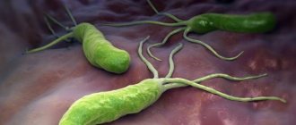
Helicobacter pylori is a pathogenic bacterium that lives mainly in the pylorus (antrum) of the stomach.
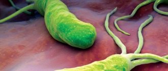
The photo below shows that the microorganism has the shape of a spiral to which flagella are attached. This structure helps it hold tightly to the walls of the digestive organ, move along it along with mucus and exist in an acidic environment, which many pathogenic microorganisms cannot tolerate and die.
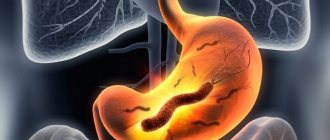
Once in the human body, Helicobacter pylori causes a dangerous disease - Helicobacteriosis. Bacteria multiply rapidly, and in the process of their vital activity they produce a lot of toxins that corrode the mucous membrane of the stomach (duodenum), and then the walls of the digestive organ themselves. Such exposure is dangerous because it creates a favorable environment for gastritis, ulcers, and malignant neoplasms.
What it is?
Helicobacter pylori is just a bacterium that is found in patients with various diseases of the stomach and intestines, the duodenum in particular.
As for the name of the bacterium Helicobacter pylori, it is not at all accidental. One part of it, “pylori”, indicates the main habitat of the bacterium - the pyloric section of the stomach, and the second part, “heliko”, characterizes the shape of the bacterium: helical, spiral.
Previously in medicine it was believed that a microorganism capable of surviving in the acidic, salty environment of the stomach did not exist in principle. But then doctors did not suspect the existence of Helicobacter. Helicobacter pylori was discovered only in 1979 by Australian scientist Robin Warren. Together with scientific colleague Dr. Barry Marshall, the “discoverers” managed to grow this Helicobacter bacterium in laboratory conditions. Then they only assumed that it was she who was the culprit of gastritis and stomach ulcers, and not unhealthy diet or stress, as previously thought.
In an attempt to confirm the correctness of his guess, Barry Marshall conducted an experiment on himself by drinking the contents of a Petri dish in which Helicobacter pylori was cultivated. Just a few days later, the scientist was diagnosed with gastritis. He was cured by taking metronidazole for two weeks. And already in 2005, the authors of this discovery, scientists, received the Nobel Prize in the field of medicine for their discovery. The whole world has recognized that ulcers and gastritis, with all the resulting and associated diseases, appear precisely because of Helicobacter.
Causes of Helicobacter pylori infection
Helicobacter bacteria cannot exist in air; with active air intake, these pathogenic organisms die. They are transmitted mainly through human mucus and saliva. Thus, most often infection occurs in the following ways:
- Sharing utensils;
- Using only personal hygiene products;
- Kisses;
- From mother to child.
Photo: How is the Helicobacter bacteria transmitted?
Thus, friends, family and cohabitants of the patient may be at risk.
In general, infection is facilitated by a low standard of living and neglect of hygiene rules. Very often, helicobacteriosis occurs in people living in communal apartments and dormitories, orphanages, as well as in medical workers. It is worth noting that in third world countries the disease occurs much more often than in developed countries. In Russia, in recent years, the incidence of gastritis and ulcers due to the influence of Helicobacter has begun to be noted among people from wealthy segments of the population.
You can protect yourself, and it is better to take care of prevention in advance than to suffer from an illness and look for ways to fight Helicobacter urgently.
How can you get infected?
Infection occurs when bacteria are transmitted from one person to another through the fecal-oral or oral-oral route. In addition, there are hypotheses about the transmission of this bacteria from cats to humans, as well as about its mechanical transfer by flies.
Most often, infection occurs in childhood. The most likely route of infection is transmission of Helicobacter pylori from person to person, which can occur in three ways:
- Iatrogenic (caused by medical procedures) pathway. In this case, infection is caused by the use of an endoscopic or other medical instrument that came into contact with the gastric mucosa of an infected patient in another person.
- Fecal-oral route. H. pylori is shed in the stool of infected people. The source of infection can be water or food contaminated with feces.
- Oral-oral route. There is evidence that Helicobacter pylori can be present in the oral cavity. Therefore, transmission of bacteria is possible through sharing cutlery and toothbrushes, or kissing.
How is Helicobacter pylori transmitted?
Both adults and children can become infected with Helicobacter pylori. The main route of transmission is fecal-oral , so the disease is classified as an intestinal infection, along with typhoid fever or dysentery. Food and water contaminated with the patient's feces can become sources of infection.
Another important route of infection is oral-oral , that is, through saliva. Previously, gastritis caused by a bacterium was called the “kissing disease,” which emphasizes the importance of maintaining good personal hygiene and avoiding the use of other people's toothbrushes or lipsticks.
A rare route of transmission of infection is iatrogenic (literally “provoked by a doctor”) or contact. Insufficient sterilization of fiber gastroscopes, which are intended for the FGDS procedure, can lead to colonization of Helicobacter pylori in the body of a previously healthy person.
What happens in the body?
At the initial stage, after entering the stomach, H. pylori, quickly moving with the help of flagella, overcomes the protective layer of mucus and colonizes the gastric mucosa. Having established itself on the surface of the mucous membrane, the bacterium begins to produce urease, due to which the concentration of ammonia increases in the mucous membrane and the layer of protective mucus near the growing colony and the pH rises. By a negative feedback mechanism, this causes an increase in the secretion of gastrin by the cells of the gastric mucosa and a compensatory increase in the secretion of hydrochloric acid and pepsin, with a simultaneous decrease in the secretion of bicarbonates.
Mucinase, protease and lipase produced by the bacterium cause depolymerization and dissolution of the protective stomach mucus, as a result of which hydrochloric acid and pepsin gain direct access to the exposed gastric mucosa and begin to corrode it, causing a chemical burn, inflammation and ulceration of the mucous membrane.
The endotoxin VacA produced by the bacterium causes vacuolation and death of gastric epithelial cells. Products of the cagA gene cause degeneration of gastric epithelial cells, causing changes in cell phenotype (cells become elongated, acquiring the so-called “hummingbird phenotype”). Attracted by inflammation (in particular, the secretion of interleukin-8 by cells of the gastric mucosa), leukocytes produce various inflammatory mediators, which leads to the progression of inflammation and ulceration of the mucosa; the bacterium also causes oxidative stress and triggers the mechanism of programmed cell death of gastric epithelial cells.
What kind of bacteria is this?
Helicobacter pylori - translated from Latin - is a spiral-shaped bacterium that lives in the pylorus. And indeed the microscopic bacterium looks like a spiral surrounded by hairs. With the help of these hairs - flagella, it quickly moves through the internal organs to its place of permanent residence - the pylorus - the lower tier of the stomach and the initial section of the intestine - the duodenal bulb. The Helicobacter bacterium has refuted the myth that hydrochloric acid in the stomach kills all microbes. On the contrary, the little predator feels at home in destructive acid, thanks to the enzyme urease, which breaks down hydrochloric acid.
How does Helicobacter affect the stomach? Destroys parietal (parietal) cells of the mucous (inner) lining of the stomach, releasing toxic products - toxins. Protective blood cells - neutrophils, lymphocytes and others - pursue the pest, trying to destroy both it and the altered parietal cells - inflammation occurs. The amount of protective mucus in the area where the bacterium resides is noticeably reduced, and hydrochloric acid rapidly affects the altered area, exacerbating the constant inflammation of the mucous membrane. This is manifested by pain in the stomach “in the pit of the stomach,” heartburn, belching, coating on the tongue, bad breath, constant nausea, that is, symptoms of chronic gastritis. Chronic long-term inflammation of the gastric mucosa leads to changes in its cells, including the development of stomach cancer.
How does Helicobacter enter the body? Since Helicobacter lives in the stomach, infection is possible when the bacteria enters the human body through the mouth. This is the habit of taking food or dishes with unwashed hands, and the desire to try food on someone else’s plate or take a bite from an apple or a friend’s sandwich when offered to try - this is how children often become infected. In addition, you can become infected by sharing utensils or through kissing, which is why Helicobacter is often found among members of the same family.
So why now go to a cafe with your own dishes? Fortunately, Helicobacter is not a resistant bacterium and washing dishes in the dishwasher is enough to destroy them. Good restaurants and cafes are of course equipped with such machines, and you can visit them without the risk of infection.
Misconceptions about Helicobacter pylori
Often, when Helicobacter pylori is detected, patients begin to worry about their eradication (destruction). The mere presence of Helicobacter pylori in the gastrointestinal tract is not a reason for immediate treatment with antibiotics or other agents. In Russia, the number of Helicobacter pylori carriers reaches 70% of the population and the vast majority of them do not suffer from any diseases of the gastrointestinal tract. The eradication procedure involves taking two antibiotics (for example, clarithromycin and amoxicillin).
In patients with hypersensitivity to antibiotics, allergic reactions are possible - from antibiotic-associated diarrhea (not a serious disease) to pseudomembranous colitis, the likelihood of which is low, but the percentage of deaths is high. In addition, taking antibiotics negatively affects the “friendly” microflora of the intestine and genitourinary tract and contributes to the development of resistance to this type of antibiotic. There is evidence that after successful eradication of Helicobacter pylori, reinfection of the gastric mucosa is most often observed over the next few years, which after 3 years is 32±11%, after 5 years - 82–87%, and after 7 years - 90.9% ( Zimmerman Ya.S.).
Until pain appears, helicobacteriosis should not be treated. Moreover, it is generally not recommended to carry out eradication therapy in children under eight years of age, because their immunity has not yet been formed and antibodies to Helicobacter pylori are not produced. If they are eradicated before the age of 8, then a day later, after briefly interacting with other children, they will “catch” these bacteria (P.L. Shcherbakov).
Helicobacter pylori clearly requires eradication if the patient has a stomach or duodenal ulcer, MALTOMA, or if he has had a gastric resection for cancer. Many reputable gastroenterologists (not all) also include atrophic gastritis in this list. Helicobacter pylori eradication may be recommended to reduce the risk of developing stomach cancer. It is known that at least 90% of cases of stomach cancer are associated with H. pylori infection (Starostin B.D.).
List of drugs required for therapy
The list of medicines includes several groups:
- Drug therapy involves the prescription of bismuth preparations (for example, De-nol). However, there are treatment regimens where these drugs can be dispensed with.
- The antibiotics themselves. Most often, domestic doctors prescribe: Augmentin, Flemoxin, Klacid, Ormax. Combination treatment with two or more types of antibiotics is possible (usually if bismuth preparations are not prescribed).
- Antisecretory drugs: Ranitidine, Omez, Lansoprazole, Gastrocepin and others.
- Antimicrobial medications (if necessary) - for example, Macmiror.
- Enzyme preparations (Mezim, Pangrol, Creon) are prescribed to improve digestion and relieve excessive stress on the pancreas. Most often, treatment of gastrointestinal diseases accompanied by the presence of Helicobacter pylori infection cannot be done without them.
- Antibiotic therapy is necessarily accompanied by the use of lacto- and bifidobacteria (Bifiform, Linex, Acepol) to normalize intestinal function and (or) means to prevent the occurrence of candidiasis (Fluconazole, Nystatin, Pimafucin, etc.).
In most cases, the outcome of drug therapy cannot be predicted. This is explained by the bacteria's resistance to drugs. After all, many people uncontrollably take antibiotics to treat all kinds of diseases. However, when combined therapy is prescribed, the result is positive in most cases. In difficult situations, it is possible to take tests to determine the sensitivity of the H. Pylori bacterium to certain antibacterial agents.
There is currently no vaccine against Helicobacter pylori. Nevertheless, several years earlier, the best scientists in the world began to develop a vaccine. Experts wanted to ensure that the drug could be taken during meals. However, identifying a product that would be effective in acidic conditions has proven problematic.
Example of a treatment plan
Since the most common antibiotic treatment regimens for Helicobacter pylori infection include taking two main medications, the doctor will most likely suggest you take De-nol in combination with another antibiotic he prefers or the Klamox + Claricin regimen.
Only your attending physician can recommend the exact types of medications that are right for you, the dosage and duration of treatment. Don't self-medicate!
Bismuth preparations
This treatment regimen for Helicobacter pylori with antibiotics is most often used. The doctor prescribes one antibacterial drug and its complementary agent.
Clarithromycin or Tetracycline. Both medications must be taken twice daily. The average dosage for an adult is 0.5 g. A proton pump inhibitor or a histamine receptor blocker (Cimetidine or Roxatidine) is also taken. The exact dosage is prescribed by the attending physician.
Antibiotic therapy
Usually one type is prescribed (Amoxicillin, Tetracycline, etc.) and two additional drugs (Famotidine, Quamatel or Ranitidine). The course of antibacterial medications according to this regimen lasts at least 10 days.
In this case, the specialist has the right to increase the dosage of the funds and change the two-time dose to four times a day.
Additional medications
Analyzing the third treatment regimen for H. Pylori with antibiotics, it is worth noting that it is combined. That is, it implies the simultaneous use of two antibacterial and two additional drugs. In addition, proton pump inhibitors and histamine receptor blockers are used.
A specialist may also prescribe Venter or Misoprostol. These medications protect the walls of the stomach from the aggressive effects of hydrochloric acid.
During the treatment period for Helicobacter, the doctor prescribes vitamins and prebiotics. After all, it has long been known that antibacterial drugs kill both pathogenic microorganisms and beneficial ones.
Symptoms and first signs
The development of infection in the digestive tract for a long time is practically asymptomatic. Bacteria attach to the mucous membrane of the intestine and duodenum, producing a toxic enzyme that gradually eats away the cells of epithelial tissues.
Only when erosions and ulcers appear on the walls of the organ does the patient begin to be bothered by the unpleasant symptoms of Helicobacter pylori:
- feeling of bloating and fullness in the stomach after eating;
- frequent belching with an acidic taste in the mouth;
- stomach pain regularly;
- there is a burning sensation in the esophagus, a bitter taste in the mouth;
- regular attacks of nausea, vomiting;
- increased gas formation, which provokes colic and discomfort.
In adults, unpleasant signs of Helicobacter pylori bacteria appear most often after eating and do not go away even after bowel movements. The patient is overcome by lethargy, loss of strength, drowsiness, and irritability. The presence of helicobacter pylori in the stomach or duodenum may be accompanied by a small skin rash, particularly on the face. With gastritis or ulcers caused by Helicobacter pylori, the patient complains of changes in stool (constipation or diarrhea), bad breath, brittle nail plate and constant general malaise.
Helicobacteriosis (Helicobacter pylori, Helicobacter pylori infection, Helicobacter pylori, H. pylori)
Gastritis
Ulcer
Iron deficiency
Vomit
Diarrhea
12503 January 28
IMPORTANT!
The information in this section cannot be used for self-diagnosis and self-treatment.
In case of pain or other exacerbation of the disease, diagnostic tests should be prescribed only by the attending physician. To make a diagnosis and properly prescribe treatment, you should contact your doctor. Helicobacteriosis: causes, symptoms, diagnosis and treatment methods.
Definition
Helicobacteriosis is an infectious disease that affects the pylorus of the stomach, or pylorus, and duodenum. Its causative agent is a unique pathogenic microaerophilic gram-negative bacterium Helicobacter pylori (H. pylori). The bacterium got its name from the section of the stomach in which it lives - the pyloric.
As a result of its vital activity, H. pylori forms a “cloud” of alkaline environment around itself, which allows this bacterium to survive in the aggressive acidic environment of the stomach, causing accelerated secretion of hydrochloric acid, as well as a decrease in alkali secretion in the duodenum.
As a result, the microorganism further colonizes the mucous membrane, forms its increased susceptibility to hydrochloric acid and provokes inflammation, leading to the development of ulcerative defects.
Helicobacter pylori is a spiral-shaped bacterium 3.5 microns long and 0.5 microns wide. It has flagella, with the help of which it moves freely along the wall of the stomach or is securely attached to it. The bacterium H. pylori is very variable; its strains (varieties) differ from each other in their ability to attach to the gastric mucosa, cause an inflammatory process, and have varying degrees of pathogenicity.
Helicobacter pylori, which colonizes the gastric mucosa, is a common cause of its inflammatory changes; it is recognized as the etiological factor of gastritis, and gastritis itself is an infectious disease. Depending on the state of the protective factors of the stomach, the resulting infectious process can occur latently or with severe clinical symptoms of inflammation. According to modern concepts, H. pylori causes chronic gastritis in all infected individuals. This can lead to peptic ulcer disease, atrophic gastritis, gastric adenocarcinoma or low-grade gastric lymphoma. H. pylori is a first-order carcinogen.
The results of numerous studies suggest a possible pathogenetic or indirect role of H. pylori infection in the development and/or course of diseases not related to digestion: cardio-, cerebrovascular, autoimmune diseases, diseases of the blood, skin, nervous system and many others.
The pathogen is relatively resistant to the environment: when boiled, Helicobacter bacteria die almost instantly, and when treated with disinfectant compounds, they die within a few minutes.
Causes of helicobacteriosis
You can become infected with the bacteria through contact with contaminated water or food. Infection is possible during endoscopy and when using other poorly sterilized medical instruments that have had direct contact with the patient’s gastric mucosa.
Household transmission (for example, through kisses, personal belongings, etc.) is also possible, as evidenced by the release of bacteria from saliva and dental plaque.
The prevalence of infection varies depending on the geographic region, patient age, ethnicity, and socioeconomic status. According to the Moscow Department of Health (2019), the prevalence of this infection in Moscow is 60.7–88%, in St. Petersburg - 63.6%, in Eastern Siberia reaches 90%.
Classification of diseases
Diseases associated with H. pylori:
- gastritis,
- duodenitis,
- gastroduodenitis,
- esophagitis,
- stomach ulcer,
- duodenal ulcer,
- iron deficiency anemia of unknown origin,
- stomach cancer,
- duodenal cancer.
Symptoms of Helicobacteriosis
The main manifestation of Helicobacter pylori is chronic inflammation of the stomach. In the case of rapid primary manifestation of the pathogen, the acute period of helicobacteriosis lasts about 10 days. The general condition of patients usually remains satisfactory, sometimes there is a short-term low-grade fever and decreased performance.
The main complaint with which patients with signs of Helicobacter infection consult a doctor is stomach pain. The localization of the symptom may change and move to the area of the duodenum.
The pain can be sharp, aching, dull, and occurs in the upper abdomen on the left and in the center in the umbilical region. Discomfort can occur during prolonged fasting, on an empty stomach, or after a certain time after eating.
Symptoms of helicobacteriosis depend on the clinical form of the disease and may include:
- feeling of heaviness in the stomach after eating;
- loss of appetite associated with sudden attacks of nausea (if the gastric mucosa is severely injured);
- causeless vomiting against the background of normal body temperature;
- heartburn (burning sensations in the esophagus and even larynx) and belching with an unpleasant sour or bitter taste;
- chronic constipation (lack of bowel movements for three days or more);
- liquefaction of stool, the appearance of a foamy or watery consistency;
- intestinal cramps and bloating.
If there is a strong contamination with Helicobacter, a number of atypical symptoms may occur, which indicate significant infection and progression of the disease:
- loss of appetite to its complete absence;
- nausea may be replaced by vomiting with blood clots;
- a sharp decrease in body weight that is not normal;
- dry mouth and metallic taste;
- the appearance of a white coating on the tongue;
- bad breath in the absence of caries;
- seizures in the corners of the mouth;
- bleeding gums.
In the chronic course of the disease and atrophic damage to the gastric mucosa, signs of iron deficiency anemia appear - frequent headaches and dizziness, pallor of the skin, decreased blood pressure and tachycardia.
Diagnosis of helicobacteriosis
For a long time, helicobacteriosis may not manifest itself in any way, while provoking the development of ulcers, adenocarcinoma or gastric maltoma. People whose relatives have a history of these diseases are at particular risk.
Diagnosis can be invasive (endoscopy followed by biopsy of gastric tissue) and non-invasive (laboratory tests).
According to international recommendations, the methods of choice for diagnosing the bacterium and assessing the effectiveness of treatment for H. pylori are a breath test with 13C-labeled urea and determination of specific H. pylori antigens in stool using an immunochromatographic method.
What diseases can be caused by H. pylori
The presence of H. pylori in the stomach is not a disease in itself. However, these bacteria increase the risk of developing various diseases of the digestive tract.
Although colonization of the gastric mucosa by Helicobacter causes histological gastritis in all infected individuals, only a small proportion of them develop clinical symptoms of this disease. Scientists estimate that 10-20% of people infected with Helicobacter pylori develop ulcers, and 1-2% develop stomach cancer.
Diseases the development of which is associated with Helicobacter pylori infection:
- Gastritis is an inflammation of the gastric mucosa. Soon after infection with H. pylori, a person develops acute gastritis, sometimes associated with dyspepsia or nausea. An acute inflammatory process affects the entire stomach and leads to a decrease in acid secretion. After a certain period of time after acute gastritis, chronic gastritis develops.
- Ulcer of the stomach and duodenum. According to scientific data, 70-85% of all stomach ulcers and 90-95% of all duodenal ulcers are caused by bacteria
- Functional dyspepsia is pain in the upper abdomen that is not caused by an ulcer or other stomach lesions. Scientific research has shown that some types of dyspepsia are associated with infection. Treatment to eradicate the bacteria provides relief for many patients with functional dyspepsia and also reduces the risk of developing stomach ulcers and cancer in the future.
- Stomach cancer. Helicobacter pylori is a scientifically recognized etiological factor in the development of stomach cancer. According to one hypothesis, bacteria promote the production of free radicals and increase the risk of mutations in stomach cells.
- MALT lymphoma of the stomach. The infection's association with this disease was first reported in 1991. This bacterium is believed to cause 92-98% of gastric MALT lymphomas.
Effective methods of treating Helicobacter pylori
One of the methods of treating Helicobacter is eradication therapy. Eradication involves the use of a combination of several types of drugs with different pharmacological properties. There are three lines of eradication therapy. The indication for such treatment regimens is the lack of results from the use of medication courses. Eradication should be applied in stages. First, the first line of treatment is used, then (if there is no effect) - the second, and only as a radical measure - the third.
First line eradication therapy
The first line of eradication therapy is presented in two versions.
According to the classical scheme, a combination of Clarithromycin and penicillin antibacterial agents is used. As a supplement, drugs are prescribed whose action is aimed at regulating the secretory function of the digestive organs. Antibiotics belong to the category of potent drugs. To reduce their negative impact on the body, eradication is supplemented with Enterol. The drug well compensates for the aggressive properties of antibacterial medications.
If there is no result of therapy with the standard regimen, the second option is prescribed:
- Tetracycline;
- Omeprazole;
- Metronidazole;
- De-Nol.
Second line eradication therapy
The failure of treatment with the first line of eradication is the basis for the use of second line of therapy. The combination of drugs is changing. The basis of therapy is a penicillin antibiotic (Amoxicillin). The medication is supplemented with Omeprazole and De-Nol. To reduce the aggressive effect of the antibiotic on the body, Levofloxacin is used.
Third line eradication therapy
The indication for the third line of eradication is not only the failure of therapy by other methods, but also the presence of severe dyspeptic disorders in the patient that arose during long-term use of antibiotics. The main set of drugs is selected from the first or second line, but drugs based on bifidobacteria are a mandatory addition. Medicines reduce the load on the digestive tract, restore microflora and protect the mucous membranes of the gastrointestinal tract.
Examples of drugs:
- Linux;
- Normobact;
- Bifiform.
Diagnostics
To detect infection in the body, various examination methods are used, each of which has its own advantages, disadvantages and limitations. Traditionally, all methods are divided into non-invasive and invasive.
Invasive detection methods:
- Histological examination - examination of specially stained samples of stomach tissue obtained through biopsy during endoscopic examination under a microscope.
- Microbiological seeding and isolation of Helicobacter bacteria. To obtain material for culture, a biopsy or a sample of gastric juice is used, which is obtained during an endoscopic examination.
- Polymerase chain reaction (PCR) - can detect infection in small tissue samples obtained through biopsy.
- Rapid urease test - this method uses the ability of bacteria to process urea. The tissue sample obtained by biopsy is placed in a medium containing urea and a pH indicator. Bacteria break down urea into carbon dioxide and ammonia, which increases the pH of the medium and changes the color of the indicator.
Non-invasive detection methods:
- Serological blood tests that can detect antibodies to Helicobacter.
- Urea breath test. During this examination, the patient is given a solution with urea, the molecule of which contains a labeled carbon isotope, to drink. Helicobacter breaks down urea into ammonia and carbon dioxide, which contains a labeled carbon atom. This gas enters the bloodstream and is expelled through the lungs with air. Half an hour after consuming the urea solution, the patient exhales into a special bag, in which a labeled carbon atom is detected using spectrometry.
- Detection of H. pylori antigens in stool.
Analysis for Helicobacter pylori
To identify Helicobacter pylori, you need to undergo a series of tests. Diagnosis is quite simple and includes several methods.
ELISA for bacteria
The research method is used to assess the level of concentration of existing antibodies to H. pylori in the patient’s blood. The immune response is formed 1-2 weeks after the microorganism enters the human body. In some cases, a false negative response is recorded, which may be due to the specifics of the immune system.
A positive test indicates the need to undergo a full examination. Your doctor will tell you how to treat the disease.
Immunological blood test for Helicobacter pylori for IgG, IgM, IgA
In response to infections entering the body, special proteins are produced - immunoglobulins igg, which try to resist the foreign agent. Detection of immunoglobulins IgG, IgM, IgA indicates infection with H. Pylori.
The main feature of immunoglobulins is that they are able to overtake pathogenic cells anywhere in a person. Proteins appear in the blood after damage to the gastrointestinal tract.
Blood test interpretation:
- A negative result or low IgG content indicates the absence of bacteria in the body and a low risk of developing gastritis associated with H. pylori and gastrointestinal ulcers. This is the norm for Helicobacter pylori in a blood test. If 3-4 weeks have passed since the infection, the result may also be negative. For epigastric pain, it is recommended to donate blood again. The price of the analysis is 700 rubles for each immunoglobulin.
- A positive result or an increased level of IgG may indicate the presence of infection in the body and a high risk of developing gastrointestinal ulcers or cancer. A positive result can also be recorded in patients who have recently recovered from pathology; a reduced IgM content is recorded immediately after infection.
- Elevated IgM levels indicate an early stage of infection.
Urease test
The analysis is carried out after taking a biopsy of the mucous membrane during an endoscopic examination. The resulting material is placed in a carrier gel that contains urea, a bacteriostatic agent and a pH indicator. Depending on the change in color of the biopsy sample, a conclusion is drawn.
Breath test for Helicobacter pylori
The examination requires the collection of a fasting background sample of exhaled air. Preparation consists of drinking an aqueous solution of urea labeled with 13C (test solution) after a light breakfast. The final 4 samples are taken every 15 minutes.
A normal breath test for Helicobacter pylori is 1%. The price of the test depends on the region. You can take an in vitro breathing test in Moscow and St. Petersburg for 2,250 rubles.
Hemotest
The study is based on determining the amount of antibody titer - IgG. Immunoglobulins are able to bind pathogens, blocking their negative effects. How to get tested for Helicobacter pylori: on the eve of the test, you should not drink alcoholic beverages or coffee, and smoking is prohibited. You need to take the test on an empty stomach - the fasting period is 8 hours. The study is an enzyme immunoassay of blood taken from a vein into a sterile tube. The analysis is repeated several times throughout the entire period of therapy. The price of in vitro analysis is 800 rubles.
Stool analysis
Today, there are 3 special research methods that can detect Helicobacter pylori:
- Cultural Research . The resulting biomaterial is placed in a nutrient medium, which creates favorable conditions for the growth and reproduction of colonial organisms. Additionally, chemical dyes or medications can be used to assess the degree of sensitivity of microorganisms to various drugs.
- Stool PCR . Due to special enzymes, “enemy” DNA is isolated with further doubling of its particle. Activities for artificial DNA replication are carried out until a complete genetic code is created, which is subsequently compared with an existing Helicobacter pylori sample.
- Immunological method . A solution with antibodies that react violently to the presence of Helicobacter pylori antigens is added to the resulting biological material. An active reaction reaction indicates contamination of the sample with pathogenic microorganisms.
Express test
This test allows you to detect antibodies to infection in whole blood. You can do the test at home, provided you follow all the rules.
The test system includes:
- indicator tablet packed in a special vacuum bag;
- pipette for applying a drop of blood to the tablet;
- a reagent used to dilute the resulting blood sample;
- antiseptic wipe;
- scarifier;
- instructions.
How to get rid of Helicobacter pylori?
In 2021, an acceptable regimen for Helicobacter pylori eradication in adults is considered to be a treatment regimen that provides at least 80% of effective re-examination-confirmed cure for Helicobacter pylori infection and healing of ulcers or gastritis, lasting no more than 14 days and having acceptably low toxicity (side effects should develop in no more than 10-15% of patients and in most cases not be so serious as to require early cessation of treatment).
New schemes and protocols for Helicobacter eradication are constantly being developed. This serves several purposes:
- increasing the convenience of treatment for patients and the degree of their compliance with the treatment regimen: eliminating the need for a strict “anti-ulcer” diet
- thanks to the use of powerful proton pump inhibitors;
- reducing the duration of treatment (from 14 to 10, then 7 days);
- reducing the number of drugs taken simultaneously through the use of combination drugs;
- reducing the number of doses per day due to the use of prolonged forms of drugs or drugs with a long half-life (T1/2);
- reducing the likelihood of unwanted side effects;
- overcoming the growing resistance of Helicobacter to antibiotics;
- meeting the need for alternative treatment regimens if there is an allergy to any of the components of the standard regimen or if the initial treatment regimen has failed.
In 2021, Maastricht-IV experts recommended the following Helicobacter pylori eradication regimens:
Treatment regimen recommended at the Maastricht-IV conference
Triple therapy was proposed at the first Maastricht conference and has become a universal treatment regimen for H. pylori infection. It is recommended by all world conciliation conferences.
The regimen includes drugs:
- one of the proton pump inhibitors (PPIs) in a “standard dosage” (omeprazole 20 mg, lansoprazole 30 mg, pantoprazole 40 mg, esomeprazole 20 mg, or rabeprazole 20 mg twice a day) for at least 7 days
- Clarithromycin (500 mg 2 times a day) 7 days
- amoxicillin (1000 mg 2 times a day) or metronidazole (500 mg 2 times a day) 7 days.
It has been shown that the regimens of PPI + clarithromycin + metronidazole (tinidazole) and PPI + clarithromycin + amoxicillin are equivalent. It has been established that the effectiveness of triple therapy increases when its duration is increased to 10 or 14 days (depending on the degree of Helicobacter pylori contamination and the patient’s tolerance to therapy).
Treatment regimen recommended by the Russian Society of Gastroenterologists
Due to different resistance to antibiotics in different regions of the world, the prevalence of different strains of Hp, and the genetic characteristics of the population, different countries or groups of countries develop their own recommendations regarding the eradication of Hp. Some of these parameters, in particular HP resistance to certain antibiotics, change over time. The choice of a specific regimen is also determined by the patient’s individual intolerance to the drugs, as well as the sensitivity of the HP strains with which the patient is infected.
At the congress of the Scientific Society of Gastroenterologists of Russia, the following HP eradication schemes were adopted; they are relevant for 2019:
1) First option. Three-component therapy, including the following drugs, which are taken for 10-14 days:
- one of the PPIs in a “standard dosage” 2 times a day +
- amoxicillin (500 mg 4 times a day or 1000 mg 2 times a day) +
- clarithromycin (500 mg 2 times a day), or josamycin (1000 mg 2 times a day) or nifuratel (400 mg 2 times a day).
2) Second option. Four-component therapy, including in addition to the drugs of option 1 bismuth drug, its duration is also 10-14 days:
- one of the PPIs in a “standard dosage” +
- amoxicillin (500 mg 4 times a day or 1000 mg 2 times a day) +
- clarithromycin (500 mg 2 times a day), or josamycin (1000 mg 2 times a day), or nifuratel (400 mg 2 times a day) +
- bismuth tripotassium dicitrate 120 mg 4 times a day or 240 mg 2 times.
3) Third option. If the patient has atrophy of the gastric mucosa with achlorhydria confirmed by intragastric pH-metry and it is therefore inappropriate to prescribe acid-suppressing drugs (PPN or H2 blockers), the third option is used (lasting 10-14 days):
- amoxicillin (500 mg 4 times a day or 1000 mg 2 times a day) +
- clarithromycin (500 mg 2 times a day), or josamycin (1000 mg 2 times a day), or nifuratel (400 mg 2 times a day) +
- bismuth tripotassium dicitrate (120 mg 4 times a day or 240 mg 2 times a day).
4) Fourth option. If full eradication therapy is not possible for elderly patients, truncated regimens are used:
- one of the PPIs in a “standard dosage” +
- amoxicillin (500 mg 4 times a day or 1000 mg 2 times a day) +
- bismuth tripotassium dicitrate (120 mg 4 times a day or 240 mg 2 times a day).
Another way: bismuth tripotassium dicitrate 120 mg 4 times a day for 28 days. If you have pain in the stomach, take a short course of PPI.
How do I know if I have Helicobacter pylori infection?
The presence of insidious bacteria in the body can be detected in different ways. First of all, this is a histological method, when during FGDS (fibrogastroduodenoscopy) - an examination of the stomach, which can be carried out in Chelyabinsk at the All Medicine Clinic - cells of the gastric mucosa are taken for examination. Not only the bacterium Helicobacter pylori is detected, but the condition of the inner lining of the stomach is also assessed - the severity of inflammation and the degree of change in mucosal cells against the background of inflammation. The following methods are breath tests. Helicobacter produces the enzyme urease, which is capable of decomposing urea to form ammonia and carbon dioxide. The essence of the methods is to determine either the composition of carbon dioxide in the patient’s exhaled air (urease breath test) or ammonia (Helic test). The Helic test is quite widely used, especially in pediatric practice, as a simple, non-invasive diagnostic method for the patient. Another common diagnostic method is the detection of specific antibodies of class G immunoglobulins - IgG in the patient’s blood. Helicobacter pylori infection is accompanied by the development of an immune response - the production of antibodies, of which IgG occurs in 95-100% of cases. This is a reliable diagnostic method that allows you to assess the degree of Helicobacter contamination, but not the best way to control the treatment of Helicobacter pylori infection, since the level of IgG antibodies can remain high after the complete death of the microorganism for a long time - 1-1.5 years. In this case, if an antibody test is nevertheless performed, a decrease in their level is assessed no earlier than 2-3 months after the end of treatment. More often, to monitor the effectiveness of treatment for Helicobacter pylori infection, a stool test is performed. It is based on the detection of Helicobacter DNA in feces and is qualitative - it shows whether Helicobacter is detected or not, without assessing the degree of contamination. The method can also be used to diagnose the presence of infection before treatment, for example, in children, as it is simple and painless.
Possible complications from antibiotic treatment
Factors that increase the risk of side effects during eradication therapy:
- Individual intolerance to drugs;
- Presence of somatic pathologies;
- Negative state of intestinal microflora during the initial period of treatment.
Complications of eradication therapy – side effects:
- An allergic reaction to drug components that disappears after discontinuation;
- Dyspeptic symptoms of the gastrointestinal tract (discomfort in the stomach and intestines, taste of bitterness and metal, nausea and vomiting, diarrhea, flatulence). Usually all these phenomena disappear spontaneously after a short time. In rare cases (5-8%), the doctor prescribes medications against vomiting or diarrhea, or cancels the course.
- Dysbacteriosis. It more often manifests itself in patients who previously had gastrointestinal dysfunction, and develops during treatment with tetracycline drugs or during therapy with macrolides. A short-term course is not able to upset the balance of intestinal microflora; to prevent dysbiosis, you need to consume fermented milk products more often: yogurt, kefir.
Nutrition and diet
Of course, the main point in the treatment of this pathology is taking medications, but proper nutrition plays an equally important role. To easily get rid of helicobacteriosis, you should follow the following recommendations:
- do not take long intervals between meals;
- eat food in small portions;
- follow a diet of 5-6 meals a day, eating slowly, chewing food well and drinking it with enough liquid;
- the patient should avoid excessively fatty, fried or spicy foods, carbonated drinks, pickled foods, and alcohol.
In reality, these are only general recommendations; in each individual case, nutrition should be calculated based on the level of acidity (low, high) and prescribed only by the specialist conducting the treatment.
Prevention
You can completely recover from helicobacteriosis if, in addition to therapy, you adhere to preventive measures:
- Maintaining hygiene. Wash your hands before eating, do not consume dirty vegetables and fruits, or questionable water. Do not use other people's household items.
- Timely detection of the disease. If you feel unwell or suspect the presence of a pathogenic bacterium in the body, it is important to immediately consult a doctor and undergo the necessary tests.
- Strengthening the immune system. A healthy lifestyle (swimming, running, walking) increases your defenses and prevents pathogens from entering the body.
- Proper nutrition. Small meals, small doses and avoidance of fried, salty, spicy, smoked foods, alcohol and smoking.
The main danger of helicobacter pylori is that it can provoke gastritis, ulcers, even malignant neoplasms. It is impossible to get rid of harmful bacteria without antibiotics. Therefore, it is important to strictly adhere to special treatment regimens and observe preventive measures.
Gastritis and Helicobacter. Is there a connection?
This bacterium is one of the main causes of gastritis. Just a few years ago, doctors did not suspect the presence of such a microorganism and treated gastritis with other methods. In some cases this was effective, in others the means used did not produce results. Why did this happen?
Gastritis is a pathological disease of the stomach, characterized by the presence of inflammation on the walls of the stomach. This definition implies that the causative agents of the disease can be different.
In fact, Helicobacter is only a factor predisposing to the occurrence of the disease. The bacterium feels like a “fish in water” in the aggressive environment of the stomach and lives in the gastric juice of 90% of the planet’s inhabitants.
Helicobacter pylori multiplies by secreting urease, which destroys the gastric walls, and also breaks down the protective lining of the stomach. Bacterial toxins cause inflammation of the immune system.