Biological basis of the technique
Helicobacter pylori is a spiral-shaped bacterium that affects the mucous membrane of the stomach and duodenum. Persistence of the microorganism leads to the development of gastritis, gastroduodenitis, gastric and duodenal ulcers.
Bacteria on the stomach lining
Once in the stomach cavity, the bacterium penetrates the mucous membrane. There, having secured itself with the help of flagella, it begins to produce the enzyme urease. The pH around the bacterial colony increases due to the enzyme, which leads to the activation of compensatory mechanisms for the production of hydrochloric acid by stomach cells. Gastrin and pepsin are also produced in increased quantities.
In addition to urease, Helicobacter secretes mucinase, lipase and protease - enzymes that break down the upper protective layer of the gastric mucosa. As a result, hydrochloric acid, pepsin and gastrin enter unprotected areas of the gastric epithelium and begin to corrode it. At the site of exposure, inflammation appears first, then erosion and eventually an ulcer.
The bacterium itself is not exposed to the aggressive gastric environment, as it produces urease, which metabolizes urea to form ammonia. NH3 locally increases the pH to 6, which creates favorable conditions for life for Helicobacter pylori.
The urease test for Helicobacter pylori is based on the ability of the bacterium to break down urea.
Indications
The following categories of patients are recommended to undergo the diagnostic procedure:
- with epigastric pain on an empty stomach and immediately after eating;
- having relatives infected with Helicobacter pylori;
- receiving treatment for gastritis and duodenitis.
Types of tests
There are several types of diagnostic methods for detecting Helicobacter by the presence of the enzyme urease:
- urease breath test (UBT and HELIC test);
- rapid urease test with gastric biopsy.
Helpil test positive for gastritis
Not so long ago, it was believed that gastritis was caused by poor diet, dry food, stress and lack of hot soup for lunch. Then scientists discovered a connection between this disease and the bacterium Helicobacter pylori - and since then, the approach to the treatment of gastrointestinal diseases has changed dramatically.
Is it possible to become infected through dishes in a restaurant, does Helicobacter cause cancer and
is it necessary to get rid of the bacterium, which lives in the body of 80% of Belarusians, said Georgy Matveevich Usov, a gastroenterologist with 40 years of experience.
Georgy Usov,
Gastroenterologist of the highest category at the Minsk Clinical Consultative and Diagnostic Center, Candidate of Medical Sciences
— What is Helicobacter? Is this a pathogenic bacterium or an opportunistic one?
— Helicobacter pylori is a bacterium that was discovered by Australian scientists Robin Warren and Barry Marshall. For this discovery they received the Nobel Prize in 2005.
Helicobacter is a pathogenic bacterium. Its presence in the human body leads to damage to the mucous membrane of the stomach and duodenum and the development of diseases such as chronic gastritis, gastric and duodenal ulcers, stomach cancer, and gastric maltoma.
Today Helicobacter is the most common chronic infection in the world.
— What factors in this case provoke the development of gastritis, ulcers, cancer? And can these diseases occur without the participation of Helicobacter?
— If Helicobacter is present, all people are diagnosed with some kind of stomach disease. Identification of a specific disease depends on the Helicobacter strain. Strains of H. pylori, which contain certain genotypes, predispose to the development of peptic ulcers and gastric cancer. And if not all infected people get stomach and duodenal ulcers or cancer, then everyone gets gastritis.
It is also true that these diseases can occur without Helicobacter pylori.
- In addition to gastritis associated with Helicobacter, there are gastritis associated with the reflux of the contents of the duodenum into the stomach, so-called reflux (chemical) gastritis, gastritis associated with the formation of antibodies to the gastric mucosa (autoimmune gastritis).
- The cause of ulcers may be the use of non-steroidal anti-inflammatory drugs, acetylsalicylic acid, occasionally nervous factors, and eating errors.
- The cause of cancer can be errors in nutrition, carcinogenic substances in food, smoking, hereditary predisposition, etc.
— How is this bacterium transmitted to humans? Can you get infected from animals? And in a restaurant through dishes?
— The route of transmission of Helicobacter is through contact and household contact. The bacterium is transmitted:
- through insufficiently clean dishes, when people eat from the same plate or drink from the same cup, eat each other’s food;
- in the family, when parents taste food before giving it to the child, or lick the baby’s pacifier;
- in the case of people using one toothbrush;
- through lipstick;
- failure to wash hands after using the toilet and before eating;
- through sexy kisses.
Helicobacter has also been identified in cats and dogs, as a result of which there is a possibility of human infection from them. As for the restaurant, if dirty dishes are poorly handled, there is a high possibility of Helicobacter infection.
— What percentage of people in Belarus live with Helicobacter? Why is this figure higher than in Western Europe and the USA?
— There have been no high-quality epidemiological studies on the spread of Helicobacter among the population of Belarus. Only relatively small studies have been conducted among the population of certain regions and certain population groups.
It is believed that the prevalence of Helicobacter among Belarusians is about 70-80%.
Among the population of the Russian Federation, according to a number of studies in individual Russian regions, the prevalence ranges from 65 to 90%. This figure is higher than in Western Europe and the USA, due to insufficient hygiene in our country.
— What symptoms should force a person to get tested for Helicobacter? Which of them are not obvious, not directly related to the gastrointestinal tract?
— Dyspeptic disorders (pain, burning in the epigastric region - upper abdomen, nausea, poor appetite, heaviness in the epigastric region, vomiting, etc.) require examination for Helicobacter.
Breath test for Helicobacter pylori
The research method is non-invasive. It is presented in several variations.
UBT is based on the ingestion of urea labeled with the 13C isotope. Bacterial urease decomposes urea with radionuclide to ammonia and carbon dioxide, which retains the labeled carbon 13C. Next, carbon dioxide is absorbed in the stomach, delivered to the lungs and leaves the body with exhaled air. The patient exhales into a special container, which is examined using a gas spectrometer or laser device.
The HELIC breath test is based on measuring a baseline level of ammonia in the exhaled air, followed by the ingestion of carbamide (urea) and a repeat test of the ammonia level.
Preparation
Before the study, you should follow the following recommendations:
- the technique is carried out on an empty stomach, the last meal no later than 8 hours ago, the last fluid intake an hour before the procedure;
- two weeks before testing, the use of antibiotics, proton pump inhibitors (Omeprazole, Pantoprozole) and H2-histamine receptor blockers (Ranitidine) is discontinued;
- for two days it is prohibited to take bismuth preparations (De-Nol), antacids, painkillers;
- it is necessary to limit alcohol intake for several days;
- You should follow dietary recommendations: give up legumes and chewing gum the day before the test;
- do not smoke on the day of the method;
- In the morning you should thoroughly brush your teeth and rinse your mouth.
Methodology
The 13C breath test is carried out according to the following instructions:
- the patient exhales unforcedly into a special container bag, after which it is immediately hermetically sealed;
- Next, you should drink a solution of labeled urea;
- After 30 minutes, you need to re-exhale unforcedly into a sealed bag.
The HELIC breathing test is carried out using an indicator tube or spectrometer with results processed on a computer. In the first case, the patient breathes into an indicator tube, and in the second into a special device.
The research algorithm is similar in both cases:
- a tube is placed in the patient’s mouth; one must breathe not into it, but with the mouth slightly open;
- You should carefully ensure that no saliva gets into the tube; it is better to pull it out and swallow it in time;
- after six minutes the tube is pulled out;
- the patient is asked to drink a solution of carbamide (urea) 500 mg dissolved in 20 ml of water;
- then you should rinse your mouth thoroughly;
- The tube is reinserted into the mouth and the examination is repeated within six minutes.
Breath test technique
Decoding indicators
What is a positive urease test? This is when the ammonia level after consuming 500 mg urea increases by more than 4 mm, and the increase compared to the pre-load test is more than 2 mm.
Advantages
The advantages of these studies are as follows:
- high information content and specificity;
- non-invasive, can be performed on patients for whom gastroscopy is contraindicated;
- quick results;
- can be used to assess the effectiveness of treatment;
- The entire gastric mucosa is examined, and not its local areas.
Flaws
The disadvantages are as follows:
- high cost of equipment;
- false positive results due to persistence of urease-producing microbes in the mouth.
Rapid urease test
During gastroscopy, the endoscopist performs a biopsy of the gastric mucosa in the area of the pylorus and altered areas. To detect urease in a piece of stomach tissue, it is necessary to place the material in a special medium that contains urea and an indicator. In the presence of Helicobacter pylori enzyme, urea breaks down into carbon dioxide and ammonium ions. The pH of the environment will deviate towards the alkaline side and the indicator will display this by changing color.
Preparation
Two weeks before the diagnosis, you should stop taking bismuth drugs (De-Nol), proton pump inhibitors (Omeprazole, Pantoprozole, Omez, Kvamatel), and antibiotics.
On the eve of the lung examination, have dinner 8 hours before the gastroscopy. Do not drink water after midnight.
Methodology
The quick urease test or “CLO-test”, “De-Nol-test”, “HELPIL-test” is carried out according to the following algorithm:
- using forceps at the end of the endoscope, a tissue biopsy is performed - pinching off a piece from the area of the fundus and body of the stomach;
- the material is placed in a medium containing urea and phenol red indicator;
- a positive result is recorded if the color of the indicator changes from yellow to bright red.
Decoding indicators
The result is based on the color intensity of the indicator. The endoscopist evaluates the coloring as a plus.
+ — 1 degree of urease activity, low level of Helicobacter, color appears within 24 hours.
++ - degree 2 of urease activity, moderate level of bacterial infection, the indicator turns red within two hours.
+++ - 3 degree of urease activity, significant level of infection, bright red color of the indicator within an hour of the test.
What is a negative urease test? This is when the indicator does not change its color within 24 hours from the start of the study.
An example of a urease test
Advantages
The rapid urease test with gastric biopsy has the following advantages:
- quick diagnostics - on average about 30 minutes;
- low cost;
- does not require specific expensive equipment.
Flaws
The rapid urease test has several significant disadvantages that do not allow this method to be considered the “gold standard”:
- if the biopsy sample is not sufficiently contaminated with bacteria, it may take about two hours for the indicator color to change;
- the method provides information about the condition of the gastric mucosa in only one place;
- the material obtained during endoscopy is not subject to further histological examination, due to this it is not possible to compare the result of the urease test with microscopic changes in the mucosa;
- high level of false positive results due to the persistence of gram-negative flora in the stomach, which can also produce urease in small quantities;
- in the case of ulcerative bleeding in the stomach, the method is not informative.
Science does not stand still; more advanced diagnostic methods are being invented every day. There is no ideal study with optimal price-quality ratio. You need to choose a convenient way to get examined. In this case, it is necessary to rely on the opinion of a specialist, since this article is not a guide to action.
The video provides information on diagnosing stomach diseases.
Helicobacter pylori is a gram-negative, spiral-shaped bacterium that can infect areas of the mucous membrane of the duodenum and stomach and thereby provoke the development of gastritis, ulcers, duodenitis, cancer and lymphomas. But infection with this bacterium does not always cause the listed diseases. In 90% of cases, carriage of Helicobacter does not cause any pathology.
Symptoms of helicobacteriosis
Frequent pain on an empty stomach that disappears after eating may indicate the presence of a stomach or duodenal ulcer, and therefore infection with Helicobacter pylori. Pain in the epigastric region may bother you at night. Sometimes they subside after drinking an alkaline drink, such as a glass of milk.
In addition, the presence of this infection in the body may be indicated by heaviness in the stomach, frequent heartburn or nausea. Vomiting, as a rule, does not occur. Sometimes patients note changes in taste preferences. You may develop an aversion to meat dishes. Fatty meat foods are poorly digested in such patients.
What is Helicobacter pylori?
The pathogenic microorganism Helicobacter pylori (H. pylori) causes the following diseases:
- chronic gastritis - inflammation of the gastric mucosa
- chronic duodenitis - inflammation of the duodenum
- peptic ulcer of the stomach (in 70% of cases) and duodenal ulcer (in 90% of cases)
- helicobacteriosis
- stomach cancer
- lymphoma of the stomach
70% of the population is infected, every third!
The constant presence of bacteria in the stomach is accompanied by the following symptoms:
- pain in the stomach after eating or before eating
- occasional nausea and even vomiting
- feeling of heaviness in the stomach
- heartburn and sour taste in the mouth
- bad breath
- bloating
These symptoms not only reduce the quality of life and force you to take medications for a long time, but can also lead to stomach cancer!
Helicobacter pylori can also “trigger” other diseases that at first glance are not associated with the stomach, the permanent habitat of the bacterium. For example, idiopathic thrombocytopenic purpura is a serious disease with a decrease in the number of platelets in the blood and a high likelihood of bleeding.
Therefore, correct diagnosis of a current H. pylori infection is extremely important!
What studies confirm the presence of Helicobacter in the body?
There are several methods for determining the presence of this infection in the body.
- Blood test for antibodies to Helicobacter bacteria.
- Examination of stool for pathogen antigen.
- The breath test is not Helicobacter.
- Cytological examination of material obtained during fibrogastroduodenoscopy (FGDS).
As a rule, a diagnosis is made only after two tests for Helicobacter pylori are positive. Today, the most reliable and effective method for detecting this pathogen is the examination of material obtained during endoscopic examination. But it is not always possible to carry out an invasive method to detect this infection; for example, this is contraindicated for pregnant women and children. It is appropriate for these categories of patients to do a breath test for Helicobacter. It is quite easy and has no undesirable consequences.
Urease breath test for Helicobacter pylori
This research method is based on measuring the concentration of urea in the air exhaled by the patient after ingestion. The bacterium Helicobacter pylori is capable of synthesizing a special enzyme - urease. This enzyme has a degrading effect on urea. In the intestines, under the influence of urease secreted by bacteria, it is broken down into components - ammonia and carbon dioxide, which are released by the lungs during breathing. Its concentration is assessed by performing a breath test for Helicobacter. The concentration of carbon dioxide is determined before the patient takes urea and after in several samples.
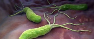
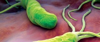
What does Helicobacter pylori look like?
Bacteria Helicobacter pylori, Helicobacter pylori, Helicobacter, HP is a gram-negative microaerophilic bacterium of a curved or spiral shape with many (4 to 6) flagella. Helicobacter pylori (HP) is about 2.9 µm long and 0.8 µm in diameter. Helicobacter pylori , Helicobacter pylori (HP) is found deep in the gastric pits and on the surface of epithelial cells, mainly under the protective layer of mucus lining the gastric mucosa. It can exist in areas of gastric dysplasia with frequent acidification of the duodenal bulb. Directly around the bacterium, the pH is about 7, and the nutrient content is quite sufficient for the life of the microorganism. Helicobacter pylori (HP) is able to exist under conditions of varying concentrations of hydrochloric acid in the gastric cavity. Helicobacter pylori (HP) survives at a pH of 4 to 8. It reproduces successfully at a pH of 6 to 8. The most favorable habitat is the antrum of the stomach. Under unfavorable conditions, Helicobacter pylori (HP) can develop into a coccal form. Helicobacter infection is very dangerous.
How to prepare for research?
In order for doctors to correctly evaluate a breath test for Helicobacter pylori, they need to prepare for it. Otherwise, the analysis results may provide incorrect information. If you are taking some medications, it may take up to three weeks to prepare the patient for this study.
- Three weeks before the study, you must stop taking antibiotics, bismuth preparations and antacids - these are drugs that reduce the acidity of gastric juice.
- Three days before the study, drinking any alcoholic beverages is prohibited.
- The day before the analysis, you should not eat foods that increase gas formation (legumes, cabbage, brown bread, potatoes, etc.).
- The night before dinner should be light and not too late.
- On the morning of the study, you should not have breakfast or smoke.
In the morning, you just need to brush your teeth - this is mandatory, but freshening your breath with chewing gum is prohibited. If you are really thirsty in the morning, you can take a couple of sips of clean boiled water, but no later than an hour before the test.
A false-positive test can be triggered by a previous gastric resection or achlorhydria, a condition in which there is a complete absence of hydrochloric acid in the gastric juice (it is not produced by gastric cells).
How is the Helicobacter breath test performed?
First, the medical professional asks the patient to breathe through a special tube. You need to breathe calmly, as a person would do in a normal situation. At this stage, two samples of exhaled air are taken.
Next, the patient is asked to drink a 5% urea solution. After 5 minutes, take a sample of exhaled air by turning the indicator tube over to the other end. In this way, three more samples are taken. The increase in ammonia concentration in the air exhaled by the patient is assessed.
If the ammonia concentration exceeds 0.5 mg/ml, the test for Helicobacter pylori respiratory is considered positive.
The procedure does not cause any negative feelings. Only the secreted saliva can cause inconvenience. To correctly evaluate the result, it should not get into the tube, otherwise the test may be spoiled. If you cannot swallow it, then you can periodically take short breaks and remove the tube. After swallowing saliva, the study continues. If, however, saliva gets into the indicator tube and the test does not work, it can be repeated after 50-60 minutes.
How is the urease test performed?
This study is a laboratory study. It involves sampling exhaled air, in which the presence and concentration of labeled carbon isotopes is then determined using spectrographic techniques. To do this, the patient exhales air into a special bag after 5 minutes of quiet breathing, after which the bag is tightly sealed with a special stopper. Then the patient is given a solution with urea to drink, and after 20 minutes (the time required for the breakdown of urea by bacteria, if present), the exhaled air is taken again into another bag. Both samples are sent to a specialized laboratory where they are examined. In general, the collection of material for research is carried out within 20-25 minutes, it is painless and does not cause any discomfort for the patient.
How to choose a laboratory?
Modern test systems are automated, and the test is assessed not by a person, but by a machine. In addition, there are systems whose indicator tubes are protected from saliva entering them. This makes the procedure more comfortable. And the research itself takes less time.
Before choosing a laboratory where you are going to do a breath test for Helicobacter, you should find out what method is used for this and what equipment will be used for the study.
The cost of the test can be quite high. This depends on the comfort of the patient and the accuracy of the study. Hardware tests are more accurate.
How to evaluate the results?
So, I passed the Helicobacter breath test. The results have been received. How to evaluate them? The evaluation of this study can be qualitative or quantitative.
The qualitative reaction is positive when the urease activity of these bacteria is detected, and negative if it could not be detected.
Quantitative research results are obtained using a special apparatus called a mass spectrometer. The result is assessed as a percentage. These numbers show the percentage of a stabilized isotope in the patient’s exhaled air, which can be used to assess the degree of infection of the gastric mucosa with Helicobacter pylori bacteria. There are four degrees of infection:
- Light - from 1 to 3.4%.
- Average - from 3.5 to 6.4%.
- Severe - from 6.4 to 9.5%.
- Extremely severe - more than 9.5%.
What is the norm when assessing the results of such a study as a breath test for Helicobacter? It is considered an indicator when only traces of labeled carbon dioxide are detected in the exhaled air. If urease activity is not detected, it means that the patient’s body is not infected with harmful bacteria. This is the norm.
Decoding the results
The result is assessed by the presence of labeled carbon isotopes in the exhaled air (air sample after taking a urea solution). A positive result is characterized by the appearance of carbon dioxide with labeled carbon isotopes, which indicates the presence of Helicobacter in the stomach. In the absence of isotopes, the result is considered negative, there is no infection. Detection of an infectious process in the stomach caused by Helicobacter pylori is an indication for prescribing etiotropic therapy aimed at destroying the pathogen using antibiotic therapy.
The test is positive. What to do?
If a breath test for Helicobacter pylori gives a positive result, as a rule, additional tests are prescribed that can confirm the presence of this bacterium in the patient’s body. This may be a stool test for the antigen of this bacterium or a blood test confirming the presence of antibodies to Helicobacter pylori. If additional studies are positive, the doctor will prescribe the necessary therapy.
Helicobacter pylori is the main etiological factor in the development of gastrointestinal diseases.
When is a urease test performed?
The main indication for this study is the suspicion of the development of an infectious process in the stomach caused by Helicobacter pylori. This is usually manifested by inflammatory changes in the mucous membrane with the appearance of the following signs:
- The appearance of pain in the epigastric region (upper abdomen), which can be of a different nature (tingling, squeezing, intense pain in one point). Typically, the pain may be more intense on an empty stomach or immediately after eating.
- Heartburn is a burning sensation in the esophagus (behind the breastbone), provoked by the reflux of acidic gastric juice.
- The appearance of belching sour or unpleasant-smelling air.
- Dyspeptic disorders, which are characterized by nausea, loss of appetite, flatulence, unstable stools and are a consequence of disruption of the digestive process.
- A tongue coated with white coating is a pathognomonic clinical sign of a pathological process in the stomach caused by various reasons, including infection.
Such clinical symptoms develop in several diseases, the main cause of which is an infection caused by this bacterium - peptic ulcer, gastritis with increased (more often) or decreased functional activity of the mucous membrane, erosion (small defects).
A breath test for Helicobacter is also carried out to monitor the effectiveness of etiotropic therapy for the infectious process, aimed at destroying pathogenic microorganisms.