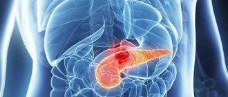
Often, in the final description of an ultrasound examination of the pancreas, many patients can read that there are hyperechoic inclusions in the pancreas. The presence of such a symptom may indicate the development of a serious pathological disorder in the organ under study. In this review, we will take a closer look at what hyperechoic inclusions are and what types of them exist.
The concept of echogenicity
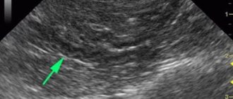
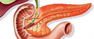
Echogenicity is a term that is used only to describe the ultrasound picture. It refers to the ability of the tissue to which ultrasound is directed (that is, high-frequency sound) to reflect it. The reflected ultrasound is recorded by the same sensor that emits the waves. Based on the difference between these two values, a picture of different shades of gray is constructed, observed on the device’s monitor screen.
Each organ has its own echogenicity index, and it may be homogeneous or not. The following dependence is observed: the denser the organ, the more echogenic it is (displayed as a lighter shade of gray). Liquids do not reflect ultrasound, but transmit it. This is called “echonegativity,” and fluid structures (cysts, hemorrhages) are called anechoic. For the urinary and gall bladder, the cavities of the heart, intestines and stomach, blood vessels, and ventricles of the brain, this “behavior” is the norm.
Thus, we have analyzed what the echogenicity of the pancreas is - this is the ability of this glandular tissue to reflect high-frequency sound emitted by an ultrasound transducer. It is compared with the properties of the liver (they should either be equal, or the pancreas should be a little lighter), and based on the resulting picture, they talk about a change in the echogenicity of the gland. The homogeneity of the organ is also assessed by this indicator.
An increase in the echogenicity of the pancreas is described when there are fewer normal glandular cells in the tissue of the organ (as we remember, fluid reduces echogenicity, and glandular cells are rich in it). Such a change can be observed both locally and diffusely. In addition, some factors may temporarily affect this indicator.
Warning! The description of echogenicity alone is not a diagnosis.
When the echogenicity of the entire gland increases
A diffuse change in the permeability of pancreatic tissue to ultrasound can be a symptom of pathology, but can also be observed normally. This cannot be said about foci with increased echogenicity - this is almost always a pathology.
The echogenicity of the pancreatic parenchyma is increased in the following pathologies:
- Glandular lipomatosis, when glandular tissue is replaced by fat cells containing almost no intracellular fluid; however, the size of the pancreas is not increased. This condition is most often asymptomatic. Read more about this disease in the article: How to recognize and treat pancreatic lipomatosis in time?
- Swelling of the gland that develops with acute pancreatitis. Accompanied by abdominal pain, diarrhea, vomiting.
- Organ tumor. If the ultrasound describes the pancreas with increased echogenicity, then there are necessarily symptoms of the disease: weight loss, pallor, weakness, lack of appetite, frequent bowel movements.
- Pancreatic necrosis, accompanied by the death of organ cells, will also appear on ultrasound as a lighter area. This disease has such signs as severe abdominal pain (up to the development of painful shock), disturbance of the general condition, uncontrollable vomiting, and diarrhea.
- As a result of diabetes mellitus, which is manifested by thirst in the absence of hot conditions, elevated temperature, active work, as well as frequent and abundant (including night) urination.
- Development of connective tissue in the gland (fibrosis) - usually as a result of inflammation or metabolic disorders. In this case, a person may remember cases of unstable stool and abdominal pain. Ultrasound shows not only an increase in echogenicity, but also a decrease in the size of the gland and the tuberosity of its contours.
The hyperechoic pancreas can also be a temporary phenomenon, manifested by:
- as a result of reactive inflammation in many infectious diseases: influenza, pneumonia, meningococcal infection. This requires treatment of the underlying disease;
- when changing the type of food consumed;
- after a lifestyle change;
- at certain times of the year (usually spring and autumn);
- after a recent heavy meal.
In such temporary conditions, the echogenicity of the pancreas is moderately increased, in contrast to pathologies, when significant hyperechogenicity is noted.
Local increase in echogenicity
What are hyperechoic inclusions in the pancreas? It can be:
- pseudocysts - liquid formations that develop as a result of acute pancreatitis; with this disease, the contour of the pancreas becomes uneven, jagged, hyperechoic;
- calcification of tissue areas - calcifications; they are also formed as a result of inflammation (usually chronic);
- areas of adipose tissue; they replace normal gland cells in case of obesity and excessive consumption of fatty foods;
- fibrous areas - where areas of normal cells have been replaced by scar tissue; usually this occurs as a result of pancreatic necrosis;
- stones in the gland ducts;
- fibrocystic degeneration of the gland is either an independent disease or an outcome of chronic pancreatitis;
- metastatic tumors.
Pathologies
Description of echogenicity, what is it and can this concept be attributed to diagnosis? No, echogenicity itself is not considered a diagnosis.
But if ultrasound reveals lesions with increased levels, this is always a consequence of pathological processes.
What kind of illnesses are we talking about:
- Lipomatosis. The disease is represented by the process of replacement of healthy tissues with fat cells. There is practically no intracellular fluid in it. The size of the organ may not change upward. The disease is characterized by asymptomatic manifestations.
- Edema. It is characteristic of the acute form of pancreatitis. The patient begins to vomit, diarrhea occurs, and pain appears in the abdominal area.
- Tumor formations in the organ. When examining an ultrasound scan of the pancreas, you should pay attention to echogenicity. If it is elevated, what is it and what signs may occur? This condition is very serious and requires a detailed examination for oncology. The patient may experience loss of appetite, significant weight loss, bowel movements and unhealthy complexion.
- Pancreatic necrosis. This condition is extremely dangerous because the death of healthy organ tissue occurs at the cellular level. On an ultrasound examination, such a disease will be noticeable by the signs of light areas of tissue. The disease is accompanied by unpleasant symptoms - pain in the abdominal cavity (pain shock may even develop), general condition may be impaired, frequent vomiting and symptoms of diarrhea.
- Diabetes. The disease is accompanied by a number of unpleasant and characteristic symptoms, including severe thirst, even when the conditions are not hot. The patient’s body temperature also rises, urination becomes frequent and abundant.
- Fibrosis. The disease is dangerous because it is replaced by connective tissue instead of healthy epithelium. The causes may be previous diseases in which there were severe inflammatory processes. Another reason may be a metabolic disorder. The patient's stool may change frequently, and pain may appear in the abdominal cavity. In addition to a high level of echogenicity, an ultrasound examination will show a significant reduction in the size of the organ, whose contours will be lumpy.
Hyperechoic inclusions in the organ itself may also occur. How are they expressed:
- Pseudocysts. These formations have nothing to do with malignant ones and are liquid. The cause of their formation may be a previous disease called acute pancreatitis. The disease is characterized by uneven and jagged contours of the organ.
- Calcification of epithelial fragments. They are represented by calcifications. These formations are a consequence of past inflammatory processes. As a rule, of a chronic nature.
- Fragments of adipose tissue. Due to the abuse of fatty foods or overweight or obesity, healthy cells are replaced by fat cells.
- Areas of fibrous tissue. Healthy cells are replaced by scar tissue. Often this process becomes a consequence of a disease called pancreatic necrosis.
- Stones can form in the pancreatic ducts.
- Fibrocystic formations. It may be a separate disease or result from chronic pancreatitis.
- Tumor formations. Moreover, we are talking about metastases that can spread throughout the body.
Treatment of pathological hyperechogenicity
Treatment of conditions where the echogenicity of the pancreas is increased is prescribed only by a gastroenterologist, who must find the cause of this ultrasound symptom:
- if the cause is acute pancreatitis, therapy is carried out with drugs that reduce the production of hydrochloric acid in the stomach and inhibit the enzymatic activity of the pancreas;
- if hyperechogenicity is caused by lipomatosis, a diet with a reduced amount of animal fats in the diet is prescribed;
- if the etiological factor is calcifications, fibrosis or stones in the ducts, a diet is prescribed, and the need for surgical treatment is decided;
- reactive pancreatitis requires treatment of the underlying disease and diet.
Advice! Not a single specialist proceeds from the assumption that it is necessary to treat the tests, and not the person. Increased echogenicity of the pancreas is an ultrasound symptom, not a diagnosis. It requires further examination, and therapy is prescribed only on the basis of subsequent data.
In some cases, ultrasound diagnoses increased echogenicity of the pancreas. What does the diagnosis mean, and should you panic when you see it?
Causes of pathology
What is the heterogeneous structure of the pancreas? This is a symptomatology of pathologies caused by a number of reasons, among which, as a rule, are incorrect lifestyle, poor blood circulation and failure of metabolic processes. In addition, a similar deviation from the norm is observed in patients with diabetes mellitus and chronic pancreatitis. Changes in the functioning of the gallbladder can lead to failure. Also, with age, the structure of the tissues of the internal organs changes in a person, including the formation of a heterogeneous structure of the pancreas. What it is has been described above.
Among the most common causes, scientists still put unhealthy diet in first place, diabetes mellitus in second place, and only in third place are alcohol consumption and smoking. Thus, excessive consumption of fried, fatty, salty and smoked foods is what affects the functioning of the pancreas most negatively.
What is echogenicity
With ultrasound, the doctor uses ultrasound for diagnosis. This means that the rays emitted by a special sensor penetrate the body tissues and are reflected back.
The reflected ultrasound is captured by the same sensor, although it is slightly modified. These changes are displayed on the computer monitor, which processes the readings using a special built-in program.
Echogenicity refers to the ability of body tissue to reflect ultrasound radiation. All organs of the body are different, and due to certain changes they exhibit a heterogeneous structure . If the structure of the organ being examined is homogeneous, then ultrasound passes through the tissue freely and is not reflected, so there will be no echogenicity indicator.
But in organs with a dense structure (and the pancreas is one of them) the waves will be reflected. By how the echogenicity changes, the doctor judges the presence of pathological changes. The examined organ tissue may be partially replaced by fatty tissue, and all these changes will be visible on the monitor as objects with different echogenicity.
The conclusion “isoechoicity of the pancreas” is normal, i.e. the structure is homogeneous. Often there may be mixed echogenicity, and then the tissue is heterogeneous.
If the parenchyma is changed, then this may be a temporary phenomenon. Diffuse changes in the parenchyma are caused by the following factors:
- poor diet;
- changes in a person's appetite;
- season of the year;
- chair and many others.
Norms of echogenicity of the pancreas
It should be remembered that altered echogenicity is not a sign of disease, but a characteristic of the organ. And if certain parameters do not correspond to the norm, this indicates that a painful process is occurring in the body.
So, normally, during ultrasound diagnostics, the echogenicity indicator is homogeneous. There is no hyperplasia, foreign objects, areas of fibrosis or necrosis. A high echogenicity indicator indicates that pathological processes are occurring in the gland.
What is echostructure and echogenicity
Echogenicity and echostructure are decisive in the diagnosis of diseases of the pancreas and other organs and are used to describe the structure of the objects being studied.
Normally, the gland tissue is homogeneous and of equal density. Often the echogenicity of an organ can be determined subjectively, i.e. based on the doctor's experience. This is due to the fact that in medical practice there are different examples of measuring ultrasound characteristics of the tissues in question. Novice specialists can evaluate the structure of the object being studied and its structure based on the scale.
Types of echogenicity:
- anechoicity (it is visualized as a black object);
- hypoechogenicity (the object in question will be dark gray);
- isoechoicity (this is the normal state of the organ, and it will be visible on the monitor as a light gray object);
- hyperechogenicity (the object will be white).
The degree of echogenicity depends on the acoustic properties of the tissue. The less fluid, the higher the echogenicity, and vice versa. Accordingly, anechoic objects are those that do not have fluid. Hyperechoic objects contain the largest amount of fluid.
About the most important thing PANCREAS
Lecture 18a. Large digestive glands. Pancreas and Liver
Based on the nature of the structure, a distinction is made between homogeneity (i.e. objects are colored uniformly) and heterogeneity (the organ in the image is colored non-uniformly).
What you need to know about increased echogenicity
Increased echogenicity in the pancreas indicates pathologies such as chronic pancreatitis and tumors. Local hyperechogenicity indicates that the gland may contain stones, salt accumulation or tumors.
All such patients are referred for additional diagnostic examination.
Causes of hyperechogenicity
Increased echogenicity occurs for the following reasons:
- unbalanced diet;
- unfavorable heredity;
- stress;
- smoking and alcohol abuse;
- pathologies of other gastrointestinal organs;
- incorrect medication use.
What does hyperechogenicity indicate?
A diffuse increase in echogenicity indicates a tumor or pancreatitis. With tumors, the following symptoms attract attention:
- digestive disorders;
- bowel dysfunction (most often diarrhea);
- flatulence;
- loss of weight and sometimes appetite;
- general weakness.
With pancreatitis, enzymes do not digest food, as is normal, but the parenchyma. Toxins are released that enter the bloodstream, poisoning the liver, kidneys, and brain. Acute pancreatitis is considered the most dangerous.
The pathology is characterized by sharp pain in the hypochondrium, nausea, and vomiting. Blue spots sometimes appear on the stomach.
Acute pancreatitis is dangerous with the risk of death, so the patient needs urgent surgical intervention. The following signs are visible on ultrasound:
- increase in organ size;
- unclear contours and structure;
- dilation of the ducts;
- accumulation of fluid around the organ;
- lack of echogenicity in some areas (this indicates tissue death).
Diffuse changes are also observed with lipomatosis. Lipomatosis is a condition where organ tissue is replaced. This happens, for example, if the patient has diabetes. The size of the organ in diabetes remains unchanged, and echogenicity changes slightly.
Are the figures definitive?
No, moderate or even high levels of change are not permanent. The echogenicity of the organ in question can change under a variety of conditions. Often a pathological indicator appears due to poor nutrition. It’s worth correcting it, and the next study will show the norm.
That is why doctors do not focus on the results of only one ultrasound, but prescribe additional ones for the patient. A person with a history of pancreatic pathology should be constantly monitored by a therapist.
What changes occur in the pancreas
As already noted, various types of abnormalities on ultrasound indicate that pathological processes are occurring in the gland. With diffuse changes, the organ may increase or decrease.
Tissues can become denser, their structure becomes heterogeneous. Often the contours of the pancreas become unclear. Decoding the diagnostic results describes in detail all such phenomena.
This is what happens in the gland in the presence of certain pathologies:
- In acute pancreatitis, the pressure in the duct increases. The organ tissues are destroyed and the body is poisoned. Such processes signal themselves with terrible pain.
- In the first stages of chronic pancreatitis, the gland is swollen. Then it decreases and becomes sclerotized.
- With fibrosis, some areas of the organ are replaced by connective tissue.
- Replacement of organ areas with adipose tissue is an irreversible process. With a massive process, the pancreatic parenchyma is compressed.
- In pancreatitis or diabetes, ultrasound shows various signs of changes in the parenchyma, hyperechoic areas are noted in it.
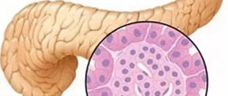
- Structural changes affect the parenchyma, since it consists of many glands.
- The formation of cysts and tumors is possible.
- Reactive changes indicate that the patient has problems with the liver and gall bladder.
- Finally, due to cell death, ultrasound shows fatty degeneration.
Treatment at home
In addition to medications prescribed by a doctor, you can take dietary supplements that heal the gastrointestinal tract and generally have a beneficial effect on the digestive processes. If pain occurs with a hyperechoic heterogeneous structure of the pancreas, you can take drugs such as Analgin and Papaverine. It is best to use an injection solution rather than tablets. For spasms they use Tavegil and Diphenhydramine. In acute pancreatitis, injections are given with Paracetamol or Baralgin. It is also recommended to use folk remedies.
Classification of hyperechoic inclusions
The following types of hyperechoic inclusions in the pancreas are distinguished:
- Pseudocysts (these are liquid formations that appear due to acute pancreatitis). The outline of the fabric becomes unclear.
- Calcifications, or calcified objects. Appear if a person has suffered a chronic disease of the organ in question (most often pancreatitis).
- Fatty objects replace normal areas. This occurs if a person eats too much fatty food.
- Fibrosis, in which normal areas of tissue are replaced by scars. It is diagnosed after pancreatic necrosis.
- Stones can accumulate in the ducts of the organ.
- Fibrocystic degeneration is usually the result of chronic inflammation of the gland.
- Metastases to the pancreas.
If the diagnosis shows questionable results, the patient is sent for additional tests. This is the only way to make an accurate diagnosis.
Diagnoses associated with increased echogenicity
There are many reasons that cause increased echogenicity. This may be local inflammation or a more serious pathology associated with malignant neoplasms. Often this deviation is temporary, being a consequence of poor nutrition or a history of acute respiratory viral infection. The cause of hyperechogenicity of the gland can also be age-related changes, when the process of replacing working cells with fibrous cells occurs. In this case, patients are recommended maintenance therapy.
Changes in the form of hyperechoic inclusions in the pancreas can be a consequence of a number of pathological and physiological changes. Pathologies include:
- lipomatosis (replacement of healthy cells with fat tissue);
- acute inflammation, accompanied by swelling and enlargement of the gland;
- pancreatic necrosis – death of organ cells and its destruction;
- diabetes due to gland dysfunction;
- oncology (malignant neoplasms);
- fibrosis (uncontrolled growth of connective tissue);
- cyst;
- stones.
The etiology of a physiological nature is determined by factors such as:
- systematic overeating;
- sudden changes in the food system;
- seasonal changes;
- infections;
- lifestyle change.
These reasons cause a slight increase in the echogenicity index, while pathologies affect it to a significant extent.
Carefully! Inattention to pancreatic problems, lack of timely support and treatment, can lead to such a complex disease as pancreatic necrosis. It is characterized by the death of organ cells, severe pain, vomiting, diarrhea and, as a rule, complex surgical intervention is required.
How is diagnostics carried out?
Before the examination, patient preparation is necessary. It is carried out on an empty stomach, the last meal should be approximately 12 hours before the ultrasound. For a few days, you need to eliminate foods that lead to the formation of gas from your diet.
On the day of the procedure, the patient is prohibited from smoking, drinking alcohol or taking medications..
The examination itself is painless and takes up to 20 minutes. The subject lies on the couch on his back, then turns over onto his right and left sides. A harmless gel is applied to the stomach. If you have a tendency to flatulence, then you need to take several sorbent tablets.
After all procedures are completed, the doctor analyzes the information received and makes a diagnosis. Ultrasound is completely harmless to the patient and can be performed as many times as needed.
How is pathological hyperechogenicity of the pancreas treated?
Treatment of all conditions associated with hyperechogenicity is prescribed only by a doctor.
Therapy depends on the cause of hyperechogenicity:
- In acute pancreatitis, drugs are prescribed that reduce the production of hydrochloric acid in the gastric mucosa. Agents that can reduce pancreatic enzyme activity are also needed. It must be remembered that therapy for an acute disease is carried out in the surgical department.
- For lipomatosis, a diet low in fat, especially of animal origin, is indicated.
- In the presence of calcifications and areas with fibrosis, along with the prescription of a diet, the issue of surgical intervention is decided.
- With reactive pancreatitis, proper nutrition and therapy for the underlying disease are necessary.
- Exacerbations of chronic pancreatitis are treated in a hospital setting. Intravenous injections and infusions are indicated.
- Treatment of carcinoma is carried out surgically; often the patient may require chemotherapy.
Proper nutrition plays a vital role in reducing pancreatic hyperechogenicity. The patient should give up fried, smoked, and salty foods.
Alcohol and smoking are strictly prohibited. It is also important to limit your consumption of sweets.
It should be remembered that increased hyperechogenicity is not a disease, but a characteristic of the organ. Based on the results of the ultrasound, appropriate treatment is prescribed. It is possible that the patient may require additional instrumental examinations and tests.
After diagnosing the pancreas using ultrasound, an entry will appear in the medical card “increased echogenicity of the pancreas organ,” what this means for the patient and what consequences it will bring to the body. The increased echogenic effect of the organ indicates pathological changes in the gland, so approach this diagnosis responsibly and seriously.
Treatment of heterogeneous structure of the pancreas
Treatment will directly depend on the diagnosis. In the presence of acute pancreatitis, the specialist prescribes a strict diet (for several days). It is necessary to give up alcoholic beverages.
As prescribed by a doctor, medications are used to suppress the gag reflex. These are Motilak or Cerucal tablets. To reduce the severity of pain, it is necessary to take antispasmodics. In severe cases, surgical treatment of inflammatory pancreatic disease may be indicated.
Replacement therapy
For pancreatitis and other pathologies of the pancreas, accompanied by changes in the structure of the organ, replacement therapy is indicated. It consists of prescribing enzyme preparations to the patient.
For pancreatitis and other pathologies of the pancreas, accompanied by changes in the structure of the organ, replacement therapy is indicated. It consists in the fact that the patient is prescribed enzyme preparations that compensate for the lack of digestive enzymes. The most commonly used are Pancreatin or Creon. They must be taken for a long time.
Replacement therapy can also be understood as the administration of insulin for insulin-dependent diabetes. Such injections are recommended throughout life.
Diet
In the first days after an attack of acute pancreatitis, therapeutic fasting is prescribed. Then the food expands a little. In the future, if the structure is heterogeneous, diet No. 5 is indicated.
The basis of this diet is that all foods that lead to increased acid formation are completely removed from the menu. It is necessary to choose recipes that do not use spices. Alcohol is strictly contraindicated for the patient throughout his life.
Alcohol is strictly contraindicated for the patient throughout his life.
The concept of echogenicity
What does the concept of “echogenicity of the pancreas” mean, which doctors talk about after conducting research? The concept of increased or decreased echogenic factor is widely used when describing the condition of the pancreas after an ultrasound examination of the organ. The echogenicity of the pancreas indicates the physical ability of the digestive organ to reflect ultrasound signals sent by the ultrasound machine.
There are a number of standard notations and tables, according to which each organ in the human body, in the absence of pathologies, reflects the signal only in its individual form. Therefore, a change in the data of the received reflection signals indicates a serious problem with the gland, which serves as a kind of warning. So, what is an increased or decreased echogenic factor and how is it determined by ultrasound equipment?
When diagnosing the body using ultrasound, the signal is reflected from the internal organs according to the principle of returning the frequency and strength of the impulse from the device to the organ and back. Receiving an echo-like return of the impulse, a pattern is displayed on the monitor, from which the doctor determines the increased or decreased signal and the condition of the pancreas, as well as possible neoplasms. And also, using the data obtained, the compactions that have arisen are determined, which makes it possible to identify foci of pathology in certain areas and the cause of their occurrence. What is visible on the monitor of an ultrasound machine when diagnosing a patient?
The signal displayed on the screen of the ultrasound machine is a gray scale, which has its own shades. The shade obtained from the diagnosis indicates whether the ultrasound pulse return signal is increased or decreased. Based on this fact, a possible pathology of the pancreas or its condition is determined. There are also extraneous factors that affect the accuracy of receiving the return signal:
- mobile phone;
- implant in the human heart;
- electrical equipment and items, and much more.
When the echogenicity of the gland increases
What does it mean for a person when the echogenicity in the pancreas is significantly increased when indicated on an ultrasound device and what are the causes of the pathology? Diffuse changes are clearly visible during ultrasound diagnostics and indicate a pathological change in the pancreatic organ. True, these indicators are also found in normal conditions. Therefore, attention is paid to individual areas of the organ being tested, and only by the method of exclusion is one or another conclusion made about the occurrence of the pathology of the disease. What abnormalities and pathologies are associated with changes in the echogenicity of the pancreas:
- The appearance of lipomatosis of the gland. When diagnosed and the increased echogenic state of the pancreas, the glandular tissue layer is replaced by fatty contents. The process occurs without obvious symptoms, which is why the pathology is detected at random.
- An increased echogenic state indicates swelling of the gland organ. With this diagnosis, an acute form of pancreatitis usually occurs, severe pain in the peritoneum and, as a sign, the appearance of diarrhea and vomiting.
- An increase in the return signal data may also indicate the emergence of foci of neoplasms. Pathology with increased echo conductivity causes the following symptoms:
- pale skin;
- sudden weight loss;
- diarrhea;
- lack of appetite.
- Increased data on the echogenic state of the pancreas will indicate the occurrence of pancreatic necrosis. On the ultrasound machine display, the lesion area is shown in a light color, unlike other areas of the pancreas. When diagnosed, necrosis of the organ cells occurs, and with severe pathology, peritonitis occurs with the appearance of severe symptoms:
- increased temperature;
- the appearance of a painful condition with possible painful shock;
- vomit;
- diarrhea;
- intoxication of the body.
- An increased signal return rate also occurs in diabetes mellitus. Obvious signs of the disease are:
- thirst;
- frequent urination;
- increased heart rate;
- joint weakness.
- An increased return signal leads to the appearance of fibrosis of pancreatic tissue. Symptoms of the disease: diarrhea or abnormal stool, abdominal pain.
With this development, healthy tissue is replaced by a pathogenic or connective form of tissue. At the same time, the visualization contours of the pancreas organ change.
Hyperechogenicity of the pancreas occurs as a temporary phenomenon. What causes this manifestation:
- pneumonia;
- acute respiratory infections;
- ARVI;
- flu;
- meningitis;
- other bacterial or viral infections that affect the patient's body.
To eliminate the problem, the cause that caused the increased echogenic effect is treated, after which the readings of the ultrasound machine and the patient’s condition return to normal.
What can cause heterogeneity in the echostructure of the pancreas?
Increased echogenicity of the pancreatic structure is observed in diabetes.
Increased echogenicity of the pancreatic structure is observed in pathological conditions:
- Lipomatosis. This means that part of the organ is replaced by fatty tissue. The size of the pancreas is normal. In this state, a person practically does not feel any symptoms.
- Edema that develops with acute tissue inflammation. This state of the structure is always accompanied by severe pain, diarrhea, and vomiting.
- Tumors. A person experiences symptoms such as severe weight loss, weakness, lack of appetite, and stool disorders. The same signs occur when the organ is filled with cysts.
- Pancreatic necrosis is a disease accompanied by destruction and death of pancreatic tissue. On ultrasound, areas of pancreatic necrosis are visualized as having a hyperechoic structure. With pancreatic necrosis, pain in the abdominal region is severe, and patients often develop pain shock. In such cases, vomiting can be uncontrollable, and the person experiences severe diarrhea.
- Diabetes. If the causes of this disease lie in an autoimmune change in the organ, then it almost always decreases in size.
- Fibrosis is the formation of connective tissue. The structure of such tissues will be uneven. The condition develops due to inflammatory phenomena.
The disturbance in the echogenicity of the structure can also be temporary. It manifests itself due to the flu.
The disturbance in the echogenicity of the structure can also be temporary. It appears due to:
- reactive inflammation;
- a significant number of infectious pathologies - influenza, pneumonia, meningococcal infection;
- diet changes;
- sudden change in the patient’s lifestyle;
- development of gastritis, cholecystitis and other pathologies of the digestive tract;
- a hearty lunch.
A temporary increase in echogenicity is also observed with the change of seasons - spring, autumn.
Local increase in echogenicity
In some cases, local echogenicity of the pancreas occurs when diagnosing pathology. In this case, the pathology of the organ will be identified in the place where adipose tissue has replaced glandular tissue. This fact includes the following emerging pathologies:
- The formation of cysts that appeared as a result of pancreatitis. With a pseudocyst, the gland organ changes its contours and the edges become uneven and jagged. In this regard, an increased echogenicity effect appears.
- An increased moment of echogenicity will also indicate the occurrence of growth pathology - calcifications that appear as a result of chronic forms of pancreatitis.
- If the body is obese, it also shows an increased return of the ultrasound signal. This situation is caused by excessive consumption of fatty foods.
- Neoplasms – metastases.
The occurrence of any of the listed pathologies requires urgent intervention and treatment, as they will lead to serious complications and mortal danger for the affected organism.
Reduced echogenicity during ultrasound examination of the gland is observed with diffuse changes in the pancreas. This state is displayed on the monitor as a dark heel or a black screen of the device. Diffuse changes affect the entire organ and are visible evenly without sudden changes in color. What causes a decrease in the return of the ultrasonic signal:
- chronic or acute forms of pancreatitis;
- focal acute attack of the inflammatory process of the pancreas;
- neoplasms on the gland.
The pancreas, with increased echogenicity or weak signal reception, is a clear sign of pathology and requires urgent intervention by gastroenterologists.
Increasing the indicator
Increased echogenicity on ultrasound
For a doctor, a diffuse increase in the echogenicity of the pancreas is a sign of many pathologies, some of which are very serious. The reason for this conclusion is a significant increase in indicators.
Hyperechogenicity of the pancreas can be caused by other reasons:
- inflammation of tissues caused by colds;
- recent overeating;
- change of lifestyle;
- off-season;
- changing nutrition priorities.
Here, hyperechogenicity manifests itself moderately, the echostructure differs slightly from the normal value.
If the echogenicity of the pancreatic parenchyma is increased, you will have to undergo an additional examination, which will be prescribed by the attending physician, and then treat the diagnosed disease.
Treatment of pathological hyperechogenicity
Therapy for hyperechogenicity requires individual treatment by a gastroenterologist and depends on the cause. The reasons for this diagnosis are:
Severe acute pancreatitis. For treatment, agents are used that reduce the production of pancreatic juice and enzymes.
Lipomatosis. Prescribing a strict dietary norm. Complete refusal to consume fats of non-vegetable origin.
When calcifications, fibroformation, stones appear in the pancreatic fluid ducts and bile ducts, the patient adheres to diet No. 5p. With further diagnosis, the doctor makes a decision on surgical interventions.
Reactive pancreatitis. The cause of its appearance is identified and by methods of drug treatment or surgical elimination of the underlying disease that caused its manifestation, complete therapy of the disease occurs. During this period, a strict dietary diet is observed.
The pancreas and its increased echogenicity are clear signs of the occurrence of pathological changes in the gland organ. For accurate and correct treatment, diagnostic measures are required to identify the cause of this condition.
Treatment of hyperechogenicity of the pancreas
If hyperechogenicity is detected, it is necessary to determine the cause of the pathology. For this purpose, the attending physician collects anamnesis and prescribes additional studies. Blood is donated for a clinical analysis, the results of which determine whether there are foci of inflammation in the body (ESR indicator), and the sugar content is checked (the information obtained determines the presence of diabetes).
More accurate data on localizing areas of increased echo density are obtained using a puncture, the biochemistry of which determines the malignancy of the neoplasm.
If the deviations do not have serious grounds, then the condition of the gland is normalized by following a diet and giving up bad habits. During the recovery period, you need to avoid stress and hypothermia. If the examination reveals severe pathological disorders, the doctor is obliged to prescribe an appropriate course of treatment aimed at eliminating the root cause of the disease.