To accurately identify pathology, it is necessary to know the normal size of the pancreas in adults.
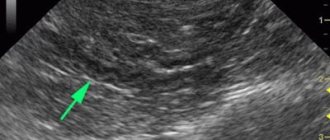
The topographic location of the pancreas (PG) does not make it possible to palpate it during an objective examination or determine its condition and size. Therefore, for the purpose of visualization and diagnosis, the most accessible method is used - ultrasound examination. Ultrasound allows you to see the organ in a three-dimensional image, determine the clarity of the boundaries, the structure and echogenicity of the tissue, pathological formations, their size and location, and the expansion of the common duct. Knowing the normal size variations of the pancreas according to ultrasound, the method can be used to clarify an unclear diagnosis.
What affects the size of the pancreas?
The size of the pancreas changes throughout life: it grows until about 18 years of age. Then it decreases from age 55, when functioning cells gradually atrophy. This is a physiological change in size. Variants of the norm include an increase in the pancreas in women during pregnancy.
The decrease in pancreas occurs:
- with age (after 55 years) with the development of tissue atrophy,
- in case of circulatory disorders in the organ,
- for viral infections.
Diffuse or local increase occurs in some pathological conditions.
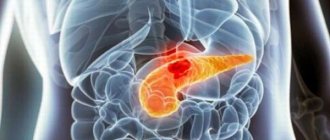
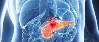
A local increase in size is observed in cases of benign or malignant neoplasm, simple cyst, pseudocyst, abscess, and calculi. Deviations from normal parameters can be significant: clinical cases of pseudocysts reaching 40 cm have been described.
In chronic pancreatitis in the stage of stable remission, the pancreas does not change its size. To verify the diagnosis, data on the condition of the Wirsung duct are used.
Diffuse enlargement of the pancreas is observed with lipomatosis, when normal cells in the pancreas parenchyma are replaced by fat cells. Ultrasound displays a heterogeneous sonographic picture; inclusions of fat can increase the echogenicity of the tissue being examined.
The size of the pancreas is changed by swelling during its acute inflammation - in most cases, the entire organ enlarges. This appears not only with inflammation in the gland itself, but also with pathology of neighboring organs: the stomach, duodenum, and gall bladder. Only at the initial stages does local swelling of a separate part of the pancreas occur: the head, body or tail. Subsequently, it completely captures the entire gland.
An increase in the pancreas with a tumor depends on the location, type and aggression of the pathological neoplasm. In 60%, pancreatic head cancer is detected: it is significantly larger than normal - more than 35 mm. In 10%, a malignant neoplasm of the body of the pancreas is diagnosed. In these cases, the size of the middle part of the organ increases.
Decoding indicators
After the diagnosis, the doctor writes a conclusion. The transcript of the study includes three parameters - acoustic density, size and condition of the central duct. It makes it possible, together with the results of laboratory tests, to make a diagnosis.
Possible diseases
Some diagnostic data may indicate diseases. A decrease in echogenicity means the acute stage of pancreatitis. The pancreas swells and the image becomes dim. A completely white gland on the monitor is a sign of an acute form of pancreatitis.
Tumors may not be visible on ultrasound; their presence is indicated by deviation of the tail of the organ. Echogenicity in malignant tumors or chronic pancreatitis is increased. You can see a change in color in some areas of the organ where neoplasms are possible.
A tumor is indicated by changes in the size of the liver and gallbladder. Taking material for histology helps determine whether a neoplasm is malignant or benign.
With pancreatic necrosis, the image shows extensive abscesses forming cavities with cloudy exudate. Inflammation of the pancreas is indicated by dilation of the Wirsung duct. The doctor visualizes stones and pancreatic abscesses.
Serious diseases of the pancreas can be asymptomatic at the initial stage and are detected as a result of a routine examination using ultrasound diagnostics. The interpretation of the results for each type of pancreatic pathology is individual.
Sizes of the gland before and after food loading
An additional examination method for pancreatitis is ultrasound with food load. Sonography is done twice: in the morning on an empty stomach and 2 hours after eating. Each time the transverse dimensions of the head, body and tail of the pancreas are measured. The increase in the sum of indicators after a physiological breakfast to the initial data is calculated. Based on it, conclusions are drawn about the condition of the organ. With enlarged pancreas:
- more than 16% is the norm,
- 6-15% - reactive pancreatitis,
- more or less than the initial data by 5% - chronic pancreatitis.
All conclusions are drawn based on a comparison of the obtained sizes with the data of normal indicators in a special table. The method allows you to prescribe adequate therapy when pathology is detected and control the process of tissue regeneration and restoration of pancreatic function.
Diet and prevention
Treatment of pancreas is impossible without following a diet. In case of severe pathology, when the functions of the pancreas sharply decrease, dietary table No. 5 according to Pevzner is prescribed. Special nutrition should reduce the load on the pancreas, so fatty, spicy, fried, pickled, salty foods are excluded. Alcohol is excluded. The basis of the diet should be cereals and lean meat. Meals should be frequent and small.
Prevention is a healthy lifestyle, proper nutrition, giving up alcohol, smoking, timely consultation with a doctor, and following all recommendations. If these conditions are met, you can maintain health and a high quality of life for many years.
Bibliography
- Parsons T. Anatomy and physiology. Handbook edited by K.S. Artyukhina. M. AST: Astrel 2003
- Sviridov A.I. Human anatomy. Textbook. K. Higher school 2001
- Fedyukovich N.I. Human anatomy and physiology: textbook. 2nd edition. Rostov-on-Don Phoenix 2002
- Maksimenkov A.N. Surgical anatomy of the abdomen. L. Medicine 1972
- Loit A.A., Zvonarev E.G. Pancreas: connection between anatomy, physiology and pathology. Clinical anatomy. No. 3 2013
Pathological deviations from the normal size of the gland
An increase in the size of the pancreas is associated with the emerging pathology and occurs gradually, in many cases asymptomatically. Since there are often no clinical manifestations, the patient is not aware of the problem until the first examination. When performing sonography, the increased size of the organ is determined and any additional formations are identified.
The following reasons lead to pathological growth of the gland:
- cystic fibrosis is a hereditary disease characterized by a thick form of pancreatic secretion produced,
- alcohol abuse (more often in men),
- inflammation in the tissues of the pancreas or in diseases of adjacent organs (stomach ulcer),
- infectious diseases,
- improper and irregular nutrition, non-compliance with the prescribed diet,
- various formations in the tissues of the pancreas,
- high level of calcium in the body, stone formation,
- long-term and unnecessary use of medications,
- inflammatory and congestive processes in neighboring organs,
- vascular diseases,
- injuries,
- diseases that reduce immunity.
What is the pathological growth of the gland associated with?
There are two main reasons why the size of the pancreas increases:
- an inflammatory process that provokes swelling;
- a malfunction that the gland tries to compensate for with its size.
Most often, an organ that is too large indicates problems such as:
- acute or chronic pancreatitis (it leads to an enlargement of one section, and extremely rarely, of the entire organ);
- blockage of the excretory duct by a gallstone, which leads to its obstruction;
- hepatitis, influenza, intestinal and other infections;
- cystic fibrosis (this is a congenital pathology in which all fluids in the body have a thicker consistency than usual);
- abscess in the head area;
- cystic adenoma;
- malignant or benign neoplasm in the gland or duodenum. With cancer, individual segments enlarge, the contours become uneven, and the aorta may shift;
- pancreatic necrosis (purulent processes in the pancreas, as a result of which toxins enter the patient’s blood and poison the body);
- lipomatosis (proliferation of fat cells in the tissues of the gland);
- duodenitis.
In addition, this organ increases in size if a person drinks heavily, has an abdominal injury (for example, hit the steering wheel in an accident), has an autoimmune disease, or takes certain medications. The gland may be larger than usual for reasons related to genetics, that is, anomalies in its development may be present initially (then it has the shape of a horseshoe).
An enlarged gland affects neighboring organs, so they are also examined. The head of the pancreas can compress the duodenum and contribute to the development of such a dangerous condition as intestinal obstruction.
Interpretation of ultrasound of the pancreas
Due to the impossibility of palpation of the pancreas, ultrasound is the only way to quickly clarify the diagnosis. The results are deciphered according to a certain scheme. It includes the following information:
- location,
- form,
- echogenicity,
- contours,
- sizes,
- structural defects or neoplasms.
The condition and size of the Wirsung duct must be indicated. According to these standards, a functionalist doctor objectively describes the picture of the pancreas. Decoding and analysis of the data obtained, verification of the diagnosis, as well as the prescription of treatment measures are carried out by the specialist who prescribed the ultrasound: gastroenterologist, therapist, surgeon or oncologist.
Sonography is based on the ability of the tissue being examined to absorb and reflect ultrasound waves (echogenicity). Liquid media conduct ultrasound, but do not reflect it - they are anechoic (for example, cysts). Dense parenchymal organs (liver, kidneys, pancreas, heart), as well as stones and tumors with high density, do not absorb, but reflect sound waves; they are echo-positive. And also normally, these organs have a homogeneous (homogeneous) granular structure. Therefore, any pathological formation appears on the ultrasound image as an area with altered echogenicity - increased or decreased.
Normal sizes of the pancreas: table
To clarify the pathology of the pancreas, all information obtained during a sonographic examination is compared with the standard indicators of a special table. If there is a significant discrepancy between the indicators, conclusions are drawn about the presence of the suspected disease.
DEPARTMENT DIMENSIONS Head 11–35 mm Body 4–30 mm Tail 7–30 mm
In adults, the normal indicators of the entire organ are expressed by the following established figures:
- length: 14−23 cm,
- width: 9 cm,
- thickness: 3 cm.
The dimensions of the pancreas, like any organ under study, are measured in millimeters - this indicates the high accuracy of the technique. Although the formations that are detected can be up to several tens of centimeters.
A slight increase in the echogenicity of the pancreas with age or the small size of a certain part of it in people over 55 years of age is not considered pathological. Deviation of the obtained indicators in other age categories indicates a hereditary pathology or acquired disease.
Gland shape
The pancreas occupies a horizontal position. Its form varies, there are no precisely defined standards. For most people, it is S-shaped or resembles a comma, but it happens:
- evenly thickened along the entire length,
- dumbbell-shaped.
With pathological damage, the shape of the pancreas changes. Occurs:
- in the form of a spiral or ring,
- horseshoe-shaped
- aberrant (additional),
- with doubling of individual parts.
Most often, deviations and anomalies are discovered by chance when the patient is examined in connection with the pathology of other organs. Some defects are not clinically significant for health and life, while some may worsen and worsen the general condition over time.
The shape anomaly is damage directly to the pancreas itself or a consequence of a severe general disease. Ultrasound does not always reflect all the details: conclusions are made on the basis of indirect data (areas of narrowing of the Wirsung duct are detected or an additional duct formed is detected). In such cases, it is recommended to undergo additional examinations to exclude a congenital anomaly, tumor or other focal changes.
Topography
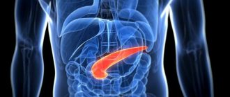
The pancreas is located horizontally, behind the stomach, almost on the posterior wall of the abdomen, 5-10 cm above the navel with a conditional projection on the anterior abdominal wall. The largest part (2/3 of the gland) is located to the left of the midline.
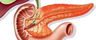
The head of the pancreas is covered by the duodenum in the form of a horseshoe, and borders the stomach on top.
The body is located behind the stomach, separated from it by the omental bursa, on the right in contact with the liver, below with the small and partially with the large intestine, behind it is adjacent the left kidney with the adrenal gland, the aorta, and the celiac plexus. The splenic vein runs along the body of the gland.
The tail is curved upward, in contact with the spleen and colon.
Wirsung duct
The Wirsung duct stretches along the entire length of the gland, connects with the bile duct, and exits into the small intestine.
With sonography, the common duct is clearly visible. The diameter is the same along the entire length - 2 mm. Dilation up to 3 mm is considered a sign of pancreatitis. In diagnosis, in addition to size, the clarity and evenness of contours, bends, narrowing and widening, and the presence of stones or other formations play a role. As a result, this also affects the size of the pancreas.
During acute inflammatory processes in the tissues of the gland, changes in the thickness of the walls and lumen of the duct also occur, but these parameters are not as pronounced as in chronic ones.
The dimensions of the common duct change when additional formations are detected: tumors, stones, cysts. If it is compressed from the outside by growing foreign tissue, this leads to narrowing in a certain area and expansion before and after the site of pathology. With an advanced process, the size of the gland increases. If obstruction by a stone occurs, its patency is also impaired, its dimensions change, including the enlargement of the gland itself. In addition to expansion or contraction, there is:
- doubling,
- split,
- deformation.
Visualization on ultrasound in different sections
For a correct sonographic examination, the glands are oriented towards large vessels that pass next to the pancreas (when viewed in a cross section) and adjacent organs adjacent to it (in a longitudinal section). Significant landmarks are the aorta and splenic vein. For this purpose, the patient is examined in various positions: on the back and right side, directing the sensor at different angles to the surface of the body.
Do you need to do such an ultrasound?
If nothing bothers you, then you shouldn’t worry about your pancreas. But if pain periodically or constantly appears in the epigastric zone or pain occurs when pressing here, and especially if the doctor finds formations during palpation, then it is advisable to conduct an ultrasound examination of this organ. This will confirm (or refute) the suspicion of complications of pancreatitis, abscess, tumors, stones, cysts and hematomas. Such diagnostics are also necessary for those diagnosed with diabetes.
During an ultrasound scan, the patient simply lies on his back, and the diagnostician uses ultrasound waves to determine the “dimensions” of the pancreas and all its parts. Of course, you can undergo an MRI, but this is a less accessible and more expensive method that not every medical institution can offer.
Although the size of this gland can change throughout life, a significant increase in even one segment, as a rule, indicates pathology. Thus, if the size of the pancreas corresponds to the norm in adults, then it copes with its duties perfectly. If deviations from normal sizes are found, then look for the cause and immediately begin treatment, because this is a vital organ.
Description of a healthy pancreas in the ultrasound protocol
The description of the pancreas of a healthy person when examined using ultrasound looks something like this: the size of the head is up to 32 mm, the body is up to 25 mm, and the tail is up to 30 mm. Diameter of the Wirsung duct: 1.5-2 mm. The contours are smooth and clear. The echogenicity is normal, the structure is homogeneous, similar to the echostructure of the liver. No additional formations were identified.
When assessing the condition of the pancreas, the following is taken into account:
- echogenicity – compared with the tissue of the unchanged liver, it increases with age,
- structural structure of the pancreas tissue: normally – fine- or coarse-grained, but at the same time homogeneous (homogeneous),
- correctness of the vascular pattern - there should be no deformations.
Size
Diagnostic ultrasound also involves measuring the parameters of the pancreas. For an adult, the size norm varies from 14 to 22 centimeters. This pancreas weighs about 75 grams. The organ consists of a head, where the uncinate process is located (the length of such a process is approximately three centimeters), a body (the length ranges from 1.5 to 1.7 centimeters) and a tail (the size is about two centimeters).
The head is surrounded by the duodenum. Localization is the level of the first and the beginning of the second lumbar vertebra. The pancreatic duct has a smooth, smooth wall. Its diameter does not exceed one millimeter in the body and two millimeters in the head. Fluctuations in parameters can occur either downward or upward.
Each type of pathological process has a different picture, which is visible during ultrasound examination. The inflammatory process, which is accompanied by swelling, is characterized by an increase in the area from head to tail. If a tumor is present, individual affected parts will enlarge. For example, in more than 55 percent of cases, a malignant tumor is located in the head of the pancreas. In this case, the head of the organ will increase by more than 4 centimeters. About 15 percent of cases are diagnosed with pancreatic cancer. The body of the organ will be enlarged.
Preparation
Preliminary preparation for ultrasound of the pancreas and liver is not required, but the examination is easier if the patient comes on an empty stomach. It is recommended to refrain from eating for 9-12 hours before the test.
In approximately 30% of cases, the examination is difficult due to flatulence, so it is recommended to keep your diet under control and exclude vegetables, fruits, brown bread, dairy products, and beans from your diet a couple of days before visiting the doctor. You can use a decoction of dill or mint seeds and drugs that reduce gas formation. It is advisable to prepare and empty your bowels before the examination or the day before; you should not resort to the use of enemas or laxatives before an ultrasound scan of the pancreas.
If the patient is specifically aimed at examining the Wirsung duct, then he should come for it after breakfast.
Indications for ultrasound
Under the influence of many factors (poor lifestyle, smoking, constant stress), the work and functions of the pancreas can be impaired. When this happens, the person begins to experience severe pain, attacks of nausea and vomiting. Since these symptoms are characteristic of many diseases of the digestive system and gastrointestinal tract, patients are prescribed an ultrasound of the pancreas and abdominal organs.
The main indications for ultrasound of the pancreas are:
- pain in the upper left hypochondrium and left side;
- painful sensations during palpation of the abdomen;
- gastric dysfunction detected during gastroscopy;
- constant attacks of nausea and vomiting;
- pathologies and liver diseases;
- digestive and stool disorders;
- abdominal trauma;
- suspected diabetes mellitus or pancreatitis;
- laboratory tests indicating organ diseases;
- jaundice.
Ultrasound is the simplest and most accessible method of examining the pancreas