It is difficult to find an analogue of the pancreas in the human body. The organ performs both an exocrine (digestive) function and is an essential part of the endocrine system. Therefore, a neoplasm on the pancreas has a serious impact on the digestive processes (lack of enzymes) and causes disruption of all types of metabolism through a primary disruption of the production of the hormone insulin.
Disappointing statistics
Of all tumors, up to 95% are cancer. According to statistical studies of Russian oncologists, among the general incidence of malignant pathology, the pancreas is in tenth place in terms of damage in men, and in thirteenth place in women.
Over the past 10 years in the Russian Federation, the annual increase in cases of pancreatic cancer was 3.5% among the male population and 15.6% among the female population. Mortality remains stubbornly at the same level and ranks fifth among both sexes.
Comparing the prevalence of the disease in countries around the world is incorrect, since the level of healthcare development is not the same. A significant number of cases are under-detected.
Diagnosis of volumetric processes in the pancreatic zone is difficult. Here, a person has several organs (stomach, duodenum, gallbladder and ducts) that are anatomically and functionally tightly connected. It is practically impossible to detect a formation emanating from the pancreas at an early stage.
Benign neoplasms
These tumors are characterized by a lack of tendency to grow and metastasize. Their cells produce hormones that enter the general bloodstream. Depending on the number of tumors, these tumors can be multiple or single. Do not forget that among benign formations there are: hemangioma (the structure resembles blood vessels), fibroma (from connective tissue), lipoma (from adipose tissue), leiomyoma (formation from muscle fibers), schwannoma (consists of Schwann cells located in the membranes ) and neuroma (from nervous tissue).
Doctors identify the following main reasons:
- heredity;
- smoking;
- drinking alcohol in large quantities;
- environmental problems;
- inflammation of the pancreas;
- excessive consumption of fatty foods.
What can lead to the development of the disease?
The reasons for the development of tumor formation in the gland have not been definitively established. It is common to talk about risk factors for the disease and the likely impact on cell degeneration. We present the most significant and studied ones.
Smoking tobacco products is associated with tumor growth in the pancreas in 20–30% of patients. Polycyclic hydrocarbons of nicotine have a negative effect. The role of poison is confirmed for every fourth patient. The risk increases as the length of the smoker's “experience” increases.
Dietary features - many scientists believe that the risk of pancreatic tumors is greater in people who eat predominantly fatty meat products. The passion for coffee and the deficiency of vegetables and fruits (plant fiber) are important. However, not everyone agrees with this hypothesis.
Obesity - the gland may not be able to withstand the load of excessive intake of carbohydrates and fats from food. With weight gain associated with hypothalamic-pituitary pathology, all types of metabolism suffer.
Age - the maximum prevalence of pancreatic neoplasia is recorded in people after 60 years of age. According to some authors, it is more common in men than in women.
Diabetes mellitus - the accumulated experience of treating and monitoring people with diabetes suggests that the risk of developing a neoplasm is increased by 60%. Especially if a person is over 50 years old and has had diabetes for more than 10 years.
Chronic pancreatitis - low-grade inflammation in the parenchyma of the pancreas causes increased fluid influx and swelling of the tissue. At the same time, the swollen acini put pressure on the pancreatic duct, making it difficult for the produced gland secretion to pass through it. As a result, internal enzymes “eat away” their own cells.
Stimulation of the epithelium of small ducts with simultaneous disruption of recovery processes leads to changes in the structure of the organ, the formation of cystic cavities, and neoplasms. Studies have shown a 10- to 20-fold increase in tumor risk if a patient has had pancreatitis for five years. This also applies to alcoholic pancreatitis.
Heredity appears in different ways. Some scientists believe that “hereditary pancreatitis” occurs. In such patients, a tumor is expected in 40% of cases. Others attribute the possibility to genetic mutations at the level of the PRSS1 gene, which inactivates trypsinogen. Inheritance occurs through autosomal dominant transmission to the next generation of the family. The risk of developing a neoplasm in family members who have already had similar cases increases from 6 to 32 times.
Infectious diseases - in particular, a connection has been identified with previous viral hepatitis B and exposure to Helicobacter pylori infection of the antrum of the stomach.
One of the versions is previous surgery on the stomach (gastrectomy), removal of the gallbladder (cholecystectomy).
Definition and classification
The cyst looks like a cavity in the pancreatic tissue and usually contains fluid: pancreatic juice, inflammatory exudate, blood. Develops as a result of an inflammatory process or injury. The internal space of the cyst can either communicate with the pancreatic duct or be a separate formation. This structural characteristic is important when choosing a treatment method and affects the prognosis of the disease. The formation of a cystic cavity occurs in several stages:
- first stage – 1.5 months;
- second stage – up to 3 months;
- third stage – up to a year;
- fourth stage – more than a year.
There are true cysts and pseudocysts. The former have a wall lined from the inside with epithelial tissue. They occur much less frequently and are easily diagnosed during instrumental studies.
There are three types of true cysts:
- congenital: diagnosed immediately after birth;
- inflammatory: are a consequence of pancreatitis;
- tumor: composed of atypical cells.
Unlike true cysts, pseudocysts do not have internal epithelium. They are usually formed by adjacent organs and the lesser omentum. Often occur as a result of inflammation of the abdominal cavity or abdominal trauma.
According to the course, pancreatic cysts are acute, subacute and chronic. The first two options have a duration of 3 and 6 months, respectively. The chronic form of the disease lasts more than six months.
Classification of tumors in the pancreas
The classification of neoplasms involves dividing them into benign tumors (in statistics, they have an ICD code of D13) and malignant tumors – C25–C25.9. They are formed in the exocrine zone, which produces digestive enzymes, and the endocrine zone, where hormones are synthesized.
It is important that exocrine neoplasms more often have a malignant course.
According to WHO recommendations, conditions related to premalignant growth are distinguished. These include mucinous (mucosal) epithelial tumors and grade 3 intraepithelial neoplasia.
Among malignant tumors, tumors are distinguished by location:
- head of the pancreas,
- bodies,
- tail,
- duct,
- islet cells,
- other parts.
Based on their prevalence, they are classified as those that go beyond the boundaries of the listed localizations.
Benign formations
The benign quality of the tumor is determined by its slow growth rate, lack of invasion into adjacent tissues and distant metastases. They are formed from different types of cells.
Epithelial benign growths have:
- adenoma is a rare tumor of glandular cells,
- papilloma - the basis is represented by the surface epithelium,
- cyst - a neoplasm with an internal cavity filled with fluid,
- insulinoma - formed from the cellular apparatus that produces insulin, is considered a neuroendocrine tumor.
Not related to epithelium in origin:
- lipoma - wen,
- leiomyoma - from smooth muscle fibers,
- fibroma - growth of scar tissue,
- hemangioma - based on a collection of blood vessels,
- neuroma - limited only to nerve nodules,
- lymphangioma - grows from the walls of lymphatic vessels.
Benign tumors have virtually no symptoms and are discovered accidentally on ultrasound during examination for pancreatitis. Signs appear with hyperplasia (increase in size), compression of neighboring organs, and the small intestine.
Treatment is not complete without surgical removal. It should be borne in mind that benign formations cause:
- chronic pancreatitis,
- internal bleeding.
In an unfavorable situation, they can transform into cancer.
Malignant tumors
Malignant neoplasms vary in cellular composition. 95% of all tumors are ductal adenocarcinomas. Their growth originates from the epithelium lining the main pancreatic ducts.
All other tumors account for a total of 5%. These include rare types of adenosquamous, colloid, hepatoid, medullary, and signet ring cell cancer. The name is due to the histological picture of the cells. When the components of the acini are identified, the cancer is called acinar cell.
The combined participation of several types of cells in tumor growth is possible; such a process is considered mixed (acinar-neuroendocrine, acinar-neuroendocrine-ductal).
There is a type of undifferentiated tumor. At the present stage, it is impossible to identify the source of its transformation.
A single TNM classification has been adopted for oncologists from different countries. It takes into account the location and extent of the tumor, damage to the lymph nodes, immediate and distant metastases. Using a combined alphanumeric designation, specialists judge various characteristics of the process and choose the optimal treatment method.
Ductal adenocarcinoma is the most aggressive form of cancer; a neoplasm occurs on the head in ¾ of cases, in the body in 18% of patients, and in the tail in 7%. Sometimes it is impossible to accurately determine the initial location of the tumor. Atypical growth occurs along the main and auxiliary ducts.
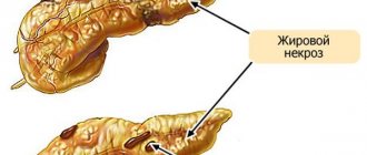
By the appearance of the removed organ, one can judge the fairly dense consistency of adenocarcinoma. On the outside, it has no clear boundaries and is sclerotic. The lobulation characteristic of the pancreas is not visible from the inside. The most typical size for a tumor located in the head is 2.5–3.5 cm, the tail and body are slightly larger. Cyst formation is possible. The color is gray-yellow. Even large nodes are not characterized by areas of necrosis and hemorrhage.
Of particular importance for the formation of relapses is the growth of adenocarcinoma along the nerve plexuses from the uncinate process on the head to the superior mesenteric ganglion, from the upper part to the perigastric ganglion. From the body and tail, cancer cells are directed towards the splenic, left perigastric ganglion, and superior mesenteric.
Ductal adenocarcinoma metastasizes most often in the liver (53–60% of cases), lung tissue and peritoneum were detected in 10–16%. Less common is the transition to:
- adrenal glands,
- kidneys,
- pleura,
- mesentery of the small intestine,
- diaphragm,
- bones.
Rarely, metastases are found in the brain, heart, and spleen.
Metastasis is typical for the location of the tumor in the tail and body of the pancreas. This is explained by the preservation of the patient’s life and the long-term supportive treatment process.
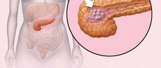
Factors provoking pathology
Oncological diseases are often difficult to diagnose, and their causes are not fully understood. This is also typical for formations that arise in the head of the pancreas. The risk of developing the disease increases due to the following reasons:
- Heredity.
- Tobacco abuse. It contains many carcinogenic components that activate the development of cancer cells.
- Chronic pancreatitis causes disruptions in blood circulation and metabolic processes at the cellular level.
- Aging of the body. Older people suffer from frequent disruptions in the immune system, as a result of which malignant tumors occur more often.
- Diabetes mellitus in a long-term form. Due to the death of beta cells, foci of tumor formation arise.
- Excess weight, which acts as a provoking factor for hyperlipidemia and hypercholesterinemia. This causes lipodystrophy of the pancreas.
- Irregular nutrition leads to overload of the gland and enzymatic deficiency.
- Excessive consumption of strong alcoholic beverages, which causes the death of groups of cells.
We also recommend viewing: Features of pancreatic cystadenoma and treatment methods
Symptoms of pathology
The initial symptoms of a pancreatic tumor are either absent or “masked” as chronic diseases of the gastrointestinal tract, cholecystitis, pancreatitis.
Clinical picture of a benign tumor
Benign tumors are asymptomatic. It has been established that only when the size reaches 5 cm do patients feel dull pain in the epigastrium and hypochondrium on the right, nausea, and stool disorders.
Signs are associated with impaired supply of digestive enzymes and compression of the duodenum.
Signs of malignant neoplasms
Symptoms of cancer are conventionally divided into manifestations of the primary tumor, its effect on neighboring organs and tissues, lymph nodes, and the presence of metastases. These include:
- pain syndrome (girdling pain, irradiation to the right in case of cancer of the head, to the left and to the back in case of damage to the tail area, in the shoulder, lower back, constant aching, sometimes severe attacks), caused by the involvement of the nerve ganglia, stretching of the gland capsule, bile ducts,
- enlarged liver with ascites,
- nodes, tubercles, growths on the dense liver, enlarged lymph nodes above the collarbone, determined by palpation,
- loss of appetite is a consequence of intoxication,
- significant reduction in body weight,
- weakness,
- the addition of infectious diseases due to severe suppression of immunity,
- Primary manifestations of diabetes mellitus are possible (thirst, itching in 20% of cases).
Signs of stagnation in the bile ducts, duodenum, due to mechanical obstruction:
- bursting pain in the hypochondrium on the right,
- jaundice,
- enlarged liver and gall bladder,
- belching, nausea, vomiting,
- hemorrhages on the skin,
- diarrhea, constipation, flatulence - caused by secretory insufficiency in the digestion of food,
- obstruction in the small intestine area.
Ductal cancer is characterized by a high rate of progression and severe symptoms. In the tumor area, spastic contraction of the arteries occurs, as a result of which additional disturbances in the nutrition of the organ appear.
In the clinic, cancer located in the head of the pancreas is divided into 2 stages (pre-icteric and icteric). In many patients, yellowing of the skin and itching are the first and only symptom of adenocarcinoma. At the same time, in 1/3 of patients the pain syndrome is absent or of low intensity.
Impaired digestion contributes to the development of mixed anemia (iron deficiency and B12 deficiency).
An increase in temperature indicates the addition of inflammatory processes in the gallbladder and ducts.
All of the above symptoms cannot be considered specific to a pancreatic tumor. No one determines with absolute accuracy the localization of the process, the size, or the stage of the flow.
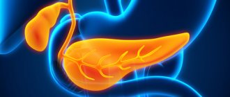
Pancreas and its role
The organ for the most part belongs to the exocrine type. Here the synthesis of enzymes that promote digestion occurs - they participate in the production of gastric juice, which is then sent to the duodenum. Inflammatory processes in this area often take a chronic form. This leads to the appearance of a focal formation of the pancreas provoked by pancreatitis.
The area of the gland where hormones are produced that regulate metabolic processes is of the endocrine type. These hormones include insulin, glucagon, somatostatin, pancreatic polypeptide, and ghrelin. They participate in glucose metabolic processes, regulate the production of glands, and influence the feeling of need for food. If pathology manifests itself in this area, the carbohydrate balance is disturbed, and tumors provoke intensive growth or suppression of hormone production.
Important. The pancreas structurally includes several parts - the head, neck, body and tail. It is in the first part that most formations arise.
Diagnosis of malignant tumors
Examination of a patient with suspected pancreatic tumor begins with general tests and biochemical tests. Only at a late stage is a moderate decrease in hemoglobin and red blood cells and an acceleration of ESR detected.
Biochemical tests can detect a low protein content (hypoalbuminemia); with jaundice, an increase in bilirubin, alkaline phosphatase, aspartic and alanine transaminase.
Of greater importance is the detection of tumor markers in the blood. It is believed that embryonic carbonic anhydrate glycoprotein (CA-19-9) has the greatest specificity (almost 90%). Normally, its content is 37 units, and in case of a malignant tumor of the pancreas it can increase from tens to hundreds of times. The disadvantage of the method is the lack of changes at an early stage.
The informativeness of the test with the CA 494 antigen for the early diagnosis of cancer is being studied. It has been found to be effective in identifying differences with chronic pancreatitis.
The main importance is attached to the development of instrumental accessible methods of research.
Using the X-ray method by introducing a barium suspension into the stomach, it is possible to identify indirect signs of a volumetric process in the pancreas. They are associated with compression of neighboring organs. The radiologist finds:
- deformation of the stomach, its protrusion forward, displacement of the lesser curvature,
- calcifications (deposits of calcium salts) with focal pancreatitis,
- “unfolding” of the horseshoe-shaped duodenum due to compression of the descending section,
- “filling defect” in the duodenum, stenosis.
On tomographic sections, the tumor is defined as a hypodense formation; it has a darker color than the parenchyma.
The use of ultrasound diagnostics has made it possible to judge the tissues of parenchymal organs by their ability to reflect waves (echogenicity). The higher the ability of the compacted tissue to reflect the wave, the lighter the visible formations become.
The transcript uses definitions for the pancreas:
- isoechoic - the entire organ normally looks gray,
- the hypoechoic area is darker,
- hyperechoic - looks light in tone, almost white, such areas are characteristic of a dense tumor,
- anechoic, also called echo-negative - the entire structure is black (for example, in the case of a cyst filled with fluid, its contours will be visible).
Various ultrasound techniques (transcutaneous, Doppler scanning, use of a signal amplifier) allow you to see not only the size of the organ:
- contrast in detail a tumor-like formation in the parenchyma up to 1 cm in size, its structure,
- identify enlarged lymph nodes,
- moderate volume of fluid in the abdominal cavity.
Oncologists receive important information from the vessels adjacent to the tumor. It is necessary to assess the degree of vascularization before surgery to predict hematogenous metastasis. Studying zones of hypervascularization from newly formed vessels allows us to judge the degree of organ damage.
The method has now been improved. Endoscopic ultrasound of the pancreas is used by introducing a microsensor into the duodenal zone, bile duct, and vessels. It is possible to examine a tumor less than a centimeter in size.
MRI - the magnetic resonance reflection method is considered more harmless and is used in diagnosing damage to the pancreas in a child. The transcript also draws attention to hypodense areas of tissue.
To definitively confirm the diagnosis, a puncture biopsy under ultrasound or computed tomography guidance is used. It is possible to accurately determine the type of tumor in 90% of cases.
Treatment
The choice of treatment method depends on the size of the cyst, its location and the presence of complications. For example, small cysts after pancreatitis are treated conservatively, with regular ultrasound examinations. On the other hand, complications such as infection or bleeding require radical surgical treatment.
Conservative therapy includes the prescription of enzyme preparations, non-steroidal anti-inflammatory drugs or antibiotics. Treatment of concomitant pathologies of the gastrointestinal tract is carried out with the help of gastroprotectors (Rebagit), antispasmodics (Meteospasmil), proton pump inhibitors (Omeprazole, Esomeprazole), etc.
Radical surgical treatment is carried out using the following methods:
- Ultrasound-guided puncture. The surgeon punctures the cavity and removes its contents, then the cyst is washed with an antiseptic solution.
- Internal drainage of the cyst. An anastomosis is created between the cavity formation and the duodenum (small intestine, stomach).
- External drainage. The outflow of the cyst contents occurs through the abdominal wall. Indicated for the inflammatory process, unformed cysts, and the patient’s serious condition.
- Removal of the cyst (cystectomy) with resection of part of the organ. This is done when the cavity is large, there are complications such as bleeding, or there is a suspicion of a tumor process.
Today, surgeons prefer minimally invasive operations performed laparoscopically or endoscopically. The choice of technique depends on the location of the cavity and its contents.
Methods of treating neoplasms
Pancreatic tumors cannot be treated either by diet or by folk recommendations. Patients should not waste time searching for fabulous advertised remedies. A combination of modern methods includes:
- surgical stage - resection of part of the pancreas or together with surrounding organs (gastropancreatoduodenal, subtotal, pancreatectomy),
- consolidation of the operation with multi-course chemotherapy.
The role of diet
The importance of therapeutic nutrition is to maximally support the functions of the pancreas. Pancreatitis necessarily develops against the background of a tumor. Therefore, it is necessary to comply with food requirements:
- give up spicy, fatty foods, fried foods,
- reduce easily digestible carbohydrates in the diet (sweets, honey, jam),
- eat at least 8 times a day, but reduce portions by half,
- all dishes should be steamed or boiled,
- Fresh vegetables, fruits, salads are prohibited,
- salt is limited, seasonings, ketchups, and mayonnaise are excluded.
To improve absorption, food should be in a semi-liquid state, pureed, meat in the form of minced meat.
Surgical intervention
The extent of the operation is determined during the examination. The location of the tumor, size, and growth into nearby vessels and lymph nodes are important. The result should minimize the risk of relapse and digestive disorders. Surgeons resect the surrounding vascular branches very carefully. Excessive removal of blood vessels causes disruption of the intestinal innervation. The development of diarrhea does not make it possible to carry out subsequent full-fledged chemotherapy.
When removing ductal adenocarcinoma, it is usually necessary to simultaneously remove adjacent organs (spleen, colon, part of the stomach, left adrenal gland, half of the diaphragm).
Other tumors are removed if there is compression of neighboring organs or bleeding. The method of choice is partial resection of the gland and angioplasty. The doctor will tell you whether it is necessary to be treated with chemotherapy after a complete cytological analysis.
Chemotherapy
Chemotherapy is used to prepare the patient for radiation. Its goal is to bring the tumor to an operable state. The drugs used are administered intravenously (Gemcitabine, Erlotinib, Capecitabine, 5-fluorouracil), they increase the sensitivity of tumor cells to radiation therapy.
There is ongoing debate among scientists about whether it is worth treating a patient with chemotherapy drugs only. It is believed that the patient can be prepared for surgery without subsequent radiation. Typically, a course of chemotherapy is carried out for 2–3 months before surgery and the same amount after. It has been proven that the survival prognosis is the same.
The drugs are often difficult to tolerate and have negative effects (joint pain, nausea, yellowing of the skin).
Irradiation
If the disease does not progress during chemotherapy, then external radiotherapy is added. The radiation dose is calculated individually for each patient.
In addition to remote irradiation, the following forms of irradiation are used:
- fast electrons,
- Bremsstrahlung radiation.
Radiation therapy is prescribed before and after surgery, and less often - instead of surgery.
Targeted therapy
Targeted therapy is a targeted therapy aimed at a specific target of malignant cells. The created drugs primarily affect tumor cells and suppress their growth. Do not affect surrounding tissues.
Targeted therapy drugs “teach” the body to produce antibodies; they must block the mutating gene that causes the cells to transform into malignant ones. So far, few of them have been synthesized, and the price is too high for general use.
Avastin is suitable for treating pancreatic tumors. One course of treatment will cost $50,000.
Benign tumors
Such formations are characterized by slow development. In this case, the tissue is not damaged, and the tumor does not grow into nearby organs. The likelihood of metastases is practically absent.
Types of tumors
Multiple and single neoplasms localized in any part of the pancreas can form. When the epithelium of an organ is damaged, adenomas or cystadenomas appear, but connective tissues suffer from fibromas and lipomas.
Muscle tissue becomes the basis for the formation of leiomyomas, but lymphangiomas and hemangiomas are formed from tissues of the vascular type. The islets of the gland are susceptible to the action of insulinomas. In this case, due to neurogenic factors, neuromas and ganglioneurinomas appear.
The source of benign neoplasms is usually heredity. Bad habits, poor nutrition, and the environment have less influence on their origin.
Symptoms
In order not to miss the active development of a benign tumor, you should be attentive to the following signs:
- changes in hormonal balance, accompanied by lethargy, dizziness and headaches, anxiety, heavy sweating;
- pain in the left or right umbilical region, which is encircling or paroxysmal in nature with the possibility of spreading to the scapular area or arm;
- icteric manifestations, since the formations of the head compress the ducts;
- nausea and vomiting that occurs after eating and is a consequence of pressure on the duodenum.
The risk of such neoplasms should not be underestimated. They can degenerate into malignant ones or lead to bile intoxication. Against the background of the disease, pancreatitis develops and problems with the thyroid gland arise.
Important. An increase in the size of benign formations can provoke intestinal obstruction. However, if they are removed in time, a complete cure is possible.
Diagnostic features
Such tumors are detected at an early stage during regular medical examinations and examinations. They do not manifest themselves in the natural environment. To specify the diagnosis, ultrasound, CT, and MRI are performed. Blood is given for general analysis and biochemistry, and is also examined according to the criterion of an oncological marker.
We also recommend viewing: Insulin and its role in the human body
Treatment and rehabilitation
The only way to get rid of a diagnosed benign tumor is through surgery. If it occurs in the tail part of the gland, then partial resection is performed and a separate section of the organ is removed. Insulinoma can be eliminated using the desquamation method, when only the damage is removed.
The formation that has formed in the pancreas, and specifically on its head, is eliminated by pancreatoduodenal resection. During the operation, the duodenum is also removed. When the formations are small and located in the tail zone of the gland, and there is no risk of degeneration, doctors recommend performing a mini-invasive laparoscopy.
After surgery, you should follow a diet, excluding fatty, salty, sweet or starchy foods from your diet. The duration of this regime is at least 1 year. At the same time, enzymes are prescribed and physical activity is limited.
Important. To eliminate the risk of a hernia, the patient is prescribed to wear a support belt. You should undergo regular ultrasound examinations for 2 years.
It is quite difficult to eliminate the risk of developing pathology. There are no specific methods of prevention, especially since the role of heredity is high. However, a balanced diet, exclusion of alcoholic beverages from the menu and therapy for pancreatitis can reduce the risk of the disease.
Survival prognosis
Postoperative mortality during resection of the gland with surrounding organs averages from 5 to 15%. After removal of the tumor in the tail, patients live about 10 months. Combination with chemotherapy and radiation increases the patient's life to 13–16 months. The five-year survival rate of patients does not yet exceed 8%.
Unfortunately, medicine still has nothing to reassure patients with damage to the pancreas. There are insufficient methods for early diagnosis, and treatment does not provide long-term remission.
Bibliography
- Sokolov V.I. Surgical diseases of the pancreas. M. 1998
- Patyutko Yu.I., Kotelnikov A.G. Pancreatic cancer: diagnosis and surgical treatment at the present stage. Annals of surgical hepatology. 1998, volume 3, no. 1, pp. 96–111.
- Kubyshkin V.A., Vishnevsky V.A. Pancreas cancer. M., 2003
- Lubensky Yu.M., Nikhinson R.A. Pancreatobiliary cancer. Krasnoyarsk University Publishing House 1984
Brief description
A neoplasm in this gland is, first of all, a lesion of the epithelial layer of the organ. It first appears in the area of the head of the pancreas.
Those at risk of developing a pancreatic tumor are those over 55 years of age, heavy smokers and those who like to abuse alcohol. But diabetics and people suffering from chronic pancreatic diseases should also be careful.
The following types of neoplasms are usually identified in this organ:
- epithelial (benign and malignant);
- pancreatic islets;
- mixed;
- non-epithelial tumors;
- lymphoid;
- hematopoietic;
- metastatic.
The most common types are benign and malignant. Therefore, we will dwell on them in more detail.
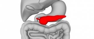
Pathologies arising in the tail of the gland
With all existing diseases of the pancreas, one can constantly observe its increase. When functional diagnostic methods are used, compression in the tail is detected.
These changes lead to disruption of the patency of the splenic vein and the formation of elevated subrenal hypertension.
Inflammation of the tail of the pancreas is observed:
- due to existing stones that block the auxiliary canal of the gland;
- development of benign neoplasms;
- suppuration of the head with transition to the parenchyma of the terminal region of the pancreas;
- pseudocyst, which complicated pancreatic necrosis;
- duodenitis;
- tumors of the small papilla of the duodenum;
- poor-quality presence of a neoplasm.
Pancreatitis can also begin with a disease of the tail zone of the gland and lead, at the initial stage of its formation, to an increase in its natural values.
duodenitis
Pathologies arising in the tail of the gland
With all known diseases of the pancreas, its uniform increase does not always occur.
Using functional research methods, expansion (thickening) of the tail is detected. These changes in tissues lead to disruption of the patency of the splenic vein and the development of portal subrenal hypertension.
The reason for this expansion of the tail of the pancreas may be:
- a calculus blocking the Wirsung duct;
- benign cystic adenoma;
- abscess of the head with spread to the tissue of the end part of the pancreas;
- pseudocyst complicating pancreatic necrosis;
- duodenitis;
- malignant neoplasm.
Pancreatitis can also begin with inflammation in the tail of the pancreas and lead, in the initial stage of its development, to exceeding its normal size.
Diagnosis and treatment of pathologies
Diagnosis begins with a detailed clarification of complaints, anamnesis, and objective examination.
Clinical signs of tail damage in all pathologies are similar to diseases of the entire pancreas. Symptoms of tail inflammation manifest themselves with the same symptoms as pancreatitis, which affects the entire gland:
- pain localized in the right hypochondrium or in the epigastric region, with irradiation to the precardiac region, lower back;
- decreased appetite;
- bowel disorders;
- temperature increase.
Tumors, cysts, lipomas, fibromas, hemangiomas do not manifest themselves clinically for a long time. Symptoms occur when these formations reach enormous sizes.
It is impossible to determine the enlargement of the tail of the pancreas during an objective examination: due to its retroperitoneal location, it is not palpable. Therefore, at the slightest suspicion of changes in the tail part, it is necessary to contact a specialist to prescribe special laboratory and functional examination methods.
Treatment is prescribed depending on the identified pathology. If the tail of the pancreas is enlarged, the underlying disease is treated. Methods also depend on changes: surgical treatment is used in the presence of neoplasms, both malignant and benign. Sometimes surgical treatment is used for abscesses, huge cysts, and pseudocysts. For inflammatory lesions, pancreatitis is treated.
Diagnostic methods
Screening methods include ultrasound examination (US), which has received positive feedback from doctors of various specialties. Sonography is convenient because it does not require complex preparation, takes little time, and is well tolerated by both children and elderly people. Using this technique, the following are determined:
- dimensions;
- presence of formations;
- clarity of boundaries;
- echogenicity (density) of tissues - increased or decreased;
- condition of the Wirsung duct.
When performing sonography, in some cases the tail of the altered pancreas is not located. It may be partially and inconsistently visualized. According to the literature, the detection rate of the pancreatic tail on echography is approximately 40-100%. It is better to locate the significantly enlarged tail part of the organ, especially when there are large echo-positive formations in it.
Therefore, additionally:
- MRI;
- radiography;
- CT scan with contrast agent;
- cholangiopancreatography (endoscopic and magnetic resonance imaging);
- EFGDS (esophagofibrogastroduodenoscopy).
Laboratory research methods include:
- a general blood test to determine the presence of inflammation (increased ESR, leukocytosis);
- sugar in blood and urine;
- during an attack of insulinoma - insulin in the blood;
- glucagon;
- fasting blood test for gastrin.
All studies are prescribed by a doctor, who analyzes what their results may mean, and based on them, medications are prescribed to treat the identified pathology.
First aid when identifying signs of disease
Tail enlargement can become acutely manifest when there is a pronounced inflammatory process and developing pancreatic necrosis. Intense pain occurs, there may be vomiting and diarrhea. Necessary:
- immediately call an ambulance;
- Before her arrival, place the patient on his side with his knees bent to his stomach - this can reduce pain;
- create complete peace and comfortable conditions;
- apply a heating pad with ice to your stomach;
- observe hunger (you can only drink warm mineral alkaline water without gas);
- give an antispasmodic tablet if there is no severe vomiting (No-Shpu, Drotaverine, Papaverine), painkillers are prohibited.
Diet and prevention
The pancreas is very sensitive to various adverse influences. To cure an already existing disease and as a preventive measure or complications, adhere to therapeutic nutrition - table No. 5. It is prescribed by a doctor, who takes into account food restrictions based on the severity of the disease. In addition to dietary nutrition, physiotherapy is included.
physiotherapy for pancreatitis
It is important to immediately go to the doctor at the initial signs of pancreatic dysfunction. This will allow timely detection of the disease and prevent further development of pathology, avoid serious complications, and improve the prognosis.
Symptoms of benign neoplasms
It is important to say that those tumors are isolated that are hormonally inactive and hormone-producing. The former do not show any signs until they reach an impressive size and begin to put pressure on nearby organs, disrupt blood flow, or enlarge the gland lining. But a hormone-producing tumor is characterized by weakness, increased sweating, tachycardia, and with low glucose levels, a hypoglycemic coma is observed.
If a benign neoplasm, due to its size, puts pressure on nearby organs, pain may develop.
If the pain is concentrated predominantly in the right hypochondrium, then this is a tumor of the head of the pancreas. When in the upper abdomen - then the body of the gland, and if in the left hypochondrium or in the lumbar region - then the tail. Compression of any part of the intestine leads to intestinal obstruction.
Obstructive jaundice can occur if the neoplasm compresses the bile or pancreatic ducts. Because of this, the sclera and skin become yellow, the urine becomes dark, and the feces become colorless.
Gastroduodenal ulcers signal gastrinoma (another type of benign tumor). Patients often complain of pain in the epigastric region. Also among the symptoms: belching with a feeling of acid, severe heartburn. With gastrinoma, a lot of hydrochloric acid enters the gastrointestinal tract, which provokes a deterioration in the state of digestive processes and the functioning of the mucous membrane.
Causes of tail enlargement
The reasons for local enlargement of the tail zone, which create a favorable environment for the development of pathologies of the tail of the pancreas, have a subject-specific and individual course.
Based on the statistical data, only in one case out of 4 with pancreatic disease, the affected area predominates directly in the tail area. Moreover, such a case does not make it possible to neglect the painful phenomenon in the tail area and the violation of the size, since the formation may have a poor quality course.
Difficult diagnostic measures will make it possible to suspect in rare cases a disease at the stage of formation, therefore the pathology can only be determined later when the formation reaches impressive values. To get closer to the tail area, they pass through the left kidney with the spleen.
In addition to the listed factors, a significant increase in the tail of the pancreas is recorded as a result of:
- cyst;
- lipomas;
- hemangiomas;
- fibroids;
- insulin.
Possible causes of pancreatic tail enlargement
Symptoms of pathologies of cysts and pseudocysts appear in rare cases. The patient may have no symptoms at all. Often, formations are detected using sonography when the patient presents with another disease.
Signs of an inflammatory process in the pancreas look like this:
- the pain is aching, dull, prolonged and not related to food consumption. Based on the severity of the pathology, pain increases in the pancreas;
- the painful syndrome covers the epigastric region, heart, chest. Unbearable pain may occur, leading to shock.
To ease their well-being, many patients with pancreatitis resort to the method of bending forward or taking a sitting position. Other symptoms of gastrointestinal pathologies.
- Changes in facial skin.
- The fingers become pale, blue.
- Stool disorder.
- Urge to vomit.
- Increase in temperature.
Since the tail contains the bulk of the islets of Langerhans, the pathology, as well as the excess size of the pancreas, is due to damage to these structures. Insulinoma develops, which is a tumor that occurs mainly in the tail of the organ.
problems with stool
Insulinoma occurs when insulin is synthesized in large quantities. When insulinoma is present, an increased tail size and a sharp decrease in blood sugar are detected.
Glucagonoma leads to the formation of diabetes and can become malignant. Glucagon breaks down glycogen in the liver and muscles, causing an increase in sugar.