Nausea and pain in the upper abdomen occur periodically in many people. Often these symptoms indicate the development of inflammation in the digestive tract. Unpleasant sensations in such cases are almost always associated with the use of certain products. Pain and nausea may indicate hardening of the gallbladder tissue. Despite the fact that this organ is small in size, the symptoms of its inflammation are very pronounced. Are women more often diagnosed with hardening of the gallbladder walls? What is it and why does it occur? It is worth noting what functions this body performs. It serves as a container for bile. During the digestion of food, this biological fluid is released into the lumen of the duodenum. This is accompanied by contraction of the gallbladder. Compaction of the walls of the organ leads to disruption of its main function. As a result, bile stagnates (cholestasis), and the digestion process slows down. In addition, the organ may increase significantly in size, which leads to pain.
Why does thickening of the gallbladder wall occur?
The tissue that makes up the organ cannot harden without a reason. This is caused by various unfavorable factors, which can be both exo- and endogenous. Hardening of the gallbladder wall occurs for the following reasons:
- Chronic inflammation of the organ - cholecystitis. This disease is considered one of the most common ailments of the digestive system. As is known, the inflammatory process causes swelling and hyperemia of the walls of the organ, and the permeability of small vessels increases. Chronic cholecystitis is characterized by phases of exacerbations and remissions. As a result, swelling of the walls is replaced by increased growth of connective tissue, which, in turn, is fraught with the development of compaction of the organ and adhesions in the gallbladder.
- Calculous cholecystitis. In addition to the chronic inflammatory process, this pathology is accompanied by the formation of stones in the lumen of the organ. Stones prevent the evacuation of bile.
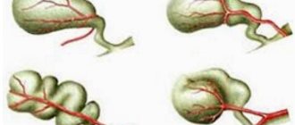
- Congenital organ deformities. During examination, many people are diagnosed with kinks and other changes in the configuration of the gallbladder. Improper structure contributes to the development of cholestasis. And it, in turn, causes chronic inflammation and hardening of the walls.
- Excessive consumption of difficult-to-digest foods. This means a large amount of fatty, bitter, salty foods.
- Diseases of the digestive system. Chronic hepatitis and pancreatitis are often combined with inflammation of the gallbladder.
- Heart failure. Long-term cardiac pathologies lead to the formation of edema both on the skin and in the internal organs.
- Polyps and other neoplasms. The growth of organ tissue is always accompanied by thickening of its walls.
All these reasons must be taken into account in order to cure chronic cholecystitis. Deformation and hardening of the walls of the gallbladder are dangerous for the development of unpleasant consequences. Among them are inflammation of other gastrointestinal organs and digestive disorders.
Treatment
Treatment for thickening of the gallbladder wall depends on the cause of the pathological process. Therapeutic measures are divided into conservative and surgical.
To obtain the necessary treatment for hardening of the gallbladder wall, you should contact a gastroenterologist, who will determine the causative disease and select the necessary therapy.
Diet
Compliance with a special diet is of great importance in the treatment of gallbladder pathologies. Experts recommend small meals up to 6 times. Fatty, hot, sour, spicy foods, processed foods, canned food, alcoholic and carbonated drinks should be excluded from the diet.
The following products are allowed:
- lean fish and meat;
- low-fat dairy products;
- eggs (2 times a week);
- porridge from any cereals;
- stale or dried bread;
- vegetarian soups;
- vegetables and fruits in any form;
- jelly, compotes, weak tea.
Dishes should be prepared with a minimum amount of fat. They should be served warm; food that is too hot or cold should be avoided. Doctors recommend following a drinking regime, that is, drinking at least 2 liters of clean water per day.
Proper nutrition is of great importance in treatment
Mode
For any pathologies of the liver and gall bladder, including hardening of its walls, it is necessary to introduce some restrictions into the lifestyle. It is recommended to reduce the intensity of physical activity. Active sports are contraindicated; it is recommended to undergo a course of therapeutic exercises.
It is important to give up bad habits - alcohol and nicotine abuse. Walking in the fresh air is beneficial.
Medications
The drug therapy regimen is selected by the attending physician taking into account the causative disease. Different groups of drugs are used.
Table. Medicines used to harden the walls of the gallbladder.
| Group of drugs | Indications for use |
| Hepatoprotectors - Phosphogliv, Ursosan, Essliver | Reduce the severity of the inflammatory process, normalize liver enzymes |
| Choleretics and cholekinetics - rose hip syrup, Allohol, Flamin | Improves the outflow and excretion of bile, prevents its stagnation |
| Antispasmodics - No-shpa, Drotaverine | Eliminate spasm of the muscle layer of the bladder, improving the outflow of bile |
| Statins - Atorvastatin, Rosuvastatin | Reduce the amount of cholesterol in tissues |
The duration of drug use is determined by the causative disease.
Surgical intervention
In some situations, the only optimal treatment method is surgery. It is indicated for acute calculous cholecystitis, polyposis, and organ tumors. Usually an operation is performed to completely remove the bladder - cholecystectomy.
Thickening of the walls of the gallbladder can be caused by various reasons. It is important to promptly identify diseases that provoke this symptom and begin treatment.
Self-medication or ignoring the symptoms of the disease can lead to serious consequences - the appearance of adhesions in the gallbladder, digestive disorders, the spread of inflammatory and infectious processes to nearby organs, perforation of the walls of the gallbladder, peritonitis.
Symptoms of tissue compaction
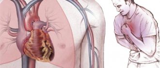
The thickening of the gallbladder wall does not appear externally. Therefore, if this process is suspected, it is necessary to undergo an examination, in particular an ultrasound. Manifestations of the disease are not always expressed, but only during the period of exacerbation of inflammation of the organ. In this case, symptoms such as nausea, a feeling of bitterness in the mouth, pain in the right abdomen, and general weakness are noted. During remission, these signs are absent. However, after eating fatty and difficult-to-digest foods, the discomfort occurs again.
Nausea and bitterness in the mouth during an exacerbation are not always associated with food. Patients often complain of discomfort in the morning, before bedtime. After eating, the symptoms intensify. Vomiting of bile is sometimes observed.
Diet No. 5
This diet, called “Healing Table No. 5,” is an integral element of successful therapy for any pathology of this internal organ. Its compliance is mandatory. Otherwise, all other therapy will be a waste of money and time.
The basic principle of this diet is fractional nutrition, which means eating small portions at regular intervals, five to seven times a day. You can prepare dishes with this diet only by steaming, boiling or baking. You will have to forget about fatty, fried, hot, spicy and smoked dishes. Also prohibited are alcoholic and carbonated drinks, sweets and baked goods, some and strong tea.
Products allowed for consumption on this diet:
| № | Helpful information |
| 1 | weak tea |
| 2 | low-fat cottage cheese |
| 3 | milk (in the absence of individual intolerance) |
| 4 | low fat fermented milk products |
| 5 | dried fruit compotes |
| 6 | decoctions, jellies, jellies and mousses based on sweet berries and fruits |
Read also: What choleretic drugs are needed when the gallbladder is bent?
- one chicken egg per week;
- lean meat (veal, chicken, rabbit, turkey);
- low-fat types of sea and river fish;
- yesterday's white bread or crackers based on it;
- vegetables (both in the form of soups and purees, and fresh);
- porridges based on buckwheat, oatmeal and semolina;
- pasta;
- low-fat and non-spicy hard cheese;
- vegetable oils (linseed, sunflower and olive);
- butter (in very limited quantities).
Inna Lavrenko
auto RU
All food should be warm, in no case cold or hot.
The same diet is indicated after surgery to remove the gallbladder.
Physical examination for cholecystitis
Having seen the conclusion of an ultrasound examination, patients begin to be interested in the question: what does thickening of the gallbladder walls mean? It is worth noting that these words mean a morphological change in the organ. Wall compaction is not an independent diagnosis. This symptom is detected during an instrumental examination. This sign almost always means that the patient has chronic inflammation of the gallbladder.
However, the doctor must make sure of this. For this purpose, a physical examination is performed. Specific signs of cholecystitis include:
- Pain when pressing on the area of the bladder (Keur's symptom).
- Increased discomfort upon palpation during inhalation.
- Pain when tapping on the right costal arch (Murphy's symptom).
All these signs are detected in both acute and chronic cholecystitis. Therefore, to find out whether there is compaction of the walls of the organ, an ultrasound examination is performed.
Why does the gastrointestinal tract thicken?
Since this condition is not normal, something must cause it to develop. So, why do the walls of the gallbladder thicken:
- acute and chronic cholecystitis;
- cholelithiasis;
- infectious diseases;
- inflammation of the bile ducts (cholangitis) and malformations;
- diseases of the pancreas and liver;
- cancer tumor.
Having undergone a comprehensive examination when thickening of the gallbladder walls appears and having discovered the causes, it will be possible to begin treatment. If therapy is not started in time, the disease will become chronic or complications will begin.
Diagnosis of gallbladder diseases
Signs of hardening of the gallbladder walls are indications for diagnostic procedures. Laboratory features of chronic cholecystitis include: increased AST and ALT. The level of these enzymes increases when bile stagnates in the ducts. During the period of exacerbation, leukocytosis and acceleration of ESR in the blood test are noted.
The thickness of the walls of the organ should not exceed 5 mm. An increase in this indicator indicates the presence of a chronic inflammatory process. Also, as the walls thicken, a change in the echo density of the organ contours is noted.
In addition to ultrasound, computed tomography of the abdominal cavity and radiography are performed. In some cases, special invasive studies are required. Among them is retrograde cholangiopancreatography.
Radiation diagnostics of pathology of the gallbladder and biliary tract
Ultrasound scanner HS50
Affordable efficiency.
A versatile ultrasound scanner with compact design and innovative capabilities.
Introduction
Gallbladder diseases are a special area in modern medicine, to which specialists pay increased attention [1-8]. On the one hand, pathology of the gallbladder (for example, polyps) can be completely asymptomatic, but has a tendency to malignancy [4-10]. On the other hand, some diseases, such as gallstones and biliary tract stones, are accompanied by severe attacks of pain and can lead to serious complications. As a result of prolonged pressure from a stone on the mucous membrane of the bladder, ulcers and bedsores may appear in it, diverticulum-like protrusions, internal and external biliary fistulas, perforations with the development of subhepatic or subphrenic abscesses, and biliary peritonitis may form. The movement of gallstones may be accompanied by blockage of the cystic duct, hydrocele of the gallbladder or empyema. When a stone obstructs the outlet of the common bile duct, obstructive jaundice occurs. Prolonged presence of stones in the bile ducts and the addition of infection lead to the development of cholangitis. The loss of a large gallstone from the gallbladder into the intestine through the formed anastomosis can lead to intestinal obstruction [1, 3, 5-8, 10-12]. Therefore, timely diagnosis of diseases of the gallbladder and ductal system is very important.
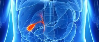
The formation of bile in the liver is a continuous process, but its entry into the intestines normally occurs mainly during the digestion process. This is ensured by the reservoir function of the gallbladder and its rhythmic contractions with sequential relaxation of the Lütkens sphincter and then the sphincter of Oddi, located at the confluence of the common bile duct into the intestine (Fig. 1).
Rice. 1.
Schematic representation of the position of the gallbladder on the visceral surface of the liver.
1 - bottom of the gallbladder; 2 - cystic duct; 3 - own hepatic artery; 4 - portal vein; 5 - gastrohepatic ligament; 6 - left lobe of the liver; 7 - caudate lobe of the liver; 8 - inferior vena cava; 9 - caudate process; 10 - neck of the gallbladder; 11 - right lobe of the liver; 12 - body of the gallbladder; 13 - square lobe of the liver.
On an empty stomach, the gallbladder contains 30-80 ml of bile, but it can concentrate 5-10 times more hepatic bile. When bile stagnates in the bladder, its amount may increase. In women, the gallbladder in a state of functional rest has a slightly larger volume than in men, but contracts faster. With age, the contractile function of the gallbladder decreases.
Ultrasonography is one of the most informative and accessible instrumental methods for diagnosing gallbladder diseases [4-10].
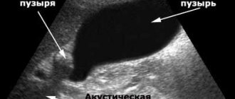
The acoustic properties of ultrasound make it possible to detect the smallest echogenic structures located in the gallbladder.
With the help of ultrasound, the diagnostic search time is significantly reduced. Unlike computed tomography (CT) and magnetic resonance imaging (MRI), with ultrasound, the researcher himself is directly involved in obtaining the image, which has its pros and cons. Positive is the possibility of a more targeted and detailed study of the object being studied. The negative side is that the quality of the image and its interpretation largely depend on the experience of the researcher and the correctness of the techniques he uses.
Polyps in the gallbladder are present in 6% of the total population. In 80% of cases, polyps in the gallbladder are observed in women who have given birth after the age of 30 years. Since polyps do not manifest themselves clinically, their diagnosis is most often accidental and occurs during an ultrasound scan of the patient for completely different reasons [4-10].
Although the reasons for the appearance of polyps on the walls of the gallbladder have not been established, and the symptoms are not obvious, four types of such formations are known. According to statistics, the following types are most often found in the gallbladder of patients today [4, 5, 9].
Inflammatory polyp of the gallbladder is a kind of inflammatory reaction of the mucous membrane of the gallbladder itself, which manifests itself in the patient in the form of various growths of granulation internal tissue of the affected organ.
Doctors often also diagnose gallbladder adenoma. This happens because it is a kind of benign tumor in the form of a polyp-like growth of the glandular tissue of the patient’s gallbladder.
Also of note is papilloma or gallbladder polyposis in some patients. This papilloma is a benign tumor of the mucous membrane of the bladder in the form of peculiar papillomas or papillary growths of different appearance and structure.
The most common type is the so-called cholesterol polyp of the gallbladder, which is an elevation of the mucous membrane of the gallbladder with cholesterol deposits on it. Cholesterosis is quite common among patients referred for surgery with a clinical diagnosis of “polyps” or “polyposis of the gallbladder”, according to some data, from 42 to 95% of cases.
Ultrasonography is an effective means of identifying the polypoid form of cholesterosis [4-10]. The following sonographic characteristics of cholesterol polyps are considered traditional: immobile hyperechoic structures that do not provide an acoustic shadow and are attached to the wall of the gallbladder. The contours of such formations are, as a rule, smooth, and the sizes of such formations are different, most often not exceeding 10 mm (Fig. 2).
Rice. 2.
Ultrasound picture of polyps in the gallbladder.
a)
Single polyp in the gallbladder (hyperechoic parietal immobile formation, with smooth contours, without acoustic shadow).
b)
Single polyp in the gallbladder.
V)
Polypoid-reticular form of cholesterosis, polyps up to 5 mm in size, increased echogenicity.
G)
Single polyp in the gallbladder.
However, according to some reports, the size of cholesterol polyps can be more than 20 mm. In addition, large polyps (7% of the total) may have reduced echogenicity and a scalloped outline.
Small cholesterol inclusions, forming a diffuse mesh in the thickness of the submucosal layer measuring 1-2 mm, look like a local thickening or compaction of the gallbladder wall and in some cases (see Fig. 2) cause reverberation (echographic symptom “comet tail”).
With widespread cholesterosis, multiple hyperechoic formations are visualized, giving a picture of a “strawberry” gallbladder (Fig. 3).
Rice. 3.
Ultrasound picture of polyps in the gallbladder.
A)
Multiple polyps in the gallbladder, a picture of a “strawberry” gallbladder.
b)
In color Doppler mapping mode, blood flow is not recorded.
The nature of the polyp stalk is traditionally taken into account in oncological practice as a sign associated with the malignant nature of the formation. The likelihood of possible malignancy is greater if it has a wide base rather than a thin stalk. However, it is necessary to take into account the possibility of a false-positive diagnosis of a wide base for large polyps due to their limited displacement in the lumen of the gallbladder. Trembling, reminiscent of a candle flame, is observed in polyps of small size and elongated shape, and indicates their thin stalk [4-10].
Gallstone disease (cholelithiasis; calculous cholecystitis) is a disease caused by the presence of stones in the gallbladder and bile ducts. The incidence of gallstone formation increases with age, reaching 45-50% in women over 80 years of age. In men, gallstones are 3-5 times less common, in children - extremely rare. Only in 20% of cases gallstones exist asymptomatically (“silent” stones) [5].
There are two main mechanisms for the formation of gallstones: hepatic and vesical inflammatory. The hepatic-metabolic mechanism consists in the formation of gallstones due to factors such as unbalanced nutrition with a predominance of coarse animal fats (pork, lamb, beef) to the detriment of plant fats; neuroendocrine disorders, for example, associated with dysfunction of the endocrine system of an age-related nature and hypofunction of the thyroid gland; lesions of the liver parenchyma of toxic and infectious origin; physical inactivity and bile stagnation. As a result, the liver produces lithogenic bile, i.e., capable of forming cholesterol or mixed stones. With the gallbladder-inflammatory mechanism, gallstones are formed under the influence of the inflammatory process in the gallbladder, leading to physico-chemical changes in the composition of bile (dyscholia). A change in the pH of bile towards the acidic side, characteristic of any inflammation, leads to a decrease in the protective properties of colloids, in particular the protein fractions of bile, and the transition of bilirubin micelles from a suspended to a crystalline state. In this case, a primary crystallization center is formed, followed by the layering of other ingredients of bile, mucus, epithelium, etc.
Gallstones are dense formations, the number of which can range from one to several thousand, size - up to several centimeters in diameter, weight - up to 30 g or more. Round-shaped stones are more common in the gallbladder, ellipsoidal or oblong in the common bile duct, and branched in the intrahepatic ducts. Depending on the composition, cholesterol, pigment-cholesterol, cholesterol-pigment-calcareous, pigment and calcareous stones are distinguished; when cut, they have a pigment core and a layered structure.
The clinical picture of cholelithiasis is diverse [5-6, 10]. Conventionally, chronic pain, chronic recurrent, dyspeptic, angina pectoris and a number of other clinical forms are distinguished. A characteristic ultrasound sign of a stone in the gallbladder is its acoustic shadow. This shadow occurs due to the high density of the stone compared to soft tissue. The presence or absence of a shadow helps to distinguish a stone from a gallbladder polyp (Fig. 4).
Rice. 4.
Ultrasound image of stones in the gallbladder.
A)
A single gallbladder calculus (a mobile hyperechoic structure giving a clear shadow track).
b)
Multiple gall bladder stones.
V)
Multiple gall bladder stones, change in location when changing body position (motility of stones).
G)
Disconnected (completely filled with stones) gallbladder, reduction in the size of the gallbladder (shrinkage).
Choledocholithiasis. The presence of stones in the bile ducts can be assumed primarily on the basis of the classic Charcot triad - pain in the upper right or middle abdomen, chills with fever and jaundice. However, this triad occurs only in 30% of patients with choledocholithiasis. According to the literature, approximately 75% of patients with choledocholithiasis complain of pain in the right upper quadrant of the abdomen or epigastric region, and cholestatic jaundice develops in some patients; in 18-84% of cases it has a history or is present at the time of examination [1, 2, 11 -13].
The most difficult for clinical diagnosis is asymptomatic choledocholithiasis, which occurs in 19.8% of patients.
The fate of the stone in the common bile duct can be different. It can, together with the bile flow, “slip” through the sphincter of Oddi into the duodenum (duodenum) without causing any complications. This option is possible if the stone is small in size (1-3 mm). Another option is a valve stone (more than 3 mm) in the common bile duct, which does not interfere with the outflow of bile, but also does not go into the intestine. Such a calculus can remain in the common bile duct for days, months and even years, increasing in size. Eventually, it clogs the common bile duct, causing disruption of the flow of bile from the liver and/or pancreatic juice from the pancreas. Even the smallest stone can cause such a blockage. Blockage of the common hepatic duct interferes with the outflow of bile from the liver, and obstructive jaundice develops.
Well-known sonographic signs of choledocholithiasis are divided into direct and indirect. Direct ultrasound signs include expansion of the common bile duct of more than 7 mm and the presence in its lumen of hyperechoic structures of various sizes, giving a shadow track. Indirect ultrasound signs include biliary hypertension, enlargement of the head of the pancreas, and the presence of changes in the liver parenchyma in the paravesical zone. However, the localization of the stone in the intrapancreatic part of the common bile duct and in the ampulla of Vater's papilla significantly complicates its diagnosis. If the size of the stone is smaller than the diameter of the common bile duct, or it partially blocks the lumen of the duct, jaundice may be remitting. In such cases, complete obstruction can occur when the stone moves along the duct and clogging the common bile duct in places of physiological narrowing. Then persistent biliary hypertension occurs with the corresponding clinical picture of jaundice [1-3, 11, 13].
As an illustration, we present our own clinical observations.
Clinical observation 1
Patient T., 62 years old, was admitted to the clinic with complaints of pain in the right hypochondrium and yellowing of the skin.
From the anamnesis it is known that in 1991 the patient underwent cholecystectomy for cholelithiasis.
Ultrasound of the abdominal organs, performed upon admission, revealed: the liver is slightly enlarged in size, the echostructure is diffusely heterogeneous, increased echogenicity, dilated intrahepatic ducts are visualized. Portal vein 12 mm. The gallbladder has been removed. The common bile duct is dilated to 15 mm, narrowed to the terminal section. In the lumen of the common bile duct there are multiple stones with a diameter of 8 to 15 mm. The pancreas is of normal size, smooth, clear contours, heterogeneous structure, increased echogenicity, duct 1 mm. The spleen is of normal size and structurally unchanged. Splenic vein 7 mm. Conclusion: condition after cholecytectomy. Ultrasound picture of low liver block caused by choledocholithiasis (Fig. 5).
Rice. 5.
Ultrasound picture of choledocholithiasis.
a, b)
In the lumen of the common bile duct there are multiple hyperechoic structures of various diameters, giving a clear acoustic shadow.
To clarify the diagnosis, endoscopic retrograde cholangiopancreatography ( ERCP)
) with endoscopic papillosphincterotomy (
EPST
). The mouth of the BDS was cannulated with a lateral papillotome, the aspiration test was positive, and turbid bile was obtained. With the introduction of 40 ml of contrast agent, the dilated common bile duct up to 25 mm, lobar and segmental ducts, multiple rounded moving shadows of stones from 0.8 to 20 mm are contrasted. Using a lateral papillotome, an incision is made within the anatomical limits of 1.0 cm. During revision of the common bile duct using a Dormia basket, 3 stones with a diameter of 15 to 20 mm, putty-like bile with flakes were extracted. The contrast loss in the duodenum is moderate. Conclusion: choledocholithoextraction (Fig. 6).
Rice. 6.
Endoscopic retrograde cholangiopancreatography.
a, b)
Common bile duct stones. The presence of multiple filling defects against the background of a contrasting shadow of the common bile duct, manifested by round and polygonal areas of clearing along the shadow of the bile ducts.
Considering the presence of large stones and the impossibility of removing all stones during ERCP, the patient underwent choledocholithotomy and choledocholithoextraction.
The postoperative period proceeded without complications. In satisfactory condition, the patient was discharged under the supervision of a surgeon at her place of residence.
Clinical observation 2
Patient L., 74 years old, was admitted to the clinic with a diagnosis of cholelithiasis. Chronic calculous cholecystitis. Multiple choledocholithiasis. Severe obstructive jaundice. Purulent cholangitis.
Ultrasound of the abdominal organs revealed: the liver is slightly enlarged in size, the contours are smooth and clear, the structure is heterogeneous, and has increased echogenicity. Portal vein 12 mm. Dilatation of the intrahepatic ducts, lobar 5 mm, the common hepatic duct (CHD) is dilated to 20 mm, the lumen is represented by structures based on the density of small stones, putties extending into the lumen of the right lobar duct. The gallbladder measures 89x32 mm, the wall is 2 mm, it contains similar masses. The common bile duct is 15 mm, the lumen is also represented by stones and putty. The pancreas is of normal size, smooth contours, diffusely heterogeneous structure, increased echogenicity, the duct is not dilated. The spleen is of normal size, medium echogenicity, the splenic vein is 7 mm. Conclusion: gall bladder stones. Ultrasound picture of low liver block caused by choledocholithiasis (Fig. 7).
Rice. 7.
Ultrasound picture of choledocholithiasis.
A)
Enlarged gallbladder (blue arrow), common bile duct stones (red arrow).
b)
Multiple hyperechoic structures in the lumen of the common bile duct, with a clear shadow path.
When performing ERCP, it was established: the BDS is in a typical place, with a diameter of 1 cm, at the bottom of the diverticulum without signs of inflammation, with a diameter of up to 0.4 cm. The mucous membrane above it is hyperemic. The orifice is visualized. No bile is supplied. The longitudinal fold is partially visible, since it is located almost entirely in the diverticulum. Cannulation of the BDS with a lateral papillotome. The aspiration test is positive. 20 ml of contrast agent was injected. The common bile, common hepatic, lobar hepatic, and terminal parts of the segmental ducts were contrasted. In the lumen of the common bile duct, multiple filling defects ranging from 0.5 to 2 cm are detected, the common bile duct is expanded to 2.5 cm in diameter. After the revision, 4 stones of 1 cm each and many small stones up to 0.5 cm were removed with a Dormia basket. The outflow into the duodenum was restored. There is a calculus up to 2 cm in diameter located 12 cm from the papilla; it is not possible to grab the calculus with a basket and remove it. Lost drainage is installed above the calculus level. Conclusion: choledocholithiasis, choledocholithoextraction, purulent cholangitis (Fig. 8).
Rice. 8.
Endoscopic retrograde cholangiopancreatography. Multiple common bile duct stones.
A)
Common bile duct completely filled with stones.
b)
Common bile duct with stones (red arrow), contrast discharge into the intestine (blue arrow).
Considering the presence of large stones and the impossibility of removing them during ERCP, the patient underwent cholecystectomy and choledocholithotomy.
Clinical observation 3
Patient K., 85 years old, was admitted to the clinic with complaints of yellowing of the skin after a painful attack; a diagnosis of cholelithiasis was made. Chronic calculous cholecystitis. Choledocholithiasis. Mechanical jaundice of mild severity.
Ultrasound of the abdominal cavity revealed: the liver is of normal size, the structure is diffusely heterogeneous, the intrahepatic ducts are dilated, lobar up to 8 mm. Portal vein 12 mm. The gallbladder measures 100×34 mm; in the lumen there are mobile stones with a diameter of up to 8 mm. The common bile duct is 17 mm, the stone in the lumen is up to 13 mm. The pancreas is of normal size, smooth, clear contours, heterogeneous structure, increased echogenicity, duct 1 mm. The spleen is of normal size, the splenic vein is 7 mm. Conclusion: gall bladder stones. Ultrasound picture of low liver block caused by choledocholithiasis (Fig. 9-11).
Rice. 9.
Ultrasound picture of the expansion of the intrahepatic ducts, common bile duct and right lobar duct.
A)
Intrahepatic ducts.
b)
Right lobar duct.
Rice. 10.
Ultrasound picture of stones.
A)
A hyperechoic dense structure with a clear shadow path is visualized in the common bile duct.
b)
Stones in the common bile duct (red arrow) and in the gallbladder (blue arrow).
Rice. eleven.
Ultrasound picture of stones.
A)
Stones in the gall bladder.
b)
Stones in the gallbladder (blue arrow) and dilated common bile duct (red arrow).
ERCP, EPST, and choledocholithoextraction were performed for diagnostic and therapeutic purposes. The BDS is located in a typical place, size 0.5 cm. The longitudinal fold is tense, bile flows. Cannulation of the mouth of the BDS with a lateral papillotomy. The aspiration test is positive. 15.0 ml of contrast agent was injected. Common bile duct - 1.5 cm. In the lumen there is a stone measuring 0.9 cm. The longitudinal fold is dissected over 1.0 cm. During revision with a Dormia basket, the stone is removed. Contrast reduction is good. Conclusion: choledocholithiasis, choledocholithoextraction (Fig. 12).
Rice. 12.
Endoscopic retrograde cholangiopancreatography.
A)
Concretion in the common bile duct.
b)
Stones in the gallbladder (blue arrow), in the common bile duct (red arrow).
Considering the age and somatic condition of the patient, surgical treatment for cholelithiasis was postponed.
Conclusion
Thus, ultrasound is the “gold standard” in identifying polyps and stones in the gallbladder, and also allows you to always determine indirect signs of choledocholithiasis. As for the presence of direct signs of choledocholithiasis (determination of a stone in the bile ducts), endoscopic retrograde cholangiopancreatography, which is also a therapeutic manipulation, is more informative.
Literature
- Amosov V.I., Bubnova E.V., Shchetinin V.N., Mosyagina S.G., Bryzgalova CB, Putilova I.V. Radiation diagnostics of small hepaticocholedochal stones // Scientific notes of the St. Petersburg State Medical University. acad. I.P. Pavlova. 2009. T. XVI. N3.
- Amosov V.I., Bubnova E.V., Shchetinin V.N. Features of radiological diagnosis of small hepaticocholedochal stones during endoscopic retrograde cholangiopancreaticography // Materials of the 2nd All-Russian National Congress on Radiation Diagnostics and Therapy: collection. scientific works. Moscow. 2008.
- Bubnova E.V., Shchetinin V.N. Features of radiological diagnosis of small hepaticocholedochal stones during ERCP. Materials of the scientific and practical conference dedicated to the 50th anniversary of the city clinical hospital N20: collection. scientific tr. Saint Petersburg. 2008.
- Nikitina M.N., Pimanov S.I., Lud N.G. Ultrasound examination and tactics of management of patients with gallbladder polyps // Med. news. 2002. N9. pp. 62-64.
- Roshchinsky S.M., Fedoruk AM Intraluminal formations of the gallbladder: results of ultrasound studies // Materials of scientific and practical research. conf., dedicated 10th anniversary of Minsk. diagnostic center. Minsk, 1999. pp. 77-78.
- Savelyev V.S., Petukhov V.A., Boldin B.V. Gallbladder cholesterosis. M.: VEDI, 2002. 192 p.
- Sugiyama M., Atomi Y., Kuroda E. et al. Large cholesterol polyp of the gall bladder: diagnosis by means of US and endoscopic US // Radiology. 1995. V. 196. N2. P. 493-497.
- Seo DW, Kim HJ, Kim DI, Park ET, Yoo KS, Lim BC, Myung SJ, Park HJ, Min YI A new strategy to predict the neoplastic polyps of the gallbladder based on scoring system using EUS // Gastrointest Endosc. 2000. V. 52. N3. R. 372-379.
- Nikitina M.N., Pimanov S.I. Ultrasound diagnosis of gallbladder cholesterosis // SonoAce-Ultrasound. 2004. N12.
- Liu CL, Lo CM, Chan JKF, Poon RTP, Fan ST EUS for detection of occult cholelithiasis in patients with idiopathic pancreatitis // Gastrointest Endosc. 2000. V. 51. P. 28-32.
- Bubnova E.V., Bryzgalova CB, Putilova I.V., Kamenskaya O.V. Radiation diagnostics of small hepaticocholedochal stones // Materials of the IV All-Russian National Congress of Radiation Diagnosticians and Therapists “Radiology-2010”. Moscow. 2010.
- Potashov L.V., Shchetinin V.N., Kudrevatykh I.P., Polyglotgov O.V., Bubnova E.V. Endoscopic treatment of complicated forms of choledocholithiasis // Abstracts of the VI International Congress on Endoscopic Surgery: collection. scientific tr. Moscow. 2004.
- Bubnova E.V. Methodology for identifying small hepaticocholedochal stones during endoscopic retrograde cholangiopancreaticography // Materials of the Nevsky Radiological Forum: collection. scientific tr. Saint Petersburg. 2009.
Ultrasound scanner HS50
Affordable efficiency.
A versatile ultrasound scanner with compact design and innovative capabilities.
Hardening of the gallbladder walls: treatment of pathology
Treatment of cholecystitis is based on diet therapy and the use of medications. In case of calculous inflammation, an operation is performed - gallbladderectomy. Drugs that improve the flow of bile include the medications “Allohol”, “Holosas”, “Ursosan”. In case of exacerbation, antibiotics are prescribed. To reduce spasm of the organ muscles and relieve pain, the drug “No-shpa” is used.