Features of occurrence and diagnosis
Speaking about leiomyoma of the digestive tract, it should be noted that this is a rather specific type of tumor that affects exclusively the muscular layer of the wall of the esophagus, without affecting the mucous membrane.
According to medical research, this type of tumor occurs in two thirds of cases in middle-aged patients, more often in men than women. There are several types of leiomyomas, in particular epithelial and non-epithelial tumors. Each of them is a formation of muscle fibers that are larger in size compared to the others. If the esophageal tumor is in an advanced state, then its tissues may develop necrosis, hemorrhages, dystrophic changes in fibers, followed by their replacement with connective tissue. Like many other benign tumors, leiomyoma is often asymptomatic. However, with large tumor sizes (more than 5 cm), the patient may be bothered by regurgitation, vomiting, nausea, belching, loss of appetite, heartburn, drooling, coughing, shortness of breath, and palpitations. The main methods for diagnosing this tumor is examination using X-ray equipment. For further clarification, esophagoscopy is also used - a special technology for examining the esophagus using a probe. Before the procedure, the patient must abstain from food and drink for several hours. In some cases, the attending physician prescribes additional preparatory measures. Esophagoscopy allows not only to clarify the diagnosis, but also to establish the benignity or malignancy of the esophageal tumor.
In the case of leiomyoma, such a common diagnostic method as biopsy is not used. This is due to the fact that during such a procedure there is a high probability of damage to the mucous membrane of the esophagus, which can lead to further complications and surgical intervention.
When is surgical treatment indicated?
Malignant neoplasms of the digestive organs (c15-c26)
Unfortunately, there is no drug treatment for this pathology; leiomyoma can only be eliminated through surgery. However, it is worth considering the slow growth of the tumor-like neoplasm. Therefore, small leiomyomas are not removed. Surgery is necessary if there is a dysfunction of the esophagus. Therefore, surgical treatment is not used:
- The diameter of the neoplasm is up to 5 centimeters.
- There are no characteristic symptoms or discomfort.
- The results of esophagoscopy confirmed the absence of a malignant process in the esophagus.
- The patient has no cachexia (that is, the patient is not rapidly losing weight).
Diagnosis of esophageal leiomyoma
When leiomyoma is detected, specialists observe the patient for a long time using various research techniques (endoscopy and X-ray). Thanks to this, it is possible to remove tumor-like pathology in a timely manner without disrupting the functions of the esophagus.
You can watch the video about how multiple leiomyomas are removed.
Treatment and removal of esophageal leiomyoma
Surgical intervention is the leading method in the treatment of esophageal leiomyoma. The operation is rational for large nodes and the presence of dysphagia. Having detected small tumors, dynamic observation is required, which will allow them to be removed in a timely manner and to prevent complications.
After enucleation of the tumor during the operation, suturing of the esophageal defect or plastic with the diaphragmatic area or parietal pleura is performed.
In the case of a large lesion, multiple nodes or suspected degeneration into cancer, it is necessary to perform resection of the esophagus (partial removal of the organ).
After the operation, a diet and Omeprazole, Kvamatel, Ranitidine are prescribed to reduce the effect of hydrochloric acid on the esophageal mucosa, especially in the presence of esophagitis (inflammation).
Symptoms of the disease
Malignant tumors of the duodenum
The condition of patients with a benign disease does not differ from the ordinary condition of a healthy person. There may be slight fluctuations in weight due to anxiety. The infected cell grows slowly and does not appear for a long time. To determine the presence of the disease, you will need to undergo an x-ray and endoscopic procedure.
Signs of the disease
The reasons for the appearance of a tumor are alcohol abuse, frequent smoking and neglect of the rules of heat treatment of consumed food, chronic diseases of the mucous membrane, and the presence of benign formations in the body. Regardless of the location of the tumor, the symptoms are the same.
Leiomyoma of the esophagus
General features of the disease:
- Dysphagia. The process of swallowing food is difficult. The first signs of the disease occur in 90% of patients. Occurs not only due to a mechanical obstruction in the passage of food, but also due to a violation of the reflexes of the upper parts of the digestive tract. It happens that violations arise due to two of the listed factors. A mechanical obstruction manifests itself as discomfort when eating solid food passing through the esophagus. When the disease begins to progress, the patient has to drink water with each dose. So the portion size is gradually reduced and gradually reduced to zero. Moreover, there is a refusal not only from solid food, but also from liquid consistency.
- Odynophagy. The occurrence of pain in the lower part of the neck and pain in the chest. Regardless of food intake, a burning, stabbing, tearing pain is felt in the chest area, radiating to the area between the shoulder blades.
- Hypersalivation. Profuse salivation. It is a protective reflex to facilitate the passage of food. It has a thick foamy consistency.
- Bad breath. When the tissue begins to disintegrate and necrosis occurs, a purulent, unpleasant odor is felt from the mouth, which is difficult to hide.
- Regurgitation. Reverse swallowing process. Different from vomiting. Swallowed food does not have time to reach the stomach to be treated with acid, but immediately returns to the oral cavity.
- Heartburn. Increased acidity is caused by an enlarged lumen of the esophagus and improper functioning of the nervous system.
The presence of a benign tumor in the esophagus is 1% and reveals a number of varieties. These include polyp, myxoma, hemangioma, adenoma, which can be located in any part of the organ with a smooth or tuberous structure.
What indicates the need for surgery?
Leimomia of the esophagus, which is treated surgically, has a specific clinical picture and indications.
Indications for an operational solution to the problem are:
- Swellings larger than 5 cm;
- Observation of a detailed clinical picture and patient complaints of pain and dysphagia. In this case, the size of the tumor does not play a decisive role;
- The diagnosis of a benign nature of the disease is questionable. Benignity is determined by examining the damaged tissue. If the same type of cells of the formation and the organ from which it was formed is revealed, a diagnosis of a benign tumor is made.
The surgical technique is selected depending on the location of the tumor. When it is localized outside the mucosa, it is possible to remove the leiomyoma from the wall of the esophagus. The thoracotomy method is mainly used. This method of removal significantly injures the chest wall, leads to a negative cosmetic effect, and requires a long period of rehabilitation.
Modern medicine has the ability to remove esophageal leiomyoma using video-thoracoscopic and laparoscopic methods. The process is carried out without thoracotomy, through 4 punctures on the sternum or abdominal wall.
Indications for resection of the esophagus
There are cases when resection of the esophagus area is indicated, these are considered:
- The huge size of leiomyoma, which not only narrows the lumen of the esophagus, but also compresses the surrounding organs;
- Circular spread of leiomyoma in the area of the cardio-esophageal junction, in most cases with manifestations;
- Extensive trauma to the mucous membrane during removal of the formation.
Sometimes esophageal leiomyoma is combined with:
- hiatal hernia;
- chronic reflux - esophagitis.
In such cases, the surgeon performs a one-time simultaneous intervention, removing the formation and correcting the hiatal hernia.
Where to go?
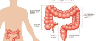
The oncologist named the early symptoms of colon cancer
Gynecological oncologists treat leiomyosarcoma of the uterine body. And, I must say, quite successfully. One of the leading scientific, treatment and preventive institutions for cancer in our country is the Herzen Cancer Center in Moscow. The clinic provides a wide range of modern methods of research and treatment of oncological diseases, including uterine cancer.
Malignant tumors of the female genital organs occupy a special place in oncology. It is these gynecological diseases that most often occur in women. What can you do, this is the scourge of modern society. Every year, the Herzen Oncology Center in Moscow provides specialized medical inpatient care to more than 11 thousand patients.
Treatment of leiomyosarcoma
The main treatment method for leiomyosarcoma is surgery. Surgery may involve limb-sparing tumor resection or amputation. The main thing is that the principle of radicalism must be observed so that not a piece of tumor remains.
Thanks to the development of modern medicine, mutilation operations have become much less common. They now account for less than 10% of patients. Amputation or disarticulation is done in extreme cases, when too much tissue and bone are involved in the oncological process or damage to the main neurovascular lines is observed.
For small pathologies located inside soft tissues, excision of the tumor may be prescribed, including 5 cm of surrounding healthy tissue. A more radical approach involves removing the leiomyosarcoma along with the musculofascial sheath in which it is located. The affected vessels and nerves are also cut out, and, if necessary, a section of skin.
Carrying out a radical operation is complicated by the lack of clear boundaries of the tumor, as well as its distribution in the thickness of the soft tissues over a large area.
If the sarcoma is in the bone, then during the operation the periosteum, a section of bone or the entire bone is also removed. Such interventions require further reconstruction. The removed fragments are replaced with prostheses or natural bone grafts taken from the patient himself or from a deceased person.
After surgery, the excised tumor and the edges of the tissue around it are examined in the laboratory. A positive outcome is the absence of sarcoma cells in the surrounding tissues. This indicates total removal of the tumor.
If the presence of cancer cells in the resection margin has been confirmed, then the chance of a relapse increases sharply.
In addition to the surgical method, for effective treatment of leiomyosarcoma it is necessary to use radiation therapy. It is prescribed preoperatively to reduce tumor burden, the risk of sarcoma dissemination during and after surgery, and the risk of recurrence. A total focal dose of 40-50 Gy is used. The irradiation zone includes the tumor and +5 cm of surrounding tissue. Neoadjuvant radiation therapy also has disadvantages. For example, there is a possibility of a wound occurring, due to which the operation will have to be postponed, wasting valuable time. Resection must be carried out no later than 3 weeks after the end of radiation therapy, since the growth of malignant cells may resume later.
Adjuvant (postoperative) radiation therapy is used much more often. Its advantage is that doctors already have accurate information about the prevalence of the tumor and its morphological structure. RT should begin 2-3 weeks after surgery, provided that the scar has already become stronger. The dose is increased to 60-70 Gy.
Chemotherapy for leiomyosarcoma is indicated for metastatic lesions. It is used for inoperable patients and those who refuse amputation. Most often, Doxorubicin is prescribed alone or in combination with other cytostatics (Vincristine, Cisplatin, Adriamycin).
For soft tissue sarcomas, Trabectedin and Pazopanib, which are classified as targeted drugs, began to be used. It is also worth noting the positive results from the drugs Sorafenib and Imatinib.
Since the symptoms of leiomyosarcoma in the initial stages are very scarce, the disease is often diagnosed in an advanced stage. Delaying treatment significantly reduces the chances of long-term survival. Very few cases of uterine leiomyosarcoma have been described, so there is no uniform standard of treatment for this pathology. Such neoplasms are treated in a similar way to sarcoma: the uterus, fallopian tube and ovaries are removed.
Radiation and chemotherapy for uterine leiomyosarcoma do not have a significant effect on patient survival. They can be used for inoperable patients, as well as in advanced cases to reduce the risk of metastasis.
Diagnosis of esophageal leiomyoma
Small esophageal leiomyomas that do not cause complaints in patients are usually diagnostic findings. If characteristic symptoms are present, consultation with a gastroenterologist suggests the presence of a neoplasm of the esophagus, but the diagnosis can only be verified based on data from radiological and endoscopic diagnostic methods.
When performing an X-ray of the esophagus, the characteristic signs of a neoplasm are determined: a filling defect in the area of the tumor; in the area of its localization, expansion of the esophagus is possible. According to the filling defect, a tumor shadow is visualized against the background of the mediastinum. With multiple nodes, intersecting contours are determined. This X-ray picture is not specific, since in addition to esophageal leiomyoma, it can occur with other tumors or cysts. Therefore, endoscopic examination is mandatory.
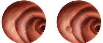
Esophagoscopy makes it possible to visualize the formation, especially if it is submucosal, assess the condition of the esophageal mucosa and perform an endoscopic biopsy. Histological evaluation of the biopsy is required to determine the benignity of the tumor, prognosis and selection of the optimal treatment method.
Therapeutic measures
Treatment of esophageal leiomyoma should be carried out in a comprehensive manner. If minor nodes are detected (the size of the nodes does not exceed 5 cm), the patient is registered and begins to be examined for the growth of the formation. Through regular endoscopic and x-ray examinations.
During this phase, drug therapy is prescribed using:
- sedatives;
- antiemetic drugs.
In medical practice, the main method of treatment for esophageal leiomyoma is surgical intervention, which is indicated in case of disruption of the esophagus and severe symptoms.
It is imperative to also take into account the likely risks of the operating method.
Traditional methods
Do not neglect herbal therapy for speedy healing of the esophagus.
The traditional medicine recipes presented in the list will help increase the effectiveness of the main treatment:
- Herbal mixture (St. John's wort, plantain, celandine, chamomile, galangal, calamus root, calendula, elecampane, tartar, chaga). The components are taken in 40 g quantities. The mixture is poured with 500 ml of water to cook for 5 minutes at medium boil. You need to drink the product 24 hours before.
- Tincture of mistletoe, periwinkle, tartar, taken 1 tbsp. l. The components are poured with 500 ml of boiling water and left for 2 hours. Course - 30 days. Only fresh tincture is accepted.
Concept of esophageal leiomyoma
Leiomyoma is a disease of the esophagus when benign tumors form on muscle tissue. There are several different types of manifestations. Tumors arise in single nodes. The contour of the formation is smooth without other manifestations. Tumors consisting of several nodes are rare. In some cases, such formations unite with each other, entwining the organ.
The localization of the tumor is in the thickness of the walls of the muscular epithelium of the esophageal tube. When the lesion enlarges, the tumor pushes the tissue apart. As the disease progresses, the mucous membrane becomes thin and elastic. If leiomyomas increase in size, then prolapse of the lumen of the esophagus occurs. The walls narrow, and the patient experiences dysphagia. Tumors consist of bundles of smooth muscle that alternate with fibrous tissue.
Most often, leiomyoma refers to a submucosal formation in the lower third of the esophagus. This is due to the fact that in the distal part of the organ the muscles are striated, which are replaced by smooth epithelium. These fibers become the main source of leiomyoma development.
Mechanisms of disease development
The formation of nodules along the wall of the esophagus is associated with the root cause of uncontrolled growth and division of epithelial cells. Because of this, smooth muscle tissue is formed in the middle layers of the esophageal tube. The process is associated with a violation of the dysfunction regulator. In this case, uncontrolled cell division occurs. It is not possible to pinpoint the exact reason for the failure of this function in medicine. However, the process is influenced by secondary factors.
The causes of the development of benign tumors are:
- chronic diseases of the esophagus;
- external factors – ecology;
- bad habits;
- hereditary predisposition;
- frequent consumption of junk food;
- chronic infections and diseases of the upper digestive tract;
- reduced immunity.
Leiomyoma of the esophagus develops as a result of prolonged esophagitis, reflux disease or gastritis. A poor environmental environment is associated with increased levels of radiation or toxic substances. This causes mutation of the cells of the human body.
When immunity decreases, the production of interleukins decreases. These cells produce substances that control the development of muscle tissue in the esophageal tube.
For the development of leiomyoma, heredity, nutrition and environmental conditions are considered significant reasons. However, regardless of the factor in the appearance of tumors of the muscle tissue of the esophagus, it is necessary to begin treatment of the disease in a timely manner. If the care is poor, then the affected areas die and hemorrhages occur. In addition, there are a number of complications of leiomyoma.
Symptoms of esophageal tumors
The specificity of manifestations is determined by the type of growth, localization and size of neoplasia; to a lesser extent, the histological structure influences the symptoms. Tumors growing into the lumen of the esophagus cause dysphagia - disruption of the passage of food masses through the esophagus: difficulty swallowing solid food, sensation of a lump behind the sternum. The severity of dysphagia increases as the tumor grows. Often with intraluminal neoplasia, moderate pain in the chest of a dull or spastic nature, a feeling of discomfort in the throat or chest, which intensifies when eating, is noted.
Symptoms may include drooling, nausea, belching, and regurgitation. Large intraluminal tumors often cause vomiting, causing patients to rapidly lose weight. Intraluminal tumors are often injured by food and ulcerate, which is accompanied by bleeding from the esophagus and anemia. Complete obstruction of the esophagus, as a rule, is not observed. Occasionally, intraluminal tumors on a long stalk migrate into the lumen of the larynx during vomiting, leading to asphyxia, sometimes with death.
Neoplasia with intramural growth is often located in the distal part of the esophagus and develops asymptomatically for a long time. Neoplasms that have reached significant sizes cause dysphagia, nausea, chest pain of moderate intensity, and loss of appetite. In the case of extraesophageal tumor growth, compression syndrome may develop, caused by compression of the mediastinal organs (vagus nerve, bronchi, large veins). Hoarseness of voice, increased chest pain, dry cough, tachycardia, and arrhythmia are noted. With esophageal cysts, suppuration and perforation can occur.
Symptoms
Leiomyoma of the esophagus develops without pronounced symptoms. Therefore, for a long time the patient may not be aware of the disease (in the absence of specialized diagnostics). Leiomyoma is characterized by slow growth. Based on this sign, the symptoms of the disease also appear slowly.
Over time, pronounced signs of leiomyoma appear:
- difficulty swallowing food;
- an uncomfortable feeling that occurs behind the sternum (a similar symptom begins to worry when the tumor reaches a diameter of up to four centimeters);
- the patient begins to notice the manifestation of frequent heartburn;
- belching is characteristic;
- pathological excessive salivation;
- a state of nausea that leads to frequent vomiting;
- arrhythmia is noted, and the heartbeat increases significantly;
- there is a lump in the throat, accompanied by a cough (dry) and a desire to clear the throat;
- appetite is significantly reduced.
Leiomyoma of the esophagus on x-ray
After eating food, swallowing becomes painful. The pain occurs behind the sternum and noticeably radiates to the back area.
When the size of the leiomyoma is small, it signals itself as a spasm of the esophagus. Sometimes discomfort occurs in the sternum area, which manifests itself as a sensation of a foreign body. It is worth noting that the general condition of the patient’s body is not disturbed, since the increase in symptoms occurs gradually.
Increasing tumor size can affect the heart. This is explained by increased pressure on the organ, which results in additional heart problems.
What is esophageal leiomyoma?
Additional symptoms
- Intensified cough of a dry nature, which occurs due to an interfering lump.
- Feeling short of breath.
- Effect on the vocal cords, leading to hoarseness.
- Cyanosis.
Additional signs of leiomyoma occur when a tumor-like formation is localized near the bifurcation of the trachea. Therefore, if the patient experiences some of the above symptoms, it is necessary to consult a gastroenterologist as soon as possible. If treatment is not started in a timely manner, there is a risk of degeneration into a malignant formation.
X-ray diagnosis of leiomyoma
Discussion
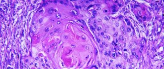
The size of the removed tumors ranged from 0.5 cm in diameter to 8.0×10.0 cm, with an average of 2.8±0.6×2.6±0.5 cm (Fig. 3). In 1 case, 2 adjacent tumors with a diameter of 0.5 and 1.5 cm were removed, creating a single filling defect of 2.0×2.0 cm on the radiograph.
Rice. 3. Leiomyoma of the esophagus (macro specimen).
Histological examination of macroscopic specimens in all operated patients confirmed a benign tumor of smooth muscle tissue (leiomyoma).
An intraoperative complication (damage to the esophagus) occurred in one patient, which was diagnosed on the operating table. Defect S.O. sewn up with an intracorporeal continuous suture with absorbent thread to restore the integrity of the muscular membrane. On the 2nd day after the operation, the sutures of the lower third of the esophagus failed, which was manifested by the X-ray picture of left-sided hydrothorax. After video-laparoscopic suturing of the perforated opening of the esophagus, video-laparotranshiatal drainage of the posterior mediastinum, video-assisted jejunostomy according to Meidl, and drainage of the left pleural cavity, scarring of the perforated opening was achieved.
In the second patient, perforation of the lower third of the thoracic esophagus occurred on the 7th day after right-sided video-assisted thoracoscopic enucleation of esophageal leiomyoma. To eliminate the complication, staged sanitary video-thoracoscopy on the right with drainage of the posterior mediastinum was performed, followed by video-laparotranshiatal drainage of the posterior mediastinum and video-assisted jejunostomy according to Meidl.
There were no deaths. The average duration of postoperative rehabilitation of patients was 18.2±7.8 days, the duration of inpatient treatment was 26.4±8 days. In 1 case, 7 months after surgery, cicatricial stenosis of the lower third of the esophagus developed. After excluding recurrence of the disease, the patency of the esophagus was restored after a single orthograde bougienage of the esophagus along the guide. No relapse of the disease was observed in any of the patients from 1 to 15 years after surgery.
Symptoms of leiomyosarcoma
The clinical picture of leiomyosarcoma depends on the location of the pathological process.
Leiomyosarcoma of the uterus
Cancerous degeneration of muscle fiber cells can be manifested by an increase in the size of the uterus, spontaneous bleeding from the female genital organs and periodic pain attacks in the pelvic area. Very often, uterine cancer develops against the background of a benign neoplasm (uterine leiomyoma).
Leiomyosarcoma of soft tissues
The disease, as a rule, is accompanied by the formation of a dense node in the thickness of the skin. The lesion has a round shape and is not connected to nearby structures. This type of tumor poses a particular danger to the patient’s life due to the formation of distant metastases without pain or any symptoms.
Leiomyosarcoma of the lung
Leiomyosarcoma of the lung is considered a fairly rare pathology. The clinical picture is practically no different from lung cancer. Signs of lung cancer do not appear at an early stage. Oncological symptoms develop as the tumor grows and include:
- coughing attacks;
- respiratory failure;
- shortness of breath;
- presence of blood particles in sputum;
- dysphagia;
- disruption of the passage of food through the esophagus;
- cancer intoxication.
Leiomyosarcoma of the abdominal cavity
A malignant neoplasm in the peritoneum is identified by a characteristic thickening of the anterior abdominal wall. During palpation of the pathological focus, doctors note the mobility of the tumor and skin. An increase in the volume of a malignant formation causes slight hyperthermia, general weakness and malaise.
Leiomyosarcoma of the liver
Liver cell sarcoma is a metastasis of a cancerous lesion of the muscle layer. The tumor process spreads through the venous system. The clinical picture consists of the following symptoms:
- frequent attacks of nausea and vomiting;
- chronic bloating;
- chills;
- decreased appetite;
- pain syndrome.
The existence of a secondary malignant neoplasm in the liver, as a rule, excludes a positive result of treatment.
Leiomyosarcoma of the stomach
Oncological formation of the muscle wall often takes the form of a limited compaction, the surface of which is covered with bleeding ulcers. For this tumor, the absence of metastases in regional lymph nodes and early secondary damage to the liver and lungs is considered typical. Symptoms include indigestion, heartburn, bloating and periodic vomiting mixed with blood.
Leiomyoma of the esophagus - tips and recommendations on News4Health.ru
Gastroenterology is a special discipline that studies the structure, physiology and pathology of the digestive system, as well as the prevention, diagnosis and treatment of diseases of the digestive system. Within gastroenterology there are narrower sections: for example, liver and gall bladder diseases are studied by hepatology, pathology of the large intestine and pararectal space - proctology. The normal functioning of the gastrointestinal tract is greatly influenced by the state of the endocrine system, oral cavity, and the presence of infectious agents in the body.
The digestive system includes a collection of many organs and glands that provide the nutrients the body needs for development and functioning. It permeates almost the entire body, starting with the oral cavity and ending with the rectum. Therefore, the pathology of even one of the digestive organs entails a disorder in the functioning of the digestive system as a whole. Modern ecology, stress, new foods and additives, as well as medications cause the occurrence or exacerbation of diseases of the gastrointestinal tract.
Diseases of the digestive system are so common that every person experiences their manifestations at least once in their life. Among diseases of the digestive tract, the undisputed leaders are inflammatory processes in its various parts, which have an acute or chronic course: inflammation of the esophagus (esophagitis), stomach (gastritis), duodenum (duodenitis), liver (hepatitis), pancreas (pancreatitis), gall bladder and ducts (cholecystitis and cholangitis), small and large intestines (enteritis and colitis), rectum (proctitis). They, in turn, cause changes in the mucous membrane of organs, their motor function and lead to the occurrence of stomach and duodenal ulcers, cholelithiasis, and ulcerative colitis. A serious problem in modern society is intestinal dysbiosis, which occurs in the vast majority of people, including newborns.
Diseases of the stomach, pancreas, and intestines are often the root cause of many skin and metabolic diseases. The symptoms of diseases of the digestive system are very diverse and depend on the affected organ. Common manifestations include loss of appetite, abdominal pain, belching, heartburn, nausea, vomiting, flatulence, upset stool, weakness, weight loss.
Modern gastroenterology has great diagnostic capabilities that make it possible to recognize diseases of the digestive system in the early stages of their occurrence. Laboratory tests include tests of blood, gastric and duodenal juice, feces, intestinal microflora, and tissue samples taken during biopsy. To examine the hollow organs of the gastrointestinal tract, X-ray and endoscopic methods are used (esophagoscopy, colonoscopy, gastroduodenoscopy, etc.), and for parenchymal organs, ultrasound diagnostics is used. Many chronic diseases of the digestive system, which subsequently require surgical intervention, are caused by neglect or undertreatment of an acute condition. Therefore, the occurrence of any pathological manifestation in the digestive system should serve as a signal for immediate contact with a gastroenterologist. When treating diseases of the digestive system, the patient will be required to strictly follow the treatment regimen and diet.
Reasons for the growth of esophageal leiomyoma
The exact causes of the disease have not yet been established. The negative influence of some factors that increase the likelihood of illness has been confirmed:
- hereditary predisposition, since a survey of patients revealed the presence of oncopathology of the digestive tract in close relatives;
- harmful addictions (alcohol, smoking);
- improper diet (consumption of mainly spicy, smoked, hot, cold foods, hot spices);
- background pathology of the esophagus (cysts, diverticula, inflammation).
Due to the influence of the above factors, the process of cell division and death is disrupted, which is why their uncontrolled reproduction begins with the appearance of nodular formations.
Classification of leiomysarcoma
Based on their origin, muscle sarcomas are divided into the following subtypes:
- soft tissue leiomyosarcoma (arising from the muscles of the lower and upper extremities, on the trunk and head);
- leiomyosarcoma of bones (bones are often involved in the tumor process with soft tissue tumors);
- leiomyosarcoma of internal organs (affects the muscle layer in the lungs, uterus, stomach, and sometimes the genitals);
- leiomyosarcoma of the skin (arises primarily from the subcutaneous muscles. A tumor from the soft tissues can spread to the skin as it grows);
- leiomyosarcoma of vascular origin (occurs primarily from the walls of arteries and veins. Vessels can also be affected secondarily when tumors of bone, soft tissue, etc. spread).
They can have different histological structures, depending on which the following types of leiomyosarcoma are distinguished:
- spindle cell;
- pleomorphic;
- epithelioid;
- myxoid;
- inflammatory.
It is also customary to subdivide tumors into 2 degrees of malignancy or differentiation (in the classification they are designated by the letters G1 and G2). They are determined depending on cell differentiation, the number of mitoses and necrosis. Mostly high-grade or low-grade leiomyosarcoma (G2) are found, which grow rapidly and spread throughout the body. For such patients, the prognosis is not very good.
If a well-differentiated grade G1 leiomyosarcoma was diagnosed, the prognosis is much better, since such tumors grow slowly and rarely form metastases.