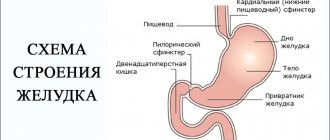
When is the procedure indicated?
The examination is carried out if diseases and pathologies are suspected that may pose a serious danger to the patient. In most cases, barium X-ray of the stomach allows you to confirm or clarify the diagnosis and prescribe the correct treatment.
Suspicion of the formation of gastric and duodenal ulcers
Peptic ulcer of the stomach and duodenum (DPC) leads to the periodic appearance of ulcers. It can take years to develop and cause serious harm to health. Using a barium X-ray of the stomach, the specialist finds the ulcer and evaluates its size, shape and other parameters, as well as establishes the stage of the disease and detects accompanying symptoms.
Detection of a malignant process in an organ
If the development of a malignant process is suspected, an X-ray of the stomach with barium is recommended without fail, since the consequences of the disease can be deadly for the patient. In the photographs, the cancerous process looks like a defect in the mucous membrane. Often, after a barium X-ray of the stomach, an endoscopic examination is performed and a sample of the tumor is taken for laboratory testing.
Diverticulosis and other deformities of the gastric walls
To diagnose wall protrusion (diverticulosis), a barium X-ray of the stomach requires additional preparation. After administration of the contrast agent, the patient alternately lifts the upper and lower parts of the body so that the contrast fills the entire organ cavity. On a barium X-ray of the stomach, small diverticulosis looks like an ulcer, but has a horizontal level and a neck.
Any inflammation of the stomach
Foci of inflammation in the stomach can appear as a result of various processes. Including peptic ulcers or oncology. An X-ray of the stomach with barium allows you to find the source of inflammation and, based on indirect signs, determine its cause.
Swallowing dysfunctions
Dysphagia, or swallowing dysfunction, is often expressed by the patient complaining of the inability to swallow food and its accumulation in the esophagus. In this case, the patient cannot always accurately indicate the location of the obstacle. Therefore, barium radiography of the stomach is used to accurately localize the pathology.
Pain in the abdominal area or navel area
Pain in the navel and abdominal area may indicate the development of malignant processes in the stomach and duodenum. Therefore, with such symptoms, the specialist often refers the patient to an X-ray of the stomach and duodenum with barium.
What is an X-ray examination?
X-ray examination is an integral component in the study of the digestive system. This procedure gives an idea of the state of the digestive organs.
The entire picture of the study is reflected on the screen of the X-ray machine. After that, the patient is given images of the organs and, if necessary, treatment is prescribed.
X-rays are performed as indicated by a gastroenterologist if there is a suspicion of damage or dysfunction of the stomach or esophagus.
The study checks the following factors:
- Detection of defects in the larynx (pharynx);
- Elimination of the formation of pathology during the transition from the pharynx to the esophagus;
- Checking the diaphragm;
- Identification of neoplasms and abnormalities in the development of the cardiac part of the stomach.
X-ray of the stomach does not require special preparation. There is no need to follow a diet here, as with other studies. True, there are small restrictions. The last meal should be no later than 8-9 hours before the start of the procedure.
If the patient is prescribed irrigoscopy of the rectum using a barium enema, then preparation for the procedure is carried out according to the following scheme: in the evening, the patient is given a cleansing enema 12 hours before the proposed examination.
In the morning, 3 hours before fluoroscopy, another additional intestinal enema is performed. Before the fluoroscopic examination, a consultation with specialists is carried out: a gastroenterologist, a radiologist, a diagnostician.
This research method is mandatory for everyone who has pathological processes in the gastrointestinal tract. Radiography makes it possible to detect diseases in the early stages, including tumors, and avoid that stage of the disease that is difficult to treat.
What can be detected with radiography?
If a barium stomach x-ray is performed correctly and with proper patient preparation, the examination can provide enough comprehensive information to make a diagnosis in most cases. At the same time, to detect a particular disease, a specialist evaluates a number of parameters.
Gastric lumen disorders
Disturbances in the lumen of the stomach may indicate the development of a malignant process or diverticulosis. If the lumen of the stomach is narrowed on a barium X-ray, diffuse fibroplastic cancer is possible. With diverticulosis, on a barium X-ray of the stomach, the lumen appears larger than it should be normally.
Abnormal placement of the stomach
Gastroptosis (prolapse of the stomach) is often diagnosed with an abdominal wall hernia or diaphragmatic hernia. The disease is clearly visible using barium X-ray of the stomach.
Niche symptom
A niche is a shadow of a contrasting mass that fills a defect during an ulcer or the development of a malignant process. Depending on the location of the defect, an X-ray of the stomach with barium distinguishes between a contour and a relief niche. In the first case, the silhouette of the defect is visible from the full face, in the second - in profile.
The lack of filling is visible in the image as a dark area
A dark area on a barium x-ray of the stomach indicates that there is a place where the contrast could not reach. As a rule, this happens with atrophic gastritis or tumor.
How to remove barium from the body after irrigoscopy
There is hardly a person on Earth who has not had a stomach ache, diarrhea, or other intestinal dysfunction at least once in his life.
Often these disorders go away on their own or with medication.
But sometimes the symptoms of intestinal problems become persistent, which indicates pathologies in the digestive system. Abdominal pain is familiar to almost all people
To identify them, various methods of examining the gastrointestinal tract (GIT), including intestinal fluoroscopy, are used.
When is fluoroscopy prescribed?
The decision to perform fluoroscopy is made by the doctor. Typically, the procedure is prescribed when a person experiences the following signs of gastrointestinal distress:
- chronic constipation,
- persistent diarrhea,
- significant weight loss over a relatively short time with the usual diet,
- change in color of stool to black in the same situation,
- vomiting blood,
- constant pain after eating or defecating.
Indications for fluoroscopy include a number of gastrointestinal diseases, including cancer.
The use of x-rays allows us to evaluate the anatomical changes in the intestine caused by pathologies or age (x-ray anatomy).
What is the essence of this research method?
The fluoroscopy technique involves irradiating the area of the body to be examined with x-rays. Radiation allows you to show a picture of what is happening inside the body, which is recorded on an x-ray. Obtaining an image using an x-ray is called radiography.
To perform an X-ray examination, a contrast agent – barium – is used. It is not barium itself that is used, but its sulfate (sulfate salt), since it is non-toxic. Contrast is necessary to increase the contrast of the photo and get a clear image.
- If a fluoroscopic examination of the upper gastrointestinal tract (esophagus, stomach and small intestine) is to be performed, the patient is given a barium suspension (a mixture prepared in the proportion of 80 grams of barium sulfate per 100 milliliters of water) to drink. If necessary, double contrast can be used - sodium citrate and sorbitol are added to the suspension.
The barium passage goes from top to bottom
The passage of barium sulfate through the intestines occurs as follows. After an hour, the contrast reaches the small intestine. After three hours it is in the transition between the small and large intestines. Within six hours, barium enters the ascending colon. After twelve hours it reaches the sigmoid colon, and after 24 hours it reaches the rectum.
Thus, the normal passage time is approximately a day. During irrigoscopy, the solution is administered through the rectum
- In this regard, for X-ray diagnostics of the lower sections - the colon and rectum (irrigoscopy, irrigography) enemas are used (according to the instructions - 750 grams of barium per liter of a half-percent tannin solution).
The resulting image is examined and analyzed by a radiologist. Based on the results of the analysis, he writes a conclusion, which is sent to the attending physician.
Some people are afraid of procedures using x-rays. They are afraid that radiation may cause cancer. There is no need to be afraid of this.
Modern radiology is at a high enough level to ensure the safety of the person being examined.
X-ray examination of the gastrointestinal tract is not performed quickly (sometimes it takes several hours). Does it hurt? No, although some discomfort is felt. However, there is much less discomfort than, for example, during an endoscopic examination. Therefore, not only an adult, but also a child can endure the procedure without any problems.
X-ray of the intestines with barium: what does the procedure show?
Fluoroscopy allows you to obtain sufficiently complete information to make a correct diagnosis and prescribe an adequate course of treatment for the disease. In particular, the doctor receives an overview of the intestine being examined:
- its shape,
- external diameter,
- lumen
What else does fluoroscopy show? It allows the doctor to see the causes that disrupt the normal functioning of the intestines (erosions, ulcers, worms, tumors).
Before proceeding with fluoroscopy, the patient takes off his outer clothing and all metal objects, and puts on a hospital gown.
Source: https://kishechnik-zhivot.ru/voprosy/kak-vyvesti-barij-iz-organizma-posle-irrigoskopii
How should a patient prepare for the procedure?
Since the patient needs time to prepare for a barium stomach x-ray, it is best to start a few days before the examination. The first step is to exclude from the diet foods that cause increased peristalsis and gas formation. 8 hours before the examination you should completely abstain from food. To relax the stomach before the x-ray and prepare it for filling with barium, the doctor may recommend taking a special drug half an hour before the examination. Contrast is administered orally a few minutes before the procedure. When a double contrast examination is required, after the patient has prepared for a stomach x-ray and taken barium, he will need to drink a gas-forming mixture.
When is x-ray prohibited?
An X-ray of the stomach with barium is a safe examination, but since it is done using ionizing radiation and a contrast agent, the method has a number of contraindications.
Hematopoietic disorders caused by pathological conditions of the bone marrow
Ionizing radiation can worsen the disease. Therefore, if hematopoiesis is disrupted, an X-ray of the stomach with barium is not performed.
Cataract
When X-raying the stomach with barium, the radiation dose does not exceed permissible limits. However, with cataracts, even a limited amount of ionizing radiation may be enough to have a negative effect on the condition of the lens.
Malignant neoplasms of the bronchi and lungs
The examination is contraindicated if the patient is undergoing radiation or chemotherapy treatment.
Pregnancy
During pregnancy, even minor radiation can have a detrimental effect on the development of the fetus. Therefore, X-rays of the stomach with barium are contraindicated for expectant mothers.
Childhood (before the onset of puberty)
In childhood, the body reacts poorly to exposure to even small doses of ionizing radiation. Therefore, doctors refer children for an X-ray of the stomach with barium only if absolutely necessary.
Indications for use
The procedure is carried out not only when alarming symptoms are detected. If a patient has been sick for a long time with a serious gastrointestinal disease, he requires regular and thorough examination for control.
Symptoms that require examination:
- severe pain of an unknown nature in the abdomen and behind the sternum;
- heartburn;
- causeless vomiting and nausea;
- discharge of gas from the mouth with a sour odor;
- belching;
- disturbance in the digestive system;
- rapid weight loss;
- reflux;
- bloody traces in stool;
- anemia with unknown causes;
- difficulty swallowing;
- abdominal pain symptom.


