Nosebleeds are bleeding from the inside of the nasal cavity or from the nasopharynx. It can appear in two places in the nasal region: in the anterior sections of the nose (this place is called Kisselbach's) and in the inferior turbinate of the anterior sections of the nose.
There is also posterior bleeding, which occurs in the back of the nose and nasopharynx (inferior turbinate or vault). This condition is most often observed in children under 10 years of age and in people over 50 years of age.
How to stop: first aid
When such a problem occurs, you need to know how to provide emergency care for a patient with nosebleeds in order to help him as much as possible. The first aid algorithm for nosebleeds will be as follows:
- Before providing 1st aid for nosebleeds, it is assessed how serious the patient’s condition is. It is necessary to immediately assess this phenomenon, whether it is possible to cope with blood loss on your own or whether you will have to wait for help from doctors.
- Then you need to initially calm down yourself and reassure the victim. Asking the person to start breathing deeply will reduce the emotional burden, lower the heart rate, and may prevent a spike in blood pressure. Since all these circumstances can worsen the situation.
- First aid for nosebleeds is performed in this way: make the person sit more comfortably. It is important that the victim’s head leans forward, so the blood fluid will flow out without obstruction.
- The nostril from which bleeding is observed should be pressed against the septum and held there for several minutes. After these actions, a blood clot forms in the area of the damaged vessel.
- You will need to drip any vasoconstrictor drops from the Naphthyzin, Galazolin, etc. series into the nasal passages. 6-8 drops in each nasal section.
- Then, a few (8-10) drops of 3% hydrogen peroxide are dripped into both nasal openings.
- Apply a wet towel or other cold object to the nose area. This compress is kept for 15-20 minutes, after which a pause is made for 3-4 minutes. The action is repeated up to 2-3 times.
- Another way to provide first aid for nosebleeds is to immerse your hands in cool water and your feet in warm water. Due to this, the walls of the blood vessels narrow, and blood fluid soon stops flowing.
During the period of nosebleeds, first medical aid is extremely important; the person’s further condition will depend on it. If the condition has been resolved, then in the near future you should not drink hot drinks or eat hot dishes, or engage in intense sports. It is advisable to see a doctor if this has not already been done.
ICD-10 in gynecology
Alphabetical index of disease names and ICD 10 codes in obstetrics and gynecology
| ICD-10 | Heading | ICD-9 |
| Infections transmitted predominantly sexually (A50 – A64) | ||
| A50 | Congenital syphilis | 090 |
| A51 | Syphilis early | 091, 092 |
| A52 | Late syphilis | 093 |
| A53 | Syphilis, other forms | 097 |
| A54 | Gonococcal infection | 098 |
| A54.0 | Gonococcal cervicitis | 098.1 |
| Gonococcal cystitis | 098.1 | |
| Gonococcal urethritis | 098.0 | |
| Gonococcal vulvovaginitis | 098.0 | |
| A54.1 | Gonococcal abscess of Bartholin's gland | 098.0 |
| A55 | Chlamydial lymphogranuloma | 099.1 |
| A56 | Other chlamydial infections | 099.8 |
| A57 | Chancroid | 099.0 |
| A58 | Granuloma inguinale | 099.2 |
| A59 | Trichomoniasis | 131 |
| A59.0 | Urogenital trichomoniasis | 131.0 |
| A60 | Anogenital herpetic viral infection | |
| A60.0 | Herpetic infection of the genital organs and genitourinary tract | 054.1 |
| A60.9 | Anogenital herpetic infection, unspecified | |
| Z21 | HIV infection is asymptomatic | |
| Malignant neoplasms | ||
| C50 | Mammary gland | 174 |
| C51 | Vulvas | 184.4 |
| C52 | Vaginas | 184.0 |
| C53 | Cervix | 180 |
| C54 | Body of the uterus | 182 |
| C55 | Uterus, unspecified | 179 |
| C56 | Ovarian | 183.0 |
| C57 | Other female genital organs | |
| C58 | Placenta | 181 |
| C67 | Bladder | 188 |
| C80 | Unspecified localization | 199 |
| Class 14 Diseases of the female genital organs Units: Inflammatory diseases of the female pelvic organs (N70-N77); Non-inflammatory diseases of the female genital organs (N80-N90); Menstrual disorders (N91-N98). | ||
| Inflammatory diseases of the female pelvic organs (N70-N77) | ||
| N70 | Adnexit | 614.2 |
| N70.0 | Adnexitis acute | 614.0 |
| N70.1 | Chronic adnexitis | 614.1 |
| Abscess: | ||
| N76.4 | —> vulvas | 616.4 |
| N75.1 | —> Bartholin gland | 616.3 |
| N71.0 | —> uterus | 615.9 |
| N70.0 | —> fallopian tube | 614.2 |
| N70.0 | —> tubo-ovarian | 614.2 |
| N73.0 | —> parametrium | 614.4 |
| N73.0 | —> broad ligament | 614.4 |
| N70.0 | —> ovary | 614.2 |
| Bartholin gland: | ||
| N75.1 | Abscess | 616.3 |
| N75.0 | Cyst | 616.2 |
| N75.8 | Bartholinitis | 616.8 |
| Vulvas | ||
| N76.4 | Abscess | 616.4 |
| N76.0 | Acute vaginitis | 616.1 |
| N76.1 | —> chronic | 616.1 |
| N95.2 | —> atrophic | 627.3 |
| N76.2 | Vulvitis acute | 616.1 |
| N76.3 | —> chronic | 616.1 |
| N76.0 | Acute vulvovaginitis | 616.1 |
| N76.1 | —> chronic | 616.1 |
| N70.1 | Hydrosalpinx | 614.2 |
| N71.0 | Acute metritis | 615.0 |
| N71.1 | —> chronic | 615.1 |
| N71.0 | Acute myometritis | 615.0 |
| N71.1 | —> chronic | 615.1 |
| N70.0 | Acute oophoritis | 614.0 |
| N70.1 | —> chronic | 614.1 |
| N73.0 | Acute parametritis | 614.3 |
| N73.1 | —> chronic | 614.4 |
| Pelvic peritonitis: | ||
| N73.3 | —> spicy | 614.5 |
| N73.4 | —> chronic | 614.7 |
| N73.4 | Pyometra | 615.9 |
| N70.0 | Pyosalpinx | 614.2 |
| N70.0 | Acute salpingitis | 614.0 |
| N70.1 | —> chronic | 614.1 |
| N70.0 | Acute salpingo-oophoritis | 614.0 |
| N70.1 | —> chronic | 614.1 |
| N73.6 | Pelvic adhesions | 614.6 |
| N73.6 | —> peritoneal | 614.6 |
| N73.6 | —> intrauterine | 614.6 |
| N73.0 | Phlegmon pelvic | 614.4 |
| N76.4 | Furuncle of the vulva | 616.4 |
| N72 | Cervicitis | 616.0 |
| N72 | Endocervicitis | 616.0 |
| N72 | —> with erosion | 616.0 |
| N72 | —> with ectropion | 616.0 |
| N72 | Exocervicitis | 616.0 |
| N71.0 | Acute endomyometritis | 615.0 |
| N71.1 | —> chronic | 615.1 |
| N76.5 | Vaginal ulcer | 616.8 |
| N76.6 | —> vulvas | 616.5 |
| Non-inflammatory diseases of the female genital organs (N80-N90) | ||
| N90.5 | Vulvar atrophy | 624.1 |
| N83.3 | —> ovary | 620.3 |
| N85.4 | Anteversion | 621.6 |
| N97 | Infertility | 628.9 |
| N85.5 | Inversion of the uterus | 621.7 |
| N81.4 | Uterine prolapse | 618.3 |
| N81.2 | Incomplete uterine prolapse | 618.2 |
| N81.3 | —> full | 618.3 |
| N83.4 | Ovarian prolapse | 620.8 |
| N85.7 | Hematometer | 621.4 |
| N83.6 | Hematosalpinx | 620.8 |
| N83.7 | Broad ligament hematoma | 620.7 |
| N85.0 | Endometrial hyperplasia | 621.3 |
| N85.2 | Uterine hypertrophy | 621.2 |
| N90.6 | Vulvar hypertrophy | 624.3 |
| N83.4 | Ovarian hernia | 620.4 |
| N87 | Cervical dysplasia | 622.1 |
| N89.3 | —> vagina | 623.0 |
| N90.3 | —> vulvas | 624.8 |
| N90.7 | Vulvar cyst | 624.8 |
| N83.0 | —> ovarian follicular | 620.0 |
| N83.1 | —> corpus luteum | 620.1 |
| N83.2 | —> ovarian retention | 620.2 |
| N88.0 | Leukoplakia of the cervix | 622.2 |
| N89.4 | —> vagina | 623.1 |
| N90.4 | —> vulvas | 624.0 atrophy 624.8 |
| N88.3 | Isthmic-cervical insufficiency | 622.5 |
| N85.4 | Incorrect position of the uterus | 621.6 |
| N85.5 | Ovarian torsion | 620.5 |
| N84.0 | Polyp of the uterine body | 621.0 |
| N84.1 | —> cervix | 622.7 |
| N84.2 | Vaginal polyp | 623.7 |
| N84.3 | —> vulvas | 624.6 |
| N96 | Habitual miscarriage | 634 |
| E28.2 | Polycystic ovary syndrome | 256.4 |
| N85.4 | Retroflexion | 621.6 |
| N85.4 | Retroversion | 621.6 |
| N81.4 | Rectocele | 618.0 |
| N88.1 | Old cervical rupture | 622.3 |
| N89.8 | —> vagina old | 623.4 |
| N83.8 | —> broad ligament | 620.6 |
| N82.0 | Vesicovaginal fistula | 619.0 |
| N82.1 | —> urethrovaginal | 619.0 |
| N85.6 | Intrauterine synechiae | 621.5 |
| N88.2 | Cervical stricture | 622.4 |
| N89.5 | —> vagina | 623.2 |
| N90.5 | —> vulvas | 624.8 |
| E28.2 | Stein-Leventhal syndrome | 256.4 |
| N81.0 | Urethrocele | 618.0 |
| N81.1 | Cystocele | 618.0 |
| N80 | Endometriosis | 617 |
| N80.0 | —> uterus | 617.0 |
| N80.1 | —> ovaries | 617.1 |
| N80.3 | —> pelvic peritoneum | 617.3 |
| N81.5 | Vaginal erythrocele | 623.8 |
| N86 | Cervical erosion | 622.0 |
| N86 | Cervical ectropion | 622.0 |
| N89.8 | Vaginal ulcer | 623.8 |
| Menstrual disorders (N91-N98) | ||
| N91.0 | Primary amenorrhea | 626.0 |
| N92.1 | —> secondary | 626.0 |
| N91.2 | NOS | 626.9 |
| N94.0 | Pain in the middle of the menstrual cycle | 625.2 |
| N91.5 | Hypomenorrhea NOS | 626.1 |
| N94.1 | Dyspareunia | 625.0 |
| N94.4 | Primary dysmenorrhea | 625.3 |
| N94.5 | —> secondary | 625.3 |
| N94.6 | —> NOS | 625.9 |
| N92 | Menstrual bleeding is heavy, frequent and irregular | |
| —> abundant and frequent | 626.2 | |
| N92.0 | —> —> with a regular cycle | 626.2 |
| N92.1 | —> —> for irregular cycles | 626.2 |
| N92.2 | —> abundant at the beginning of the cycle | 626.3 |
| N92.4 | —> —> in the premenopausal period | 627.0 |
| N92.3 | —> ovulatory | 626.5 |
| N92.6 | —> irregular NOS | 626.4 |
| N95.0 | —> in the postmenopausal period | 627.1 |
| N64.6 | False menstruation | 626.6 |
| N92.0 | Menorrhagia NOS | 626.4 |
| N92.2 | —> puberty | 626.3 |
| N92.4 | —> menopausal | 627.0 |
| N92.4 | Menorrhagia premenopausal | 627.0 |
| N92.1 | Metrorrhagia with irregular cycles | 626.6 |
| N92.4 | —> menopausal | 627.0 |
| N92.4 | —> premenopausal | 627.0 |
| N92.1 | Menometrorrhagia | 626.2 |
| N92.3 | Ovulatory bleeding | 626.5 |
| N91.3 | Oligomenorrhea primary | 626.1 |
| N91.4 | —> secondary | 626.1 |
| N91.5 | —> NOS | 626.1 |
| N91.2 | Lack of menstruation | 626.0 |
| N91.3 | Scanty menstruation | 626.1 |
| N94.3 | Premenstrual tension syndrome | 625.4 |
| N95.1 | —> menopause | 627.2 |
| N92.0 | Polymenorrhea | 626.2 |
| N92.2 | Pubertal menorrhagia | 626.3 |
| N92.2 | —> bleeding | 626.3 |
| N95.0 | Postmenopausal bleeding | 627.1 |
Pregnancy, childbirth and the postpartum period (O00 – O99) Blocks: 1) Complications of pregnancy:
2) Complications of childbirth:
3) Postpartum complications:
4) Abortion and ectopic pregnancy | ||
| Somatic complications of pregnancy | ||
| O20 | Abortion is threatening | 640.0 |
| O99.0 | Anemia – see “Somatic diseases” | |
| O29 | Anesthesia complications: | |
| O29.0 | —> Pulmonary | |
| O29.1 | —> Cardiological | |
| O29.2 | —> CNS | |
| O29.3 | —> With local anesthesia | |
| O29.4 | —> With spinal | |
| O29.5 | —> For epidural | |
| O29.6 | —> During intubation | |
| O29.8 | —> Others | |
| Pregnancy: | ||
| Z35.5 | —> Primipara old | 659.5 |
| Z35.6 | —> Very young | 659.8 |
| Z35.4 | —> Multiparous | 659.4 |
| O30 | —> Multiple | 651.9 651.0 twins 651.1 triplets 651.9 unspecified |
| Somatic diseases | ||
| O99 | Anemia | 648.2 |
| O99.1 | Other blood diseases | |
| O99.2 | Diseases of the endocrine system (except diabetes mellitus, see) | |
| O99.3 | Mental disorders | 648.4 |
| O99.4 | Diseases of the circulatory system | 648.6 |
| O99.5 | Respiratory diseases | 648.9 |
| O99.6 | Digestive diseases | 648.9 |
| O99.8 | Other diseases | 648.9 |
| O23.5 | Vaginitis | 646.6 |
| Phlebeurysm: | ||
| O22.0 | —> lower extremities | 671.0 |
| O22.1 | —> crotch | 671.1 |
| O22.4 | Haemorrhoids | 671.8 |
| O26.5 | Hypotensive syndrome | 669.2 |
| Hypertension: | 642 | |
| O10.0 | —> existing before pregnancy | 642.0 |
| O10.9 | —> existed before pregnancy without proteinuria and preeclampsia | 642.2 |
| O11 | —> existing before pregnancy with associated proteinuria and/or preeclampsia | 642.2 |
| —> caused by pregnancy | ||
| O13 | —> caused by pregnancy without proteinuria | 642.3 |
| O14 | —> caused by pregnancy with significant proteinuria | 642.4 |
| O13 | —> caused by pregnancy with preeclampsia l/st | 642.4 |
| O14 | —> caused by pregnancy with preeclampsia s/st | 642.4 |
| O14.1 | —> caused by pregnancy with preeclampsia t/st | 642.5 |
| Diabetes mellitus: | ||
| O24.4 | —> developed during pregnancy | 648.0 |
| O24.0 | —> existing before pregnancy | 648.0 |
| O24.0 | —> pre-pregnancy insulin dependent | 648.0 |
| O24.1 | —> pre-pregnancy insulin-independent | 648.0 |
| Urinary tract infection during pregnancy: | ||
| O23.0 | —> Paranephritis | 646.6 |
| O23.0 | —> Pyelonephritis | 646.6 |
| O23.1 | —> Cystitis | 646.6 |
| O23.8 | —> Cervicitis | 646.6 |
| O23.2 | —> Urethritis | 646.6 |
| Bleeding: | ||
| O20.8 | —> Bleeding in early pregnancy (up to 22 weeks) | 640.8 |
| O20.0 | —> Threatened abortion | 640.0 |
| O30 | Multiple pregnancy | 651.9 |
| O14.0 | Nephropathy in pregnancy-induced hypertension | 642.4 |
| Eating disorders: | ||
| O25 | —> Inadequate nutrition | |
| O26.0 | —>Excessive weight gain | 646.1 |
| O26.1 | —> Insufficient weight gain | |
| O12.0 | Swelling caused by pregnancy: | 646.1 |
| O12.2 | —> Edema with proteinuria | 646.2 |
| O23.0 | —> Paranephritis | 646.6 |
| O23.0 | —> Pyelonephritis | 646.6 |
| O12.1 | —> Proteinuria caused by pregnancy | 646.2 |
| O12.2 | —> Proteinuria with edema | 646.2 |
| Proteinuria with hypertension: | ||
| O13 | —> insignificant | 642.4 |
| O14 | —> significant | 642.4 |
| Preeclampsia with hypertension: | ||
| O13 | —> mild degree | 642.4 |
| O14.0 | —> medium degree | 642.4 |
| O14.1 | —> severe | 642.3 |
| Vomiting of pregnant women: | ||
| O21.0 | —> easy | 643.0 |
| O21.1 | —> heavy (up to 22 weeks) | 643.1 |
| O21.2 | —> late (after 22 weeks) | 643.2 |
| Diabetes mellitus – see “Diabetes” | ||
| O22.2 | Superficial thrombophlebitis of the lower extremities | 671.2 |
| O22.5 | Cerebral vein thrombosis | 671.5 |
| O22.0 | Threatened abortion | 640.0 |
| O22.3 | Phlebothrombosis of the deep veins of the lower limb | 671.3 |
| O15.0 | Eclampsia during pregnancy | 642.6 |
Obstetric complications of pregnancy:
| ||
| Malpresentation | ||
| O32.4 | High head position | 652.5 |
| O32.2 | Oblique position of the fetus | 652.3 |
| O32.3 | Facial presentation | 652.4 |
| O32.3 | Submental presentation | 652.4 |
| O32.0 | Unstable position | 652.0 |
| O32.2 | Transverse position | 652.3 |
| O32.1 | Breech presentation | 652.1 |
| O32.6 | —> Combined | 652.8 |
| Discrepancy between the size of the pelvis and the fetus | ||
| O33.0 | Deformation of the pelvic bones | 653.0 |
| O33.1 | Evenly narrowed pelvis | 653.1 |
| O33.2 | Narrowing of the pelvic inlet | 653.2 |
| O33.3 | Narrowing of the pelvic outlet | 653.3 |
| O33.4 | Disproportion of mixed maternal and fetal origin | 653.4 |
| O33.5 | Large fruit size | 653.5 |
| O33.6 | Fetal hydrocephalus | 653.6 |
| O33.7 | Other fetal anomalies: ascites, hydrops, myelomeningocele, sacral teratoma, tumor | 653.7 |
| Abnormalities of the pelvic organs | ||
| O34.0 | Congenital anomalies of the uterus | 654.0 |
| O34.1 | Tumor of the uterine body | 654.1 |
| O34.2 | Postoperative uterine scar | 654.2 |
| O34.5 | Others (pinching, prolapse, retroversion) | 654.3 |
| O34.3 | Isthmic-cervical insufficiency | 654.5 |
| O34.4 | Cervical abnormalities (polyp, tumor, postoperative scar, stenosis) | 654.6 |
| O34.6 | Vaginal abnormalities | 654.7 |
| O34.7 | Abnormalities of the perineum and vulva | 654.8 |
| O34.8 | Other anomalies (cystocele, rectocele, rigid pelvic floor) | 654.9 |
| Maternal complications of childbirth (obstetric trauma - see Trauma) | ||
| O75.4 | Anoxia cerebral | 669.4 |
| O74 | Anesthesia - complications | 668 |
| O74.0 | Aspiration pneumonia | 668.0 |
| O74.1 | Other pulmonary complications | 668.0 |
| O74.2 | From the side of the heart | 668.1 |
| O74.3 | From the side of the central nervous system | 668.2 |
| O74.4 | With local anesthesia | 668.8 |
| O74.6 | With spinal | 668.8 |
| O74.6 | With epidural | 668.8 |
| O74.7 | During intubation | 668.8 |
| O75.0 | Mother's distress | 669.0 |
| Prolonged labor | ||
| O63.0 | —> Protracted 1st period | 662.0 |
| O63.1 | —> Prolonged 2nd period | 662.2 |
| O63.2 | —> Delayed birth of the 2nd fetus | 662.3 |
| Difficult labor | ||
| O66.0 | Impacted shoulder | 660.4 |
| O66.2 | Unusually large fruit (more than 4500 g - R 08.8) | 660.9 |
| O66.3 | Other fetal anomalies: ascites, hydrops, meningomyelocele, sacral teratoma, tumor | 660.8 |
| O66.8 | Other types of obstructed labor | 660.8 |
| Bleeding during childbirth | ||
| O67.0 | Bleeding disorder | 641.3 |
| O67.8 | Other | 641.8 |
| O75.2 | Fever during labor (hyperthermia) | 659.2 |
| Labor disorders | ||
| O62.0 | Primary weakness | 661.0 |
| O62.1 | Secondary weakness | 661.1 |
| O62.2 | Others (uterine atony, irregular contractions, weak contractions, weak labor NOS) | 661.2 |
| O62.3 | Rapid labor | 661.3 |
| O62.4 | Uncoordinated uterine contractions | 661.4 |
| O62.4 | Hypertensive uterine contractions | 661.4 |
| O75.3 | Septicemia during childbirth | 659.3 |
| O75.4 | Heart failure during childbirth | 669.4 |
| Obstetric trauma: | ||
| O71.2 | Postpartum uterine inversion | 665.2 |
| O71.0 | Uterine rupture before labor begins | 665.0 |
| O71.1 | —> —> during childbirth | 665.1 |
| O71.3 | —> cervix | 665.3 |
| O71.4 | —> upper vagina | 665.4 |
| O71.5 | —> bladder | 665.5 |
| O71.5 | -> urethra | 665.5 |
| O71.6 | Injuries to the pelvic joints and ligaments | 665.6 |
| Perineal tears: | ||
| O70.0 | 1st degree rupture: posterior commissure, labia, skin, superficial | 664.0 |
| O70.1 | —> 2nd degree (pelvic floor, vaginal muscles, perineal muscles) | 664.1 |
| O70.2 | —> 3rd degree (sphincter of the anus, rectovaginal septum) | 664.2 |
| O70.3 | —> 4th degree (mucous membrane of the anus or rectum) | 664.3 |
| O71.7 | Hematoma of the pelvis (perineum, vagina, vulva) | 664.5 |
| O75.1 | Mother's shock during childbirth | 669.1 |
| O88 | Embolism | 673 673.0 air 673.1 amniotic fluid 673.8 other |
| Complications of childbirth | ||
| Fetus | ||
| O36.4 | Intrauterine fetal death | 656.4 |
| O36.3 | —> Hypoxia | 656.3 |
| O40 | Hydramnios (polyhydramnios) | 657 |
| O41.1 | Infection of the amniotic cavity and membranes (amnionitis, chorioamnionitis, membranitis, placentitis) | 658.4 |
| O41.0 | Oligohydramnios (no mention of rupture of membranes) | 658.0 |
| O45 | Placental abruption premature | 641.2 |
| O43 | Placental disorders | |
| O44.0 | Placenta previa without bleeding | 641.0 |
| O44.1 | —> —> with bleeding | 641.1 |
| O69 | Umbilical cord pathology | 663 |
| O69.0 | Umbilical cord prolapse | 663.0 |
| O69.1 | Umbilical cord entwined around the neck | 663.1 |
| O69.2 | Umbilical cord entanglement | 663.2 |
| O69.3 | Short umbilical cord | 663.4 |
| O69.4 | Bleeding from the presenting vessel | 663.5 |
| O69.5 | Contusion (hematoma, thrombosis) of the umbilical cord vessel | 663.6 |
| Premature rupture of membranes | 658.1 | |
| O42.0 | Rupture followed by childbirth up to 24 hours | 658.1 |
| O42.1 | —> later than 24 hours | 658.1 |
| O68 | Stress | |
| Abortion and ectopic pregnancy | ||
| Abortion | ||
| O03 | Spontaneous abortion | 634 |
| O03 | Miscarriage | 634 |
| O02.1 | Failed miscarriage | 632 |
| O04 | Medical abortion | 635 |
| O04 | Abortion for medical reasons | 635 |
| O05 | Other types of abortion | 636 |
| O06 | Induced abortion NOS | 637 |
| O07 | Failed abortion attempt | 638 |
| Categories O03 - O06 use the following fourth characters: 5 - complete abortion, complicated by infection of the genital tract and pelvic organs 6 - complete abortion, complicated by bleeding 8 - complete abortion with other complications 9 - complete abortion without complications | ||
| Ectopic pregnancy | ||
| O00 | Abdominal | 633.0 |
| O00.1 | Pipe | 633.1 |
| O00.1 | —> with a pipe rupture | 633.1 |
| O00.2 | Ovarian | 633.2 |
| O00.8 | Other localizations | 633.8 |
| Postpartum complications | ||
| O89 | Anesthesia - complications in the postpartum period | |
| O89.0 | —> pulmonary complications | 668.0 |
| O89.1 | —> heartfelt | 668.1 |
| O89.8 | —> others | 668.8 |
| O86.1 | Vaginitis | 646.6 |
| O87.8 | Varicose veins of the genital organs | 671.1 |
| O87.2 | Haemorrhoids | 671.8 |
| O90.4 | Hepatorenal syndrome | 674.8 |
| O73.0 | Retained placenta without bleeding | 667.0 |
| O90.3 | Cardiomyopathy (in the postpartum period) | 674.8 |
| Postpartum bleeding: | ||
| O72.0 | —> in the 3rd period | 666.0 |
| O72.2 | —> later | 666.2 |
| O72.2 | —> secondary | 666.2 |
| O72.3 | —> due to afibrinogenemia | 666.3 |
| O85 | Fever (septic) | 672 |
| O86.4 | NOS | 672 |
| Mastitis: | ||
| O91.0 | Nipple abscess | 675.0 |
| O91.1 | —> subareolar | 675.1 |
| O91.1 | Purulent mastitis | 675.1 |
| O91.2 | —> non-purulent | 675.2 |
| Other breast diseases: | ||
| O92.3 | —> Agalaktia | 676.4 |
| O92.6 | —> Galactorrhea | 676.6 |
| O92.4 | —> Hypogalactia | 676.5 |
| O92.5 | —> Suppressed lactation | 676.5 |
| O92.0 | —> Inverted nipple | 676.0 |
| O92.1 | —> Cracked nipple | 676.1 |
| O85 | Postpartum peritonitis | 670 |
| O86.2 | —> Paranephritis | 646.6 |
| O86.2 | —> Pyelonephritis | 646.6 |
| O90.4 | Acute renal failure | 669.3 |
| O85 | Sepsis (septicemia) postpartum | 670 |
| O87.0 | Superficial thrombophlebitis | 671.2 |
| O90.5 | Thyroiditis | |
| O87.1 | Phlebothrombosis of deep veins | 671.4 |
| O87.1 | Phlebothrombosis pelvic | 671.4 |
| O87.9 | Phlebitis NOS | 671.9 |
| Surgical complications: | ||
| O90.0 | —> Suture dehiscence after caesarean section | 674.1 |
| O90.1 | —> —> perineum after surgery | 674.2 |
| O90.1 | —> Secondary perineal rupture | 674.2 |
| O90.2 | —> Hematoma of a surgical wound | 674.3 |
| O86.0 | —> Suppuration of a postoperative wound | 674.3 |
| O86.0 | —> —> Infiltration | 674.3 |
| O86.1 | Cervicitis | 646.6 |
| O86.2 | Cystitis | 646.6 |
| O85 | Endometritis | 670 |
| O88 | Obstetric embolism | 673.2 |
Compiled by: V.P. Kolpakov and N.N. Balikhin (edited)
,
© 2021. International classification of diseases, tenth revision online, updated version 4, 2015 (ICD-10 codes online, ICD-10 code online), ICD-10 classification. Site update 03/04/2018, cache —
source
Common causes in adults
There are many reasons for nasal discharge of blood; external circumstances, local and general factors can contribute to this. It is worth considering the most common reasons that influence the occurrence of nosebleeds.
External causes of bleeding from the nose:
- Poor indoor humidity, which causes dry air, especially in winter, when the heating system is turned on in the house.
- Overheating of the body.
- Atmospheric changes or barometric changes, this can occur when rising high or diving to depth.
- Exposure to toxic or poisonous substances on the body when working in hazardous enterprises.
- Taking certain types of medications.
- Snorting drugs, especially cocaine.
Local causes of nosebleeds:
- Nasal injuries.
- ENT diseases.
- If there is a curvature of the nasal septum, there are vascular disorders, the layer of the mucous membrane in the nasal cavity is changed.
- Tumor process in the nose - adenoids or polyps. Quite rarely, these are malignant growths such as sarcoma or carcinoma.
- Penetration of a foreign object into the nasal passage, or various insects, etc.
Head position when nosebleeds occur
General causes of frequent nosebleeds in adults:
- Fragility of blood vessels due to changes in their walls, as a result of diseases such as vasculitis, vascular atherosclerosis, various infections, lack of vitamins.
- Hormonal disorders.
- Hypertension. Diseases such as atherosclerosis, cardiac disorders, chronic pyelonephritis, adrenal diseases, etc. contribute to this condition.
- Blood pathologies. These are poor clotting, anemic condition, leukemia, low platelet levels.
- Cirrhosis of the liver.
The cause must be determined through a comprehensive examination of the body; a blood test and coagulogram are required.
The main causes of intra-abdominal bleeding
Doctors divide all factors that can provoke the described phenomenon into two large groups: traumatic and destructive.
The first includes conditions in which the following occurs:
- mechanical damage to the chest: bone fractures, disruption of the integrity of the lungs and heart;
- closed abdominal injuries resulting from a blow, a fall from a height, or compression;
- open abdominal injuries resulting from stab or gunshot wounds;
- excessive physical activity;
- strip operations: removal of appendicitis, gallbladder, kidney, resection of the liver, stomach.
In the latter case, bleeding occurs due to slipping or cutting through the ligature (threads) used to ligate blood vessels and apply sutures to the operated organ.
The second group includes conditions in which complications occur from diseases of the gastrointestinal tract, gall bladder, pancreas, liver, kidneys, and spleen. The development of bleeding in the abdomen can occur as a result of:
- germination of a cancer tumor into organs located in the designated area;
- damage to an abdominal aortic aneurysm;
- varicose veins of the food tube and stomach;
- ovarian apoplexy;
- cyst maturation;
- partial or complete rupture of the fallopian tube;
- development of Mallory-Weiss syndrome - superficial damage to the mucous layer of the esophagus and stomach;
- complications of diseases that provoke bleeding disorders;
- pathological growth of liver hemangioma.
Intraperitoneal bleeding can also be provoked by long-term use of drugs that slow down blood clotting - anticoagulants and fibrinolytics.
Why only from one nostril?
Bleeding from one nostril in adults also occurs as a result of various reasons, they can be local or general.
Local factors influencing the occurrence of bleeding from one nostril:
- trauma to the internal nasal structure;
- the need to be exposed to the scorching rays of the sun for a long time;
- inflammations that have developed in the nose;
- all kinds of tumors, such as polyps, angiomas, papillomas and granulomas, sometimes sarcomas, which are cancerous tumors.
Common reasons:
- hypertension;
- ARVI, influenza and other colds;
- hemorrhagic diathesis, hemophilia;
- due to specific working conditions, for example, this phenomenon is often observed among pilots, divers, high-altitude climbers, etc.;
- diseases of the spleen or liver.
Diagnostics
Diagnosis of abnormal uterine bleeding is aimed at identifying the cause of the pathology in order to select rational treatment tactics.
First of all, anamnesis is important: taking medications (including COCs, NSAIDs, anticoagulants, etc.), the presence of congenital or chronic diseases, the presence of bad habits.
During the gynecological history, the duration of the cycle is determined, what kind of menstruation the patient has: the amount of blood released, the nature of the discharge, the presence of clots, the number of pads to be replaced.
During laboratory diagnostics, the gynecologist excludes:
- possible pregnancy (measurement of β-hCG in the blood);
- anemia (CBC, including platelets);
- coagulopathies;
- pathology of hemostasis;
- hypothyroidism (TSH, progesterone);
- infection with chlamydial infection;
- cervical pathology (PAP test).
The gold standard for diagnosing intrauterine pathologies is considered to be diagnostic hysteroscopy with a biopsy of endometrioid tissue (precancerous and cancerous lesions of the endometrium are excluded).
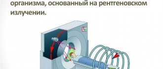
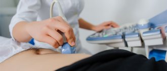
In addition, commonly used instrumental examination methods include pelvic ultrasound, Doppler ultrasound, hysterography and MRI.
When determining the etiology of AUB in postpartum women, obstetricians and gynecologists use the “5T” system:
- Tone – decreased uterine tone.
- Tissue is the remains of the placenta in the uterus after childbirth.
- Trauma - damage up to ruptures of the soft birth canal and uterine walls.
- Thrombin – blood clotting disorders and hemostatic pathologies.
- Therapy is ineffective or incorrect treatment.
Recommendation from the magazine “Deputy Chief Physician”
In accordance with the new rules of the Ministry of Health, the referral issued by the attending physician must contain a number of mandatory items: details of the organization and the patient, diagnosis according to ICD-10, additional clinical information - main symptoms, full name and position of the attending physician.
Download a visual algorithm for filling out directions in the magazine.
If your nose bleeds heavily
It happens that the bleeding is so strong that it is difficult to stop, usually as a result of damage to the vascular wall.
- profuse bleeding from the nose threatens significant blood loss and can even be fatal;
- approximately 20% of the population suffering from this pathology requires emergency medical care;
- The most dangerous bleeding is considered to be the anterior one, it occurs in 90-95% of people;
- arterial hypertension is one of the most common causes of bleeding from the nose;
- in 85% of cases, this symptom occurs against the background of general pathological causes, and only in 15% of cases, bleeding from the nose develops due to a malfunction of the organ itself.
Causes
Pathogenesis . The patient’s adaptation to blood loss is largely determined by changes in the capacity of the venous system (containing up to 75% of blood volume in a healthy person). However, the possibilities for mobilizing blood from the depot are limited: with a loss of more than 10% of the bcc, the central venous pressure begins to fall and the venous return to the heart decreases. Small output syndrome occurs, leading to decreased perfusion of tissues and organs. In response, nonspecific compensatory endocrine changes appear. The release of ACTH, aldosterone and ADH leads to the retention of sodium, chloride and water by the kidneys, while increasing potassium losses and decreasing diuresis. The result of the release of epinephrine and norepinephrine is peripheral vasoconstriction. Less important organs (skin, muscles, intestines) are switched off from the bloodstream, and the blood supply to vital organs (brain, heart, lungs) is preserved, i.e. centralization of blood circulation occurs. Vasoconstriction leads to deep tissue hypoxia and the development of acidosis. Under these conditions, proteolytic enzymes of the pancreas enter the blood and stimulate the formation of kinins. The latter increase the permeability of the vascular wall, which promotes the passage of water and electrolytes into the interstitial space. As a result, red blood cell aggregation occurs in the capillaries, creating a springboard for the formation of blood clots. This process immediately precedes the irreversibility of shock.
What does it mean: signs and symptoms
The anterior type of bleeding is characterized by the fact that blood forms in the front of the nose.
The posterior view involves the deeper parts of the nasal structure. Sometimes blood from the nose does not flow because it flows down the throat. As a result, the following symptoms occur:
- Nausea.
- Vomiting with blood.
- Hemoptysis.
- The stool is tarry, that is, black in color, this is due to the fact that the blood, under the influence of digestive enzymes, acquires a tarry hue.
Symptoms of this condition will depend on the amount of blood loss.
If the blood loss is not so significant (up to several milliliters), the person’s general well-being does not change. The exception is suspicious persons, or people who are afraid of blood; they may develop fainting or hysterics.
If the bleeding is prolonged, then over time the following signs appear:
- general weakness;
- floaters appear before the eyes;
- feeling of thirst;
- dizziness;
- rapid heartbeat;
- blanching of the skin and mucous membranes of a person;
- development of shortness of breath.
If the blood loss rate is already 20%, then hemorrhagic shock may develop, which manifests itself as follows:
- confusion in consciousness;
- frequent heart beats;
- a thread-like pulse is palpable;
- surges in blood pressure, which subsequently lead to a decrease in blood pressure;
- a decrease in the amount of urine or it is completely absent.
Intestinal bleeding ICD code 10 in adults
Version: MedElement Disease Directory
Short description
Excluded from this subheading: - Acute hemorrhagic gastritis (K29.0); — Bleeding from the anus and rectum (K62.5); - Gastrointestinal bleeding due to peptic ulcer (K25-K28); — Angiodysplasia of the stomach with bleeding (K31.8);
- Diverticulitis with bleeding (K57).
Etiology and pathogenesis
The mechanism of development of gastrointestinal bleeding depends on the cause that caused it.
Symptoms, course
Upper gastrointestinal bleeding
Direct symptoms (main clinical signs): vomiting blood (hematemesis), black tarry stools.
In the case of accelerated (less than 8 hours) transit of contents through the intestines and blood loss of more than 100 ml, scarlet blood may be released with feces (hematochezia).
Bleeding from the lower gastrointestinal tract
Often, obvious bleeding from the lower gastrointestinal tract is moderate and is not accompanied by a drop in blood pressure and other general symptoms.
In some cases, patients report the presence of periodic intestinal bleeding only after careful questioning. Massive bleeding from the lower gastrointestinal tract, which is accompanied by hypovolemia, is rare. Hypovolemia (syn.
oligemia) - reduced total amount of blood. , acute posthemorrhagic anemia, arterial hypotension, tachycardia.
The color of the blood released has diagnostic significance. With intestinal bleeding, the most common occurrence is the appearance of unchanged blood (hematochezia). Moreover, the lighter the blood released from the rectum, the more distal the source of bleeding is. Scarlet blood is released mainly during bleeding caused by damage to the sigmoid colon.
As a rule, when the source of bleeding is located in the more proximal parts of the colon, the appearance of dark red blood is noted.
In case of bleeding associated with damage to the perianal area (hemorrhoids, anal fissures), the released blood (in the form of traces on toilet paper or drops falling on the walls of the toilet) is usually not mixed with stool, which remains brown in color.
When the source of bleeding is localized proximal to the rectosigmoid region, the blood is more or less evenly mixed with feces.
Diagnostics
Diagnosis of bleeding from the upper gastrointestinal tract
1. Assessment (diagnosis) of blood loss
| % | |
| Peptic ulcer | 46-56 |
| Erosion of the stomach and duodenum | 9-12 |
| Varicose veins of the esophagus | 16-20 |
| Erosive esophagitis and peptic ulcer of the esophagus | 4-7 |
| Mallory-Weiss syndrome | 4-4,5 |
| Tumors of the esophagus and stomach | 3-5 |
| Other reasons | 4-5 |
| Severity | Volume of blood loss in liters | BCC deficit % |
| I | 1-1,5 | |
| II | 1,5-2,5 | 20-40 |
| III | >2,5 | 40-70 |
| Index | 0 points | 1 point | 2 points | 3 points |
| Age | 60 — 79 | > 80 | ||
| Shock | No shock | Pulse > 100 blood pressure > 100 systolic | Systolic blood pressure | |
| Concomitant pathology | No | Chronic heart failure, coronary heart disease | Renal failure, liver failure, cancer with metastases | |
| Endoscopic picture | Mallory-Weiss syndrome | Ulcers, erosions and other noncancerous sources of bleeding | Malignant sources of bleeding (tumors, malignant polyps) | |
| State of hemostasis | No bleeding | Blood in the lumen, a blood clot on the surface of the defect, a pulsating stream of blood |
| Number of points | Rebleeding rate (%) | Mortality of patients (%) |
| 5 | ||
| 1 | 3 | |
| 2 | 5 | 0,2 |
| 3 | 11 | 3 |
| 4 | 14 | 5 |
| 5 | 24 | 11 |
| b | 33 | 17 |
| 7 | 44 | 27 |
| >8 | 42 | 41 |
-Blatchford score is also used for the assessment and prognosis of a patient with bleeding from the upper gastrointestinal tract .
The score is calculated using the following table:
| Glasgow-Blatchford criteria | |
| Index | Evaluation score |
| Blood urea mmol\l | |
| ≥ 6,5 | 2 |
| ≥ 8,0 | 3 |
| ≥ 10,0 | 4 |
| ≥ 25 | 6 |
| Hemoglobin (g/l) for men | |
| ≥ 12,0 | 1 |
| ≥ 10,0 | 3 |
| 6 | |
| Hemoglobin (g/l) for women | |
| ≥ 10,0 | 1 |
| 6 | |
| Systolic blood pressure (mmHg) | |
| 100-109 | 1 |
| 90-99 | 2 |
| 3 | |
| Other markers | |
| Pulse ≥ 100 (per minute) | 1 |
| Melena (tarry stools) | 1 |
| Loss of consciousness | 2 |
| Liver diseases | 2 |
| Heart failure | 2 |
Diagnosis of bleeding from the lower gastrointestinal tract
1. Establishing the fact of bleeding and assessing the degree of its severity (see above).
source
Coding of gastrointestinal bleeding in the ICD
Diagnoses of any medical institutions are subject to the unified International Statistical Classification of Diseases and Related Health Problems, officially adopted by WHO.
K92.2 – according to ICD 10, code for gastrointestinal bleeding, unspecified.
These figures are displayed on the title page of the medical history and processed by statistical authorities. Thus, data on morbidity and mortality due to various nosological units is structured.
The ICD also includes a division of all pathological diseases into classes.
In particular, gastrointestinal bleeding belongs to class XI - “Diseases of the digestive organs (K 00-K 93)” and to the section “Other diseases of the digestive organs (K 90-K93)”.
Gastrointestinal bleeding
Gastrointestinal bleeding is a serious pathology associated with damage to the blood vessels in the cavity of the gastrointestinal tract and the leakage of blood from them.
In such cases, the loss of blood can be significant, sometimes leading to shock and can pose a serious threat to the patient's life.
Intestinal bleeding in ICD 10 has the same code as gastrointestinal unspecified - K 92.2 .
In any case, this condition is extremely dangerous and requires urgent medical attention. Etiological reasons leading to gastrointestinal tract:
- peptic ulcer of the stomach or duodenum in the acute stage;
- gastroesophageal reflux disease (corrosion of the walls of blood vessels by aggressive gastric juice);
- chronic or acute hemorrhagic erosive gastritis;
- nonspecific ulcerative colitis, Crohn's disease;
- chronic inflammation of the esophagus;
- long-term use of non-steroidal anti-inflammatory drugs, glucocorticosteroids, acetylsalicylic acid;
- acute stress and the occurrence of ulcers in the gastrointestinal tract under the influence of ischemia and stress neurotransmitters and hormones;
- hypersecretion of gastrin as a result of Zollinger-Ellison syndrome;
- with severe, uncontrollable vomiting, ruptures occur in the esophagus, which can bleed;
- enterocolitis and colitis of bacterial origin;
- benign and malignant neoplasms in the gastrointestinal tract;
- portal hypertension.
To find the cause of the bleeding, it is necessary to understand the department that is affected. If there is scarlet blood from the oral cavity, then the esophagus is damaged; if it is black, then this is bleeding from the stomach.
Unchanged blood from the anus indicates damage to the lower sections of the intestine, if mixed with mucus, feces, or clots, it is from the upper sections.
In any case, regardless of the etiology of bleeding, the gastrointestinal tract code is set according to ICD 10 - K92.2.
Save the link, or share useful information on social media. networks
source
During pregnancy
Nosebleeds in pregnant women can occur both at the beginning and at the end of pregnancy, but the reasons for this circumstance may be different. In the first trimester, this condition can be caused by physiological changes in the woman’s body. There is a relationship with increased progesterone, a hormone that is responsible for the preservation and normal development of pregnancy.
Due to the action of progesterone, blood flow increases throughout the entire system of a pregnant woman. Sometimes small capillaries cannot withstand such pressure and rupture, for this reason nosebleeds may develop.
After the 20th week of pregnancy, a complication such as gestosis may develop, in which pressure on the arteries increases, which leads to blood from the nose. Other factors influencing the frequent discharge of blood from the nose are characterized by a lack of vitamins and micronutrients, injuries, drying of the nasal mucous membranes, and poor blood clotting.
Symptoms
Common early signs of internal bleeding are general weakness, drowsiness, pale skin and mucous membranes, dizziness, cold sweat, thirst, darkening of the eyes. Fainting is possible. The intensity of blood loss can be judged both by changes in pulse and blood pressure, and by other clinical signs. With low blood loss, a slight increase in heart rate (up to 80 beats/min) and a slight decrease in blood pressure are observed; in some cases, clinical symptoms may be absent.
Moderate internal bleeding is indicated by a drop in systolic pressure to 90-80 and an increase in heart rate (tachycardia) to 90-100 beats/min.
The skin is pale, there is coldness in the extremities and a slight increase in breathing.
Possible dry mouth, fainting, dizziness, nausea, adynamia, severe weakness, slow reaction. In severe cases, there is a decrease in systolic pressure to 80 or lower, an increase in heart rate to 110 or higher beats/min.
There is a strong increase and disturbance in the rhythm of breathing, sticky cold sweat, yawning, pathological drowsiness, tremors of the hands, darkening of the eyes, indifference, apathy, nausea and vomiting, a decrease in the amount of urine excreted, excruciating thirst, blackouts, severe pallor of the skin and mucous membranes, cyanosis limbs, lips and nasolabial triangle.
With massive internal bleeding, the pressure drops to 60 mm, and the heart rate increases to 140-160 beats/min.
Characterized by periodic breathing (Cheyne-Stokes), absence or confusion of consciousness, delirium, severe pallor, sometimes with a bluish-gray tint, cold sweat.
The look is indifferent, the eyes are sunken, the facial features are pointed. With fatal blood loss, coma develops.
Systolic pressure drops to 60 mm or is not detected. Agonal breathing, sharp bradycardia with a heart rate of 2-10 beats/min. , convulsions, dilated pupils, involuntary release of feces and urine. The skin is cold, dry, “marbled”. Then comes agony and death. Nausea and vomiting of dark blood (“coffee grounds”) indicate bleeding into the cavity of the stomach or esophagus. Tarry stools can occur when there is internal bleeding in the upper digestive tract or small intestine. The discharge of unchanged scarlet blood from the anus indicates hemorrhoids or bleeding from the lower parts of the large intestine. If blood enters the abdominal cavity, dullness of sound in flat areas during percussion and symptoms of peritoneal irritation during palpation are detected. When pulmonary bleeding occurs, a cough with bright foamy blood occurs; when blood accumulates in the pleural cavity, severe shortness of breath, difficulty breathing, and lack of air occur. The flow of blood from the female genital organs indicates bleeding into the uterine cavity, or less often, into the vagina. When bleeding occurs in the kidneys or urinary tract, hematuria is observed. Thirst. Cough. Lack of air. Low body temperature. Dyspnea. Vomit. Vomiting blood. Reticulocytosis. Intense thirst. Cramps. Dry mouth. Nausea. Tremor. Cold sweat.
The child has
The children's body is extremely susceptible to nosebleeds, the reasons for this are as follows:
- A blow to the nose or mechanical impact on the mucous membrane of the organ. The child often puts his fingers in his nose or tries to push any small object into his nostrils.
- Defects in the structure of the nose of an anatomical nature.
- Bacterial or viral infections.
- Weakening of the immune system, vitamin deficiency.
- Thermal or chemical burns.
- Overheat.
- Various pathologies, often hemophilia, abnormalities of the liver and spleen, tumor process in the nasal cavity.
- Dry indoors.
In a teenager
During adolescence, the child’s body undergoes changes, both anatomical and physiological. The appearance of blood from the nose is often not associated with illness. After adolescence and puberty are over, everything will return to normal.
Regular nosebleeds in a child should not leave his parents indifferent; you should definitely consult a doctor for advice.
Causes of nosebleeds during adolescence:
- getting an injury or bruise to the nose as a result of a fight, game or accident;
- various growths, for example, cystic formations, polyps and adenoids;
- the nasal septum may be deviated from birth or acquired;
- weakening of capillary walls due to increased physical load, overheating, hypothermia, etc.
Gastrointestinal bleeding: causes, classification and symptoms, treatment
One of the severe complications of various diseases is gastrointestinal bleeding, which is the leakage of blood into the lumen of the stomach or intestines from the vessels passing under the mucous membrane. The pathology is dangerous because it cannot always be immediately recognized; blood loss is often severe and can lead to death.
It is necessary to know what ailments this complication can arise from and how it manifests itself in order to suspect it in time and take the necessary measures.
According to the international classification of diseases ICD-10, gastrointestinal bleeding has a general code of K92, with the exception of bleeding in newborns with code P54.
Causes of gastrointestinal bleeding
All causes that lead to gastrointestinal bleeding can be divided into 2 groups:
- associated with pathology of the digestive organs;
- not related to diseases of the digestive system.
Group 1 includes:
- peptic ulcer of the stomach and duodenum;
- erosions in the stomach and intestines;
- Mallory-Weiss syndrome (cracks in the mucous membrane);
- abnormalities and tumors of blood vessels ( angiomas );
- tumors , polyps ;
- foreign bodies and injuries to the digestive organs;
- intestinal diverticula;
- Crohn's disease , ulcerative colitis ;
- acute intestinal infections;
- rectal fissure;
- haemorrhoids;
- varicose veins of the esophagus , stomach with cirrhosis of the liver.
With ulcers and erosion, when the defect is located near large vessels, their wall is destroyed under the influence of hydrochloric acid and enzymes.
The reason may be long-term use of aspirin and its analogues, hormonal drugs.
The 2nd group consists of pathology of other organs:
- bleeding disorders (hemophilia, thrombocytopenia, taking anticoagulants, disseminated intravascular coagulation syndrome);
- diseases of blood vessels (capillary toxicosis, vasculitis, atherosclerosis);
- diseases of the circulatory system (hypertension, heart failure);
- severe intoxication;
- renal and liver failure;
- traumatic brain injury;
- stressful situation.
The etiology of low coagulability, vascular pathology, intoxication, liver and kidney failure is not associated with vascular rupture, but with an increase in their permeability.
With hypertension and atherosclerosis in older people, arterial rupture may occur, and with cardiac venous congestion, veins may overflow and rupture.
Severe brain injuries and stress can be accompanied by the formation of acute deep ulcers in the stomach and intestines.
Anatomically
There are 2 groups of bleeding:
- From the upper part of the digestive tract, which includes the esophagus, stomach, duodenum. From the lower part - jejunum, ileum, colon (colon, sigmoid, rectum).
- From the lower section - jejunum, ileum, colon (colon, sigmoid, rectum).
According to the clinical course
There are 3 types of bleeding:
- Acute – with a sudden onset and severe symptoms, typical for ulcers, varicose veins of the esophagus, Mallory-Weiss syndrome.
- Chronic - with periodic minor blood loss, characteristic of polyps, diverticulum, Crohn's disease, inflammatory process.
- Recurrent – occurring repeatedly, may have various causes.
By intensity
There are 2 types of bleeding:
- Explicit, when its signs are obvious - vomiting with blood, feces with blood.
- Hidden, without external manifestations, when blood in the stool is determined only by a laboratory method.
Severity of gastrointestinal bleeding
Depending on the amount of blood loss and the patient’s condition, there are 4 degrees of severity:
- Mild : blood loss no more than 5% of the total volume, general condition is satisfactory, blood pressure is within normal limits, slight tachycardia - up to 100 beats. per minute, hemoglobin 100 or more g/l.
- Moderate : blood loss 6-15%, moderate condition, pressure reduced to 80 mm Hg. Art., hemoglobin 90-80 g/l.
- Severe : blood volume deficit 16-30%, severe condition, pressure 70-60 mm Hg. Art., hemoglobin is reduced to 50 g/l;
- Extremely severe : blood deficiency more than 30%, pressure below 60 mm Hg. Art., thread-like pulse, can only be detected in the carotid arteries, the patient is in a state of hemorrhagic shock, coma, unconscious, on the verge of agony.
Symptoms
Clinical manifestations are accompanied by obvious bleeding, when blood loss is noticeable to the body. A syndrome develops, consisting of local and general signs of gastrointestinal bleeding.
Blood in stool can also look different. When the source is located in the upper part of the tract, the blood is exposed to gastric juice and digestive enzymes, hemoglobin is converted into hydrochloric acid hematin, which has a gray-black color. In these cases, feces have the appearance of tar and a foul odor.
From the lower intestines, blood in the stool will appear in the form of clots, bloody impurities in the form of stripes, or excreted fresh if the object is located in the rectum. It can be scarlet or dark, depending on which vessels supply the blood - arteries or veins. A characteristic symptom is a decrease or disappearance of abdominal pain if it was present before bleeding (for example, with an ulcer, gastritis).
Common symptoms of bleeding are:
- pale skin;
- general weakness, dizziness, fainting;
- decreased blood pressure, increased heart rate;
- in severe cases – cold sticky sweat,
- lethargy, loss of consciousness.
Diagnostic methods
During the examination, the general condition of the patient, skin color, pulse, pressure, presence and nature of vomit and stool are taken into account. If the patient does not recover, a digital examination of the rectum is performed. Palpation of the abdomen is carried out with caution so as not to cause additional trauma.
The diagnosis is based mainly on additional research methods to determine the source and severity of the pathology. These methods include:
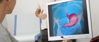
- fibrogastroduodenoscopy - allows you to examine the esophagus, stomach, duodenum, detect varicose veins, erosions and ulcerative defects, cracks, tumors, foreign bodies;
- Ultrasound - reveals the condition of the abdominal organs, accumulation of blood near a bleeding source;
- laboratory blood test - determine the level of hemoglobin, red blood cells, platelets, hematocrit (the ratio of blood elements to the liquid part), the state of coagulation (coagulogram);
- examination of stool for the presence of hidden blood - for hidden bleeding;
- endoscopy of the lower intestine - sigmoidoscopy, colonoscopy, can identify hemorrhoids, polyps, fissures, tumors, ulcerative colitis.
Differential diagnosis is aimed at identifying the nature and cause of gastrointestinal bleeding, taking into account data from anamnesis, examination and additional studies.
The totality of the results makes it possible to distinguish bleeding associated with diseases of the digestive system from those caused by diseases of the blood vessels, blood system, coagulation disorders, intoxication, infections, and medications.
Urgent Care
If, based on the history and clinical manifestations, there is reason to suspect bleeding, you should immediately call an ambulance and begin the following emergency actions:
- lay the patient on a flat surface, unfasten the belt, collar, provide access to fresh air;
- put cold on the abdominal area, this can be ice in a plastic bag, a bubble or a heating pad with cold water;
- turn your head to the side in case of vomiting to avoid asphyxia;
- measure your pulse, blood pressure and monitor them every 10-15 minutes before the ambulance arrives;
- if the pulse disappears, begin closed cardiac massage and artificial respiration.
Actions that should not be taken:
- leaving the patient alone, because the pressure may drop sharply, cardiac activity may stop when resuscitation measures are required;
- allow the patient to get up, provide him with a toilet in bed - a vessel for urine, a bedpan;
- rinse the stomach, give drink, food, medicine.
Patients with bleeding are urgently hospitalized in the surgical department of the hospital.
Treatment of gastrointestinal bleeding
Medical tactics for gastrointestinal bleeding depend on their nature and severity; it can be conservative or surgical.
Conservative treatment
If the bleeding is not severe and does not progress, drug therapy is prescribed: hemostatic drugs, antianemic drugs - iron preparations, vitamin B12, folic acid, transfusion of blood components - platelets, red blood cells, replenishing the volume of circulating blood.
The underlying disease is treated: peptic ulcer, vascular pathology, disorders of the coagulation system and organ function.
Surgery
If this procedure is ineffective, surgical intervention is performed according to vital indications - by laparotomy (traditional incision) or laparoscopy (through a probe). The bleeding area is eliminated by suturing it, resection, removing a polyp, diverticulum, or tumor.
How to recognize gastrointestinal tract infections and what actions to take can be found in this video.
Features of bleeding in children
In infants, the most common causes of blood in the digestive tract are congenital pathologies: hemorrhagic disease, anomalies (duplication of the stomach and intestines), Dieulafoy's disease and Randu-Osler syndrome (vascular abnormalities), internal angiomas, Peutz-Jeghers syndrome (intestinal polyposis), diaphragmatic hernia, Meckel's diverticulum.
Severe vomiting may result in Mallory-Weiss syndrome. In older age, the causes are acute erosions and ulcers, portal hypertension, intestinal obstruction, inflammation, and foreign bodies.
A feature of bleeding in children is often the absence of severe symptoms, up to a loss of 15% of the circulating blood volume, and then a sudden loss of consciousness occurs.
Therefore, you need to be extremely attentive to the child and always inspect the stool.
The principles of diagnosis and treatment in children are the same as in adults, but the leading method is surgical, since most of the causes are based on gross anatomical changes of a congenital nature.
Consequences of gastrointestinal bleeding
Acute blood loss can lead to the development of severe complications:
- acute anemia;
- acute failure of internal organs (heart, kidneys, liver);
- hemorrhagic shock;
- coma, death.
The consequences of small but repeated blood loss are chronic anemia, hypoxia of internal organs with the development of dystrophic changes in the heart, liver, kidneys, and dysfunction of the central nervous system.