More about the operation
Rectal tumors are neoplasms that arise as a result of degeneration of the cells of its mucous layer. The main danger of such conditions is that tumor formations may not be detected for a very long time. In addition, the benign nature of the tumor does not exclude the risk of its malignancy (malignancy).
Rectal tumors can be single or multiple and differ in location and cellular structure. Removal of benign rectal tumors is carried out transanal, open and laparoscopic. In the first case, the operation is performed through the anus, the open method - through a small incision in the peritoneum, in the second - through several micropunctures of the abdominal wall.
Coloproctological surgeons at the GMS Clinic prefer low-traumatic transanal and laparoscopic surgical methods. However, in large rectal lesions or in severe complex cases, open surgery may be required.
Bowel resection technique
Bowel resection surgery can be performed through laparotomy or laparoscopy. In the first case, the surgeon makes a longitudinal incision in the abdominal wall; the operation is performed in an open manner. The advantages of laparotomy are a good overview during all manipulations, as well as the absence of the need for expensive equipment and trained personnel.
With laparoscopy, only a few puncture holes are needed to insert laparoscopic instruments. Laparoscopy has many advantages, but is not always technically feasible, and for some diseases it is safer to resort to laparotomy access. The undoubted advantage of laparoscopy is not only the absence of a wide incision, but also a shorter rehabilitation period and faster recovery of the patient after the intervention.
After processing the surgical field, the surgeon makes a longitudinal incision in the anterior abdominal wall, examines the abdominal cavity from the inside and looks for the altered section of the intestine. Clamps are applied to isolate the portion of intestine that will be removed, and then the affected area is cut off. Immediately after dissection of the intestinal wall, it is necessary to remove part of its mesentery. Vessels feeding the intestine pass through the mesentery, so the surgeon carefully ties them up, and excises the mesentery itself in the shape of a wedge, with its apex facing the root of the mesentery.
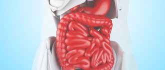
Removal of the intestine is carried out within the healthy tissue, as carefully as possible, in order to prevent damage to the ends of the organ by instruments and not provoke their necrosis. This is important for the further healing of the postoperative suture on the intestine. When the entire small or large intestine is removed, it is called a total resection; subtotal resection involves excision of part of one of the sections.
subtotal resection of the large intestine
To reduce the risk of infection with intestinal contents during surgery, tissues are isolated with napkins and tampons, and surgeons practice changing instruments when moving from a more “dirty” stage to subsequent ones.
After removing the affected area, the doctor faces the difficult task of creating an anastomosis (connection) between the ends of the intestine. Although the intestine is long, it cannot always be stretched to the required length; the diameter of the opposite ends may differ, so technical difficulties in restoring the integrity of the intestine are inevitable. In some cases, this cannot be done; then the patient has an outlet hole placed on the abdominal wall.
Types of intestinal connections after resection:
End to end is the most physiological and involves connecting the lumens in the same way as they were located before the operation. Disadvantage: possible scarring;- Side to side - the opposite ends of the intestine are connected by their lateral surfaces;
- Side to end - used when connecting parts of the intestine with different anatomical characteristics.
If it is technically impossible to restore the movement of intestinal contents as physiologically as possible, or the distal end needs to be given time to recover, surgeons resort to placing an outlet on the anterior wall of the abdomen. It can be permanent, when large sections of the intestine are removed, or temporary, to speed up and facilitate the regeneration of the remaining intestine.
A colostomy is a proximal (near) segment of the intestine, removed and fixed to the abdominal wall, through which feces are evacuated. The distal fragment is sutured tightly. With a temporary colostomy, a second operation is performed after a few months, in which the integrity of the organ is restored using one of the methods described above.
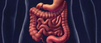
Resection of the small intestine is most often performed due to necrosis. The main type of blood supply, when blood flows to the organ through one large vessel, then branches into smaller branches, explains the significant extent of gangrene. This happens with atherosclerosis of the superior mesenteric artery, and the surgeon in this case is forced to excise a large fragment of the intestine.
If it is impossible to connect the ends of the small intestine immediately after resection, an ileostomy is fixed to the surface of the abdomen to remove feces, which either remains permanently or is removed after several months with the restoration of a continuous flow of the intestine.
Resection of the small intestine can also be performed laparoscopically, when instruments are inserted into the abdomen through punctures, carbon dioxide is injected for better visibility, then the intestine is clamped above and below the site of injury, the mesenteric vessels are sutured and the intestine is excised.
Resection of the colon has some peculiarities, and it is most often indicated for neoplasms. In such patients, all, part of the colon, or half of it is removed (hemicolectomy). The operation lasts several hours and requires general anesthesia.
With an open approach, the surgeon makes an incision of about 25 cm, examines the colon, finds the affected area and removes it after ligating the mesenteric vessels. After excision of the large intestine, one of the types of end joining is performed or a colostomy is performed. Removal of the cecum is called a cecectomy, the ascending colon and half transverse or the descending colon and half transverse is hemicolectomy. Resection of the sigmoid colon - sigmoidectomy.
The operation for resection of the colon is completed by washing the abdominal cavity, layer-by-layer suturing of the abdominal tissue and installing drainage tubes into its cavity for the outflow of discharge.
Laparoscopic resection for lesions of the colon is possible and has a number of advantages, but is not always feasible due to severe damage to the organ. Often there is a need to switch from laparoscopy to open access right during surgery.
Operations on the rectum differ from those on other parts, which is associated not only with the structural features and location of the organ (strong fixation in the pelvis, proximity of the genitourinary system), but also with the nature of the function performed (accumulation of feces), which is unlikely to be possible. take over another part of the colon.
Resections of the rectum are technically complex and result in much more complications and unfavorable outcomes than those performed on the thin or thick sections. The main reason for interventions is cancer.
Resection of the rectum when the disease is located in the upper two-thirds of the organ makes it possible to preserve the anal sphincter. During the operation, the surgeon excises part of the intestine, ties the vessels of the mesentery and cuts it off, and then forms a connection that is as close as possible to the anatomical course of the terminal intestine - anterior resection of the rectum.
Tumors of the lower segment of the rectum require the removal of components of the anal canal, including the sphincter, therefore such resections are accompanied by all kinds of plastics in order to somehow ensure the exit of feces out in the most natural way. The most radical and traumatic abdominoperineal extirpation is performed less and less often and is indicated for those patients whose intestines, sphincter, and pelvic floor tissues are affected. After removal of these formations, the only option for fecal drainage is a permanent colostomy.
Sphincter-preserving resections are feasible in the absence of cancerous tissue growing into the anal sphincter and allow preserving the physiological act of defecation. Interventions on the rectum are performed under general anesthesia, in an open manner, and are completed by installing drains in the pelvis.
Even with impeccable surgical technique and compliance with all preventive measures, avoiding complications during intestinal operations is problematic. The contents of this organ contain a lot of microorganisms that can become a source of infection. Among the most common negative consequences after intestinal resection are:
- Suppuration in the area of postoperative sutures;
- Bleeding;
- Peritonitis due to suture failure;
- Stenosis (narrowing) of the intestinal section in the anastomosis area;
- Dyspeptic disorders.
Why choose us
At the Center for Operative Proctology at GMS Hospital, operations to remove rectal tumors are performed by experienced coloproctologists with many years of practice. The main advantages of treatment at the GMS clinic:
- the use of advanced diagnostic equipment makes it possible to detect intestinal tumors at an early stage;
- For surgical treatment, the latest low-traumatic techniques are used, aimed at speedy rehabilitation and recovery;
- the presence of a modern histological laboratory allows one to immediately conduct a histological analysis of a remote lesion at the world level;
- We successfully treat complex and advanced forms of tumors;
- medical support of the patient in the postoperative period;
- individual rehabilitation and recovery programs;
- minimal risk of postoperative complications;
- reduces the risk and need for stoma formation
Most operations are performed using endoscopic and laparoscopic technologies, which reduces the time spent in hospital and the recovery period by several times. You can make an appointment for a consultation with a coloprotologist at GMS Hospital around the clock - by phone or online.
Cost of removing benign rectal tumors
The prices indicated in the price list may differ from the actual prices. Please check the current cost by calling +7 495 104 8605 (24 hours a day) or at the GMS Hospital clinic at the address: Moscow, st. Kalanchevskaya, 45.
| Name | Common price | Price with 30% discount |
| Transanal endoscopic microsurgical removal of rectal tumors | RUB 365,720 | RUB 256,004 |
Prices for the most popular services are indicated with a 30% discount, which is valid when paying in cash or by credit card. You can be served under a VHI policy, pay separately for each visit, sign an agreement for an annual medical program, or make a deposit and receive services at a discount. On weekends and holidays, the clinic reserves the right to charge additional payments according to the current price list. Services are provided on the basis of a concluded contract.
Plastic cards MasterCard, VISA, Maestro, MIR are accepted for payment. Contactless payment with Apple Pay, Google Pay and Android Pay cards is also available.
Make an appointment We will be happy to answer any questions Coordinator Oksana
In what cases is it necessary to contact a specialist?
A direct indication for surgery is any diagnosed benign formation of the rectum. Surgical treatment should not be delayed if:
- the tumor has begun to grow rapidly and is blocking the intestinal lumen;
- the tumor is too large to be removed by less traumatic methods.
The danger of such pathologies lies in the prolonged absence of symptoms of the disease. Very often, a neoplasm is detected by chance, during an examination, due to treatment of other problems.
Be sure to make an appointment with a coloproctologist if you are concerned about the following symptoms:
- periodic pain in the lower abdomen (abdominal pain);
- bowel disorders (constipation alternating with diarrhea);
- the appearance of mucus, blood, and pus in the stool;
- frequent false urge to evacuate (tenesmus);
- decreased appetite;
- difficulty retaining intestinal gases;
- flatulence;
- sensation of a foreign body in the rectum;
- intestinal obstruction.
Such manifestations may indicate the presence of a neoplasm in the intestines, so you should not ignore them. Remember, any benign tumor has a tendency to become malignant. There is no need to be afraid of surgery - modern proctology in most cases uses minimally invasive methods to minimize surgical trauma and quickly recover, without the need to form a stoma.
Therapeutic methods
The initial stages of a tumor located in the rectum can be cured with surgery. The use of radiation and chemotherapy will not help get rid of the disease. Therefore, treatment is carried out comprehensively. Special therapy is carried out before and after surgery to remove rectal cancer.
Surgical procedures for the treatment of oncology
Intervention to remove a tumor in the rectum is not performed if the patient is admitted to the surgeon in serious condition. In addition, for certain diseases, surgery for rectal cancer is not performed due to a weakened body. In other cases, depending on the diagnostic indications, cancer treatment methods are carried out with complete or partial removal of the rectum.
With anterior resection, doctors remove the growth when the growth is located in the upper part of the rectum. The intervention occurs in the form of an incision in the lower abdomen and the connection between the sigmoid colon and the rectum is removed. Low resection is done when the tumor is in the middle or lower parts of the rectum. The surgeon performs a complete removal of the rectum.
Extirpation of the patient occurs with two incisions, located in the abdomen and perineum, when the cancer affects most of the rectum. The patient's rectum, areas with growths in the anal canal, and nearby tissue are removed.
Local removal is performed for grade 1 tumors using an endoscope. The procedure allows you to remove small growths in the rectum. If the formations are located near the anus, then the endoscope is not used. The tumor is removed by inserting surgical instruments through the anus.
In addition to resection, surgery uses methods to remove the tumor while preserving the sphincter. In this case, transanal excision may be performed. The indication for surgery is the appearance of tumors in the lower part of the rectum. Special surgical instruments are used for the procedure. In this case, the lymph nodes are not removed.
The surgeon may use open laparoscopy. This requires making several incisions in the abdominal wall. A laparoscope is inserted into one hole, surgical instruments are inserted into the others and growths or the affected area are removed. Laparoscopic resection allows the patient to recover quickly after surgery.
Preparation, diagnostics
Preoperative preparation is carried out according to the standard scheme for surgical interventions. Comprehensive preoperative examination includes:
- consultation with a coloproctologist;
- laboratory tests: blood tests, stool tests, histological examination of biopsy material;
- instrumental studies to determine the localization of the pathological area of the intestine and assess the condition of nearby organs (colonoscopy or sigmoidoscopy with biopsy, if necessary - ultrasound, CT, MRI, etc.);
- consultation with a therapist.
You can undergo a preoperative examination at the GMS clinic in 1 day.
In addition to diagnostic procedures and consultations with specialists, preoperative preparation includes the following steps:
- 3-5 days before surgery you need to switch to a slag-free diet, eliminate all foods and drinks that contribute to constipation and flatulence;
- on the eve of the operation, you need to prepare the intestines (cleanse with an enema or medications prescribed by the doctor);
- last meal no later than 10-12 hours before the intervention;
- On the day of surgery, do not drink, eat, or smoke.
Warn your doctor about the medications and dietary supplements you are taking; they may need to be temporarily discontinued. The coloproctologist will tell you more about the preparatory stage during the consultation.
How to diagnose the stage of disease development
Only an oncologist can diagnose cancer and the stage of its development, who prescribes clinical tests and conducts a study of the patient’s condition. First of all, anamnesis is collected, complaints are recorded, and the condition of the rectum is studied. Particular attention should be paid to factors that cause concern in the intestinal area.
Next, patients undergo a complete blood count and stool test to detect occult blood. And only then a colonoscopy or irrigoscopy is done. In difficult cases, an ultrasound scan of the abdomen and pelvic organs is prescribed, and a tumor biopsy is also performed.
Symptoms are characterized by the following features:
- monotonous nature of signs;
- the presence of one or more symptoms;
- tumors are located in different parts of the intestine, so a digital examination of the rectum is performed.
Ultrasound helps detect tumors and metastases that can reach the liver, causing perifocal inflammation. Typically, 4 different types of examination are used: endoscopic, intraoperative, percutaneous, endorectal.
In very difficult situations, tomography or nuclear magnetic resonance is prescribed. Diagnosis must be carried out in order to avoid complications, such as problems with intestinal patency, the presence of bleeding (including hidden), inflammation, and anemia. Such problems that accompany cancer need to be urgently treated, including surgery.
How is the operation performed?
Removal of benign formations of the rectum is carried out in a hospital, under general anesthesia. Surgical tactics are determined by the doctor individually, depending on the clinical characteristics of your case. The essence of the operation is to isolate the affected area of the intestine and targeted excision of the tumor within healthy tissue.
For a local process, the optimal procedure is TEM - transanal endoscopic microsurgery (or TEM).
Treatment of rectal cancer using the TEM technique allows for surgical intervention through the anus. This approach can be called revolutionary in the practice of world surgery.
As a result of this operation, access to the required organ is provided using a special instrument - a rectoscope. This type of complex external surgical intervention has become a fairly common way to remove large polyps and cancerous tumors.
Since surgery using TEM is performed directly in the anal lumen of the intestine, it is less traumatic for the patient’s body and has a sufficient number of advantages.
The main advantages of the TEM method:
- the minimum percentage of the likelihood of severe complications;
- an order of magnitude less pain and discomfort;
- short-term hospitalization;
- low percentage of unsuccessful operations, which result in the formation of a so-called “colostomy” (that is, removal of the colon to the anterior abdominal wall).
Finally, with TEM, there is absolutely no need to excise the rectal sphincter muscles during the operation, and thus, this method is also suitable for those people whose abdominal surgery is accompanied by risk (heart disease, hypertension, etc.) .
In case of extensive damage, rectal resection is performed - removal of the damaged segment or almost complete excision of the intestine, followed by restoration of the integrity and continuity of the large intestine. There are two main methods of surgical intervention:
- open (laparotomy) operation - performed through a midline incision in the anterior abdominal wall;
- laparoscopic intervention - performed through several mini-punctures in the anterior abdominal wall, through which the surgeon inserts microsurgical instruments connected to a video camera.
At the GMS clinic, removal of benign rectal tumors is performed by experienced coloproctologist surgeons and oncologists using modern, safe anesthesia. Surgical treatment is carried out in most cases using low-traumatic technologies in the most comfortable conditions for the patient.
You have questions? We will be happy to answer any questions Coordinator Tatyana
Symptoms of an intestinal tumor
Depending on how the intestinal tumor grows and develops, the signs make themselves felt.
The most common of them:
- A mixture of blood and excreted feces. It depends on the stage of the disease whether the stool will be completely stained or blood will come out in the form of small streaks.
- In addition to blood, pus and mucus are observed in the stool. The accumulation of pus causes an unpleasant odor.
- A frequent occurrence is the change from constipation to diarrhea; this is accompanied by pain, and constipation lasts about a week.
- If the tumor affects the cecum, the skin becomes pale, blood pressure rises, sometimes the temperature rises, and “cold sweat” also appears. This appears in the later stages.
- Frequent nausea caused by fever, vomiting that does not bring relief.
- Pressure occurs on the abdominal wall of the tumor projection, causing aching, prolonged pain.
- After defecation (often false), complete bowel movement is not felt.
- Delayed bowel movements that last more than a week cause pain. The abdomen becomes heavy and often hard.
- The general condition of the patient: fatigue, lethargy, the skin sometimes acquires a yellow tint.
Whatever the type of tumor in the intestine, the symptoms appear in aggregate, but in the early stages it is quite difficult to determine them.
Symptoms directly depend on the location of the original tumor, the intensity of its spread, size, and state of health of the person.
Features of the rehabilitation period
After the operation, you will be transferred to the intensive care ward, where you will remain under the supervision of a doctor for a day. The very next day you are allowed to take liquid food and drink. On average, recovery takes 4-6 weeks, during which time you must carefully adhere to medical recommendations regarding physical activity, lifestyle and diet.
The Department of Operative Proctology at GMS Hospital has a large arsenal of modern methods of restorative therapy, which allows patients to quickly restore their health and return to an active life. You can make an appointment with a coloproctologist by phone or by filling out the form on the website.