Bleeding from dilated veins of the esophagus is a dangerous condition. Most often, the cause of these bleedings is a complication of cirrhosis of the liver (the result of progressive proliferation of connective tissue, resulting from portal hypertension and varicose veins of the esophageal veins with their subsequent rupture).
In gastroenterology, the search for means and methods for the effective treatment of portal hypertension has always been a priority. New methods of emergency care for patients with bleeding are now being developed, and attempts are being made to stop the proliferation of connective tissue. Although bleeding from varicose veins still remains a serious condition, the prognosis for life is not as bleak and clear as it was 15-20 years ago.
ICD 10 I85.0
Bleeding from the esophagus: causes and consequences
URVP is not the only cause of esophageal bleeding. Almost any pathology that can cause damage to the blood vessels of an organ can cause hemorrhages.
Bleeding from the esophagus can be a complication:
- esophageal cancer;
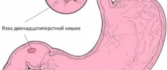
- peptic ulcer;
- deep erosion with esophagitis.
Hemorrhages of the esophagus occur due to diverticula of the esophagus, trauma, or radiation damage.
Bleeding from the esophagus can manifest itself with various symptoms, the most important of which are:
- vomiting with blood;
- change in stool character.
Depending on the cause and intensity of the bleeding, vomiting may be:
- dark, almost black masses;
- single bloody streaks in the vomit;
- a mouth full of scarlet blood.
When bleeding, the stool becomes dark, semi-liquid, and foul-smelling.
Depending on the amount of blood loss, the severity of the patient’s condition is distinguished:
- mild degree – blood loss up to one liter;
- medium degree – blood loss up to 1.5 liters;
- severe degree - blood loss up to 2 liters.
The consequences of esophageal bleeding depend on:
- from the reason that caused it;
- on the intensity of blood loss;
- depends on how correctly and promptly the assistance is provided.
Bleeding from the vessels of the esophagus. Stomach and intestinal bleeding - symptoms and first aid
The reasons for the development of bleeding from the esophagus are as follows:
- Varicose veins of the esophagus. It is this pathology that causes most severe bleeding. It is formed as a manifestation of the late stage of various diseases, leading to an increase in pressure in the portal vein system. This leads to stagnation of blood in the veins of the esophagus, their enlargement, stretching, tortuosity, and the formation of varicose nodes. The wall above the varicose veins stretches, thins, and becomes easily wounded. It can be damaged even with careful examination with an endoscope, consumption of even relatively hard food, vomiting, straining, and surges in blood pressure. In addition, her inflammation can quickly set in, which makes her even more vulnerable.
In 80% of cases, esophageal varicose veins form as a complication of liver cirrhosis and liver failure. The insidiousness of this pathology is that dilated veins do not make themselves felt for a long time and the first manifestation is sudden massive bleeding.
Varicose veins in the esophagus can be the result of a congenital abnormality of their structure. But this is rare and, as a rule, does not lead to such a significant increase in them as with acquired varicose veins.
With cirrhosis of the liver, the veins in the lower part of the esophagus, where the portocaval anastomoses are located, are affected. In heart failure, varicose veins also form. As a rule, veins change along the entire length of the esophagus.
With malignant goiter, changes in veins form in the upper parts of the esophagus.
- Mallory-Weiss syndrome. Its essence is longitudinal ruptures of the esophagus during vomiting. About 10% of all gastrointestinal bleedings are due to this pathology.
- Injury to the esophagus due to chest injuries.
- Esophageal angioma.
- Rendu-Osler disease.
- Injury to the esophagus from rough food or a foreign body.
- A tumor of the esophagus with its growth into the wall of the vessel with its damage.
- Iatrogenic damage, possible when examined with a rigid endoscope.
- Erosion and ulcers of the esophagus involving the vascular wall.
Symptoms
Manifestations depend on the amount of blood loss and its cause:
- Feeling of tickling and soreness in the throat. These symptoms sometimes precede bleeding.
- Salty taste in the mouth. It may also precede vomiting of blood.
- Vomiting blood. Often it can be sudden, a “fountain”. The blood is scarlet. Less often dark, the color of “coffee grounds”. The dark brown color indicates that this blood has already entered the stomach and has come into contact with gastric juice.
Vomiting blood is the main symptom of esophageal bleeding.
- General symptoms of blood loss. Their severity depends on how much blood was lost and how quickly. Sudden blood loss of a large volume of blood is especially life-threatening. This is general weakness, varying degrees of impairment of consciousness (from darkening of the eyes and short-term fainting to coma), dizziness, increased heart rate, decreased blood pressure, even shock.
- The most severe consequence of blood loss is hemorrhagic shock. It is characterized by a sharp decrease in blood pressure, disruption of vital functions - cardiac activity and breathing, and impaired consciousness. The pulse quickens and becomes poorly detectable. You can feel it with your fingers only on large vessels (on the neck, in the groin folds).
- Melena. This is what black stools are called when there is bleeding. It does not appear immediately, but after some time, when the blood from the esophagus, having passed through the stomach and intestines, comes out with feces already darkened and blackened
- Anemia. Decrease in the number of red blood cells and hemoglobin in the blood.
If bleeding is chronic and a person loses a significant amount of blood, but gradually, over a long period of time, in small portions, then the body has time to relatively adapt to blood loss thanks to natural compensatory mechanisms.
Then anemia may become the only long-lasting symptom. Blood in small quantities will flow from the esophagus into the stomach, intestines and exit with feces. Its color becomes dark, almost black.
Not all patients are attentive to the color of their stool, so with chronic blood loss, anemia remains for a long time the only manifestation of esophageal bleeding.
In the blood with chronic blood loss there is a decrease in the number of red blood cells
Diagnostics
A specialist may suspect bleeding of the esophagus already when examining the patient and talking with him. It is important that the patient has pathologies of the liver, heart, and hemorrhagic complications in the past.
To determine the source of bleeding, fibroesophagoscopy and contrast radiography are used.
Laboratory methods (general and biochemical blood tests, coagulogram) make it possible to judge the degree of blood loss, the condition of the liver, and the blood coagulation system.
Hepatorenal syndrome
The human body is a single interconnected system, so failures in one organ lead to dysfunction in others. Hepatorenal syndrome develops due to a malfunction throughout the body due to loss of liver function. In addition to the medical history, renal failure is added.
There are two types of pathology:
1 type It develops within two weeks and is characterized by kidney failure and high mortality.
Type 2 Develops from a couple of months to six months. It is possible to save a person if you consult a doctor in a timely manner.
People who abuse alcohol have a high chance of liver failure at any time and who, even after diagnosis, have not stopped drinking. Signs that require you to see a doctor:
- jaundiced skin;
- malaise;
- weakness throughout the body.
In the hospital, the patient undergoes a series of tests that demonstrate characteristic changes in the body. Among them are determination of the level of urea, ammonia, other wastes with nitrogen content in the blood, sodium in the urine, and blood pressure level. Patients with hepatorenal syndrome, due to kidney problems, may walk very rarely, or not walk at all for several days in a row.
The condition of hepatorenal syndrome cannot be determined by external signs. A patient with cirrhosis should be monitored regularly to detect such complications.
Consequences and complications
The greater the volume of blood loss, the more dangerous the consequences. Acute bleeding can lead to hemorrhagic shock and rapid death. Loss of small volumes leads to the development of persistent anemia. If the cause of intraintestinal hemorrhage is not identified in time, the disease can progress to the stage when doctors are powerless.
Therefore, the first thing to do in case of gastrointestinal bleeding is to consult a doctor. Internal bleeding is dangerous because it is difficult to assess the extent of blood loss and the likelihood of certain complications.
Initial diseases
High pressure in the cavity of the portal vein, through which blood from the stomach, pancreas, spleen (digestive organs) passes to the liver, will be a factor causing varicose veins of the esophagus. The syndrome of pressure exceeding the permissible level in the portal vein system in medicine is called portal hypertension, which, as a rule, accompanies the following diseases:
- Structural changes in the blood vessels of the liver and the organ as a whole (chronic hepatitis, cirrhosis, tuberculosis, tumors, amyloidosis);
- Sclerosis;
- Thrombosis;
- Compression (narrowing of the lumen) of the portal vein: tumors of various sizes, including cysts, gallstones;
- Buddy-Chiari disease.
These diseases are identified as the main causes of varicose veins of the esophagus. In some cases, such primary sources of varicose veins are complemented by another one - chronic cardiovascular insufficiency, which causes an increase in pressure in the systemic bloodstream.
Depending on the pathology of the liver or cardiovascular system, there are differences in the parameters of the affected veins:
- If the cause of phlebectasia is liver disease, then the damaged veins are concentrated in the lower part of the esophagus or in the central part of the stomach; if the basis of the disease is a cardiac lesion, then the deformed veins are localized throughout the organ;
- In liver diseases, vascular nodes are 2-3 times larger than in cardiovascular failure.
There is also a congenital form of esophageal varicose veins, the causes of which have not been established.
The root cause is liver cirrhosis
As medical practice shows, 70% of people with cirrhosis of the liver have varicose veins of the esophagus .
The principle of interaction is simple: with cirrhosis, instead of healthy cells, scar tissue forms on the liver. This complicates the movement of blood; stagnation occurs in the liver portal vein system, which causes varicose veins in the lower (distal) zone of the esophagus. This chronic process is accompanied by disruption of the healthy structure of the liver.
In adults, the most common causes of cirrhosis are:
- Frequent consumption of alcoholic beverages;
- Viral hepatitis;
- Taking medications that negatively affect the liver parenchyma;
- Some hereditary diseases.
Liver cirrhosis in newborns is usually the result of viral infections suffered by the mother during pregnancy (rubella, herpes, hepatitis), which, having penetrated the placenta, affected the fetus in utero.
The role of liver cirrhosis in the occurrence of pathology
To enlarge the picture, click on it.
Varicose veins of the esophagus are recorded in approximately 70% of patients with cirrhosis of the liver. What explains this relationship? In the presence of liver cirrhosis, the formation of scar tissue instead of healthy cells is observed. This pathology causes obstructed blood flow.
In this case, congestion is observed within the portal vein system. It is such congestion that provokes the development of varicose veins in the area of the lower distal zone of the esophagus. With such a chronic process, a disruption of the usual structure of the liver is observed.
Source: https://stopzaraza.com/krov/krovotechenie-iz-pishhevoda-simptomy-pomoshh-skolko-zhivut.html
Bleeding from the esophagus in liver cirrhosis
One of the most severe liver diseases is cirrhosis, characterized by irreversible proliferation of connective tissue, as a result of which hepatocytes die and the structure and function of the organ are disrupted. Blood flow in the v portae system slows down, pressure in the vein increases, when it reaches critical values, excess blood is discharged into the systemic bloodstream through the gastric and esophageal veins.
The veins of the esophagus, not adapted to such loads of volume and pressure, undergo varicose changes. Constant, spasmodic rises in pressure in the portal system, changes in the veins of the esophagus, disturbances in the coagulogram - all this provokes bleeding.
Traditional medicine
In acute cases of the disease, the patient is immediately hospitalized. The primary task of the doctor is to stop the bleeding. For this purpose, the so-called Blackmore Honda is used. What this tool looks like is illustrated in the picture below. The probe can be inserted either through the nasal or oral opening. The instrument consists of tubes and two cylinders, one of which fixes the position of the instrument. In the second, air is pumped, causing the balloon to inflate and press against the mucous membrane, stopping the bleeding. So, the probe can remain in the esophagus for up to 2 days. Food and medications can be administered through one of the instrument's tubes.
Other operations can also be used:
- endoscopic sclerosis of the esophageal veins;
- esophagoscopy;
- stitching;
- endovascular embolization.
Let's take a quick look at these treatment methods.
The first procedure involves introducing a special sclerosing agent into the esophageal veins through an endoscope, which glues the walls together, thereby stopping the bleeding. Esophagoscopy involves cauterizing the bleeding area and then tightly packing it. Vein suturing means placing stitches on the bleeding areas. The last of these methods involves inserting a catheter through a small hole in the skin and placing it in the bleeding vein. Next, special spirals are inserted through it to block the vessels. The method is quite complex, but compared to others it has a very low probability of relapse.
Along with measures aimed at stopping bleeding, blood replacement therapy is prescribed. To do this, the introduction of red blood cells, plasma replacement solutions, freshly collected blood, or direct blood transfusion from the donor to the patient is carried out. Special fluids that replace blood (calcium chloride, ethamsylate) are administered intravenously.
Drug treatment is also prescribed. The patient is given large amounts of vitamin C, as well as the drug Vikasol, which is a kind of substitute for vitamin K. Pituitrin is administered along with the drip, which is necessary to reduce pressure in the veins located near the esophagus. To prevent intoxication of the body from the decomposition of blood clots in the intestines, broad-spectrum antibiotics are used.
In extreme cases, when all the measures described above do not produce results, the bleeding is stopped surgically.
There are three ways to treat the disease surgically:
- Resection of the esophagus.
- Portocaval anastomosis.
- Vein ligation.
For several days after all manipulations, the patient requires strict bed rest. Also, during the first days you should not eat; the minimum period of fasting is three days, and after surgery it can be more than five days. During this period, the patient receives nutrients intravenously.
Symptoms and signs
Bleeding is often immediately preceded by:
- lifting weights;
- straining;
- binge eating;
- stressful situation.
Bleeding from esophageal varices is often caused by drinking alcohol.
The clinical picture of bleeding depends on the amount of blood loss.
If the bleeding is internal, minor, but constant, the patient notes the following symptoms:
- constant and increasing weakness;
- loss of appetite;
- cold clammy sweat;
- pain in the epigastrium and retrosternal region;
- periodic black, foul-smelling stools;
- increasing anemia and exhaustion.
If the bleeding is massive, then the condition immediately becomes threatening:
- the patient suddenly feels severe weakness, consciousness becomes foggy;
- cold sweat appears,
- nausea appears, followed by profuse vomiting of masses of liquid and coagulated blood;
- Blood pressure drops sharply and heart palpitations appear.
Any type of bleeding from the esophagus with cirrhosis of the liver must be treated in a hospital; no folk remedies or conspiracies will help here. Even if bleeding from varicose veins of the esophagus spontaneously stops, this does not guarantee that a relapse will not occur, possibly even more severe.
First aid to the patient:
- lay the patient horizontally, turn the head so that blood does not enter the respiratory tract;
- reassure if possible;
- free from restrictive clothing, covering with a warm blanket;
- measure blood pressure.
What causes esophageal bleeding and how to treat it?
Damage to the blood vessels in the digestive system leads to dysfunction of the entire body and sometimes death of a person. Esophageal bleeding has dangerous consequences. If the patient does not act, his life is at risk. It is almost impossible to independently diagnose this pathology.
Causes
The reasons that cause bleeding from the esophagus are known to doctors. Common factors are:
- stomach ulcer;
- cirrhosis of the liver;
- pathological formations in the esophagus;
- varicose veins of the digestive system;
- neoplasms;
- burns;
- diseases associated with dysfunction of the cardiovascular system;
- constant uncontrolled use of medications;
- hemophilia;
- chronic stressful experiences;
- poisoning with alcohol-containing products.
In some cases, lymphogranulomatosis is mistaken for a malignant tumor. Only with surgery is a non-cancerous tumor diagnosed. The most common organs of the digestive system affected by lymphogranulomatosis are the stomach and small intestine.
Some medications taken by a person, if not properly monitored by medical personnel, can cause pathological conditions in the mucous membrane of the stomach and intestines. Damage leads to slow bleeding. The condition affects a person’s functioning.
In case of alcohol abuse, vomiting that occurs in this condition can lead to tearing of the longitudinal lining of the esophagus. Intoxication is accompanied by blood loss.
Treatment
In the hospital, the patient immediately undergoes an endoscopic examination for the purpose of differential diagnosis and identifying the location of damage in the venous vessel.
After this, urgent treatment of bleeding from varicose veins of the esophagus begins, aimed at:
- to stop bleeding as soon as possible;
- restoration of the bcc;
- correction of coagulopathy;
- prevention of recurrent bleeding.
Emergency care for bleeding
Methods to stop bleeding in varicose veins caused by cirrhosis of the liver include:
- drug treatment;
- tamponade of a bleeding vessel using a double-balloon Blackmore probe;
- endoscopic manipulations (ligation, thrombosis, sclerosis of a bleeding vessel)
- TIPS (minimally invasive intrahepatic bypass);
- surgery.
Conservative therapy
Urgent drug therapy is aimed at:
- Restoration of blood volume - perfusion of FFP, red blood cells, blood substitutes.
- Reducing portal hypertension. All drugs in this group are divided into venodilators and vasoconstrictors.
- Venodilators, dilate portocollateral vessels (nitroglycerin).
- Vasoconstrictors cause contraction of the arterioles of internal organs, thus reducing venous blood flow and reducing portal hypertension (direct vasoconstrictors include vasopressin, indirect vasoconstrictors include octreotide).
Tamponade of bleeding VRVP
Restoring hemostasis in VRVP using a double-balloon Blackmore probe is a temporary measure; the method is based on mechanical compression of the bleeding vessel. Despite its simplicity, this method is used only to stop massive bleeding. This is due to the fact that the procedure is poorly tolerated by patients.
Endoscopic hemostatic methods
- Endoscopic ligation. The method is based on strangulation (compression) of bleeding varicose veins with ligatures. In a compressed node, tissue ischemia occurs, and then necrosis. The outcome is a star-shaped scar. Currently, endoscopic ligation is the main method if there are no contraindications. Silicone rings are used to ligate not only varicose veins, but also varicose veins of the gastric cardia.
- Endoscopic sclerotherapy. There is a technique for introducing sclerosant into a vessel, but now paravasal administration of sclerosant is more often used. The main goal of this technique is to create swelling of the submucosal tissue, which, by squeezing the bleeding vein, would stop the bleeding. Subsequently, due to sclerotic processes, a scar frame is formed in the submucosa.
- Endoscopic use of adhesive compositions. The use of cyanoacrylate adhesive compositions is based on the fact that when they enter the blood, they quickly polymerize, obliterating (clogging) the vessel, and bleeding from the varicose veins stops.
TIPS (low-traumatic intrahepatic bypass)
A probe is inserted through the jugular vein, and an intrahepatic shunt is formed between the branches of the hepatic vein and the portal vein. As a result, high decompression of the portal system is achieved. However, the method requires expensive equipment and highly qualified surgeons. The most common early complication of surgery is stent thrombosis, requiring reoperation. Among the late complications, the authors note severe encephalopathy.
According to most authors, TIPS should be performed only in cases of profuse bleeding, when all other methods of restoring hemostasis have failed and the patient is planning a liver transplant.
Advanced surgeries
With the introduction of endoscopic treatment and TIPS into practice, advanced surgical interventions are now rarely performed. The indication for them is the ineffectiveness of endoscopic techniques and the inability to perform TIPS. This is due to the traumatic nature of these operations, high mortality and severe encephalopathy in the postoperative period.
Drug therapy
One of the first drugs used to stop variceal bleeding was vasopressin.
It causes a pronounced contraction of the arterioles of the internal organs and a decrease in portal blood flow. The use of vasopressin leads to stopping bleeding in 55% of cases, but side effects (myocardial ischemia, decreased cardiac output, cardiac arrhythmias, hypertension, hyponatremia, etc.) are observed in 20-30% of patients [1]. Due to the above side effects, the drug is currently practically not used. It is administered intravenously at a dosage of 0.2-0.4 U/min until bleeding stops and 12 hours after, then the drug is discontinued by gradually reducing the dose over 24-48 hours. In an attempt to reduce the side effects of vasopressin, nitroglycerin was given concomitantly. Thus, Tsai UT et al., comparing the results of using intravenous infusion of vasopressin (19 patients) and vasopressin in combination with sublingual administration of nitroglycerin (20 people), observed complications in 17 patients from the first group (6 severe) and 7 patients from the second group (2 severe) [21]. There were no significant differences between the number of patients in whom hemostasis was achieved.
Terlipressin is a synthetic analogue of vasopressin. Dosage 2 mg every 4-6 hours (intravenously), for 24-48 hours. In one study, the use of this drug in 80 people with variceal bleeding made it possible to achieve hemostasis in 80% of cases; complications occurred in 38.8% of patients (6.2% severe) [4].
Somatostatin increases resistance in the splanchnic arteries and reduces portal blood flow and portal pressure. It is administered as a bolus of 250 mcg followed by intravenous infusion at a rate of 250-500 mcg per hour. In the work we mentioned earlier, its introduction made it possible to achieve hemostasis in 84% of cases, and complications were observed in only 4 out of 81 people [4].
A synthetic analogue of somatostatin - octreotide, known in our country as sandostatin, is administered intravenously at a rate of 25-50 mcg per hour (sometimes first prescribed as a bolus at a dose of 50 mcg) for up to 5 days. Jenkins et al. reported that octreotide and sclerotherapy were equally effective for variceal bleeding, with hemostasis achieved in 85% and 82% of cases, respectively [11]. It should be emphasized that in both groups, the additional use of balloon tamponade for 12 hours to achieve hemostasis was not considered as a sign of ineffectiveness of the intervention. Without such additional treatment, bleeding was controlled in only 26% and 18% of patients randomized to sclerotherapy or octreotide, respectively. In another study, administration of octreotide, combination of octreotide with endoscopic therapy, and endoscopic therapy alone resulted in bleeding control in 69%, 97%, and 93% of cases, respectively [16]. The results of octreotide therapy vary widely, with Silvain, et al., reporting the achievement of hemostasis in 55% of patients receiving it [17], and according to Sung et al., the use of this drug effectively controlled bleeding in 84% of cases [19]. This is probably due to the use of different doses of the drug, as well as the different severity of the underlying pathology of esophageal varices. Based on a meta-analysis of studies on the use of octreotide in acute episodes of variceal bleeding, it was concluded that it is more effective than vasopressin or terlipressin and comparable to sclerotherapy, but it was emphasized that further research is needed to determine the dose, route of administration and duration of use of the drug [2 ].
Recovery period
After stopping the bleeding, all efforts of doctors are aimed at prevention:
- repeated bleeding;
- peritonitis;
- hepatic encephalopathy.
Patients are prescribed strict bed rest and parenteral nutrition.
To prevent rebleeding:
- non-selective beta blockers (nadolol, propranolol) or carvedilol are prescribed;
- planned sclerotherapy of varicose veins is carried out at weekly intervals until all veins are thrombosed (sclerotherapy is often replaced by ligation).
Recently, publications have appeared claiming that the least amount of re-bleeding is observed when using a complex - non-selective beta blockers + nitrates + ligation.
To prevent peritonitis, quinolones (norfloxacin, ciprofloxacin, ceftriaxone) are prescribed for a week.
Prevention of hepatic encephalopathy involves taking measures to reduce ammonia in the intestines (low protein diet, laxatives, antibiotics), as well as stimulating the processes of ammonia neutralization (hepa-merz).
After the bleeding stops, the patient is allowed to eat only after a few days. Dishes should be of semi-liquid consistency, preferably chilled (cold cream, butter, jelly, jelly, yoghurt). As the patient's condition normalizes, the diet expands. Meals should be in very small portions, but often. Animal fats increase clotting, so they should be present in the patient’s diet.
Drawing up a restorative diet for liver cirrhosis after bleeding from varicose veins is a very difficult task, it can only be done by an experienced nutritionist, since many factors must be taken into account: the possibility of re-bleeding, the condition of the liver, hepatic encephalopathy increasing after bleeding, possible complications from the heart. That is why any amateur activity is unacceptable here. The diet correction developed by a nutritionist can only be carried out by the treating gastroenterologist.
After suffering bleeding, the patient must strictly adhere to the daily routine, in no way drink alcohol in any form, and avoid stressful situations.
After discharge, the patient is transferred to outpatient supervision of a hepatologist, cardiologist, and neurologist. A gastroenterologist supervises such patients. Every year the patient must undergo a general examination, including FGES.
Liver cirrhosis is a serious, currently incurable disease. According to average statistical data, the life span of patients in the decompensation stage is no more than 5-7 years. Therefore, the efforts of doctors are aimed at preventing this disease.
To prevent the development of the disease it is necessary:
- strictly limit alcohol intake in any form (alcoholics develop cirrhosis of the liver in every third person),
- prevention of infection with viral hepatitis, be attentive to other liver diseases that contribute to the occurrence of cirrhosis (fatty liver), and if they occur, timely treatment is necessary;
- do not allow unauthorized and uncontrolled use of hepatotoxic drugs (amiodarone, methyldopa);
To prevent bleeding from varicose veins of the esophagus, it is also important to follow nutritional rules.
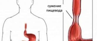
Symptoms
At the early stage of cancer, there are practically no symptoms; the first signs of the disease appear when the esophageal tube is significantly narrowed. Often, the symptom of narrowing - dysphagia - is not paid attention to, pushing stuck pieces of food with a large amount of liquid. Difficulties passing food last for several months, the patient limits himself to food and loses weight. Dysphagia is accompanied by abundant production of saliva, and later there is a putrid smell from the mouth due to stagnation of food over the constriction and vomiting of what has just been eaten. Patency may improve with the disintegration of the tumor that has narrowed the lumen.
Germination into the trachea will manifest itself as a cough with sputum containing food particles. At this stage, high fever and symptoms of inflammation of the bronchopulmonary system appear.
Causes
Esophageal varicose veins occur due to delayed drainage of venous blood into the portal vena cava system. The primary cause of esophageal varicose veins is considered to be portal hypertension - an increase in blood pressure in the portal, hepatic vessels, and inferior vena cava. Having collected waste blood from the abdominal organs, the portal vein carries it to the liver.
The pressure in the portal vein rises due to a number of completely different circumstances:
- Hepatitis and fatty liver disease - chronic viral damage to the liver (hepatitis) leads to compression of the mouth of the portal vein at the point where it enters the liver. Metabolic disorders (diabetes, obesity), constant intoxication with ethanol, drugs, harmful substances lead to fatty degeneration of hepatocytes - hepatosis. Blood circulation in the organ slows down, and the lumen of blood vessels decreases.
- Liver cirrhosis, Pick's cirrhosis - replacement of functional liver tissue with connective dense tissue, progresses against the background of infections and alcoholism. Or fibrotic degeneration is caused by extremely severe cardiovascular failure. Bleeding from the veins with cirrhosis of the liver is profuse, violent, begins at night and threatens life without urgent intervention.
- Portal fibrosis of the liver, myelofibrosis - liver diseases with the formation of connective tissue scars that destroy the organ.
- Tumors - benign and malignant, compress and narrow the blood vessels of the liver.
- Parasites - echinococci, schistosomes invade the liver, feed on its tissues, destroy the structure of the organ, and cause vascular damage.
- Chiari disease, Budd-Chiari syndrome - inflammation, thrombosis of the hepatic veins with subsequent blockage, vascular damage and hemorrhage.
- Sclerosis, thrombosis, stenosis, portal vein atresia - acquired or congenital. They narrow the lumen of blood vessels, causing blood stagnation.
In rare cases, the cause of varicose veins (VV) of the esophagus is severe general hypertension and congenital anomalies of vascular development. Portal hypertension is characterized by dilated veins in the thoracic and abdominal segments of the esophagus. Varicose veins of the cervical spine indicate diseases of the lungs and thyroid gland.