Author
Baryshnikov Vladimir Leonidovich
Leading doctor
Candidate of Medical Sciences
Radiologist
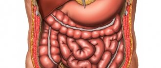
MSCT of the abdominal cavity and retroperitoneum
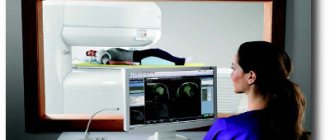
– a modern diagnostic method that provides a high degree of detail and makes visualization of internal organs as convenient as possible. Multislice computed tomography (as the abbreviation MSCT stands for) is based on the use of X-ray radiation. Tissues with different densities transmit X-rays differently. This information is read using special equipment - a tomograph. Multislice tomographs used in “Family Doctor” make it possible to obtain 64 slices with a thickness of 0.625 mm. for one revolution of the gantry (the working part of the tomograph). This information is processed using powerful computer software that reconstructs images of the area of interest in any plane, as well as in 3D (3D). MSCT examination of the abdominal cavity and retroperitoneal space is actively used to assess the condition of organs related to this area, differential diagnosis and detection of diseases in the early stages.
Description of the MSCT technique
Diagnostics is based on the effect of X-rays on the object under study, followed by analysis of the degree of absorption of radioactive radiation by tissues.
During scanning, an X-ray tube rotates around the patient and generates beams that pass through the body and are picked up by sensors.
The obtained data is converted into a planar image of the slice under study. In 0.5 sec. the doctor receives up to 64 sections, and in 15 seconds more than 2000 images.
From the images of the sections, three-dimensional models of organs are created with the possibility of subsequent reconstructions.
Indications, restrictions
Considering the potential risk of radiation damage to organs and tissues, MSCT of the OBP is prescribed over the low information content of ultrasound methods and endoscopic examinations.
The method is indicated for diagnosis:
- volumetric formations of the abdominal cavity;
- focal lesions of parenchymal organs;
- prevalence of malignant neoplasms;
- purulent destructive diseases;
- acute mesenteric ischemia;
- traumatic injuries;
- perforations;
- acute pathology of the gallbladder;
- intra-abdominal bleeding;
- obstructive jaundice;
- intestinal obstruction;
- dissection, rupture of the abdominal aorta.
The procedure is limited during periods of active growth and development in children and adolescents under 18 years of age and in pregnant women.
In patients with pathology of the kidneys, thyroid gland, or a history of allergic reactions, scanning is performed only after assessing the functional state of the organs and carrying out specific preparation.
Indications and contraindications
MSCT of the abdominal cavity, if necessary, can be prescribed to adults and children. But there are strict indications for its implementation:
- abdominal trauma (detecting damage to internal organs, bleeding and foreign bodies);
- abdominal pain of unknown origin;
- suspicion of a tumor of the stomach, intestines, liver, pancreas or bile ducts;
- assessment of the prevalence of the tumor process in malignant neoplasms of various localizations;
- differential diagnosis of non-tumor diseases of the digestive system (cysts, parasitic infestations, stones in the bile ducts, cholecystitis, cholangitis, Crohn's disease), if other research methods fail to make a diagnosis.
An MSCT certificate of the abdominal cavity is usually used to study those organs that are impossible or technically difficult to study x-ray, as well as when differential x-ray diagnostics are difficult and to clarify ultrasound data.
Carrying out multislice computed tomography is associated with radiation exposure, and with the introduction of contrast, additional side effects. Young children who require general anesthesia before the procedure have additional risks.
Therefore, for children under 14 years of age, testing is prescribed only in extreme cases, when it cannot be avoided or for health reasons.
In adults, contraindications to it are:
- pregnancy;
- too much body weight (more than 150 kg);
- restrictions associated with parenteral administration of contrast (allergy, renal failure, severe diabetes mellitus and hyperthyroidism).
It is not advisable who are breastfeeding using MSCT
How to prepare properly
MSCT of the abdominal cavity and retroperitoneal space is performed on an empty stomach, after preliminary cleansing of the intestines.
Patients with a complicated medical history are additionally prescribed premedication and adequate hydration.
In a contrast-enhanced study, patients donate blood a week before the tomography to determine the level of creatinine in the blood serum.
To reduce gas formation, a diet and carminative medications are prescribed 1-3 days before the procedure. For constipation, a cleansing enema is given on the eve of the scan.
For optimal visualization of the liver, pancreas and small intestine, a fluid intake of 1500-2000 ml is prescribed 1.5 hours before the procedure with bolus contrast.
When using oral radiocontrast agents, patients take orally 20-40 ml of urografin solution diluted in 1000-1500 ml of water:
- 1st serving 500 ml the day before the procedure;
- 2nd serving 250-500 ml on the day of scanning;
- 3rd serving 250 ml before the study.
How does MSCT differ from CT?
The modern technique of MSCT of the abdominal cavity (abdominal organs) is a multilayer tomography method. If a standard CT machine, the equipment has several rows of emitters, as well as detectors, whose task is to capture the signal. The emphasis should be placed on the fact that MSCT provides a detailed image of organs and determines pathology in the early stages.
Due to these features, the specialist receives a number of advantages during diagnosis:
- the duration of the procedure, even with the introduction of a contrast agent, is significantly reduced;
- The radiation exposure is minimal, which means that the examination will not cause harm to the body;
- obtaining a clear image of the abdominal cavity;
- in one rotation of the device, the entire abdominal organs are visualized;
- all processes that occur in organs are immediately recorded by the device;
- the number of possible image defects is reduced.
Many people are interested in the question of what MSCT of the abdominal cavity shows. A diagnostician using this examination method can see any pathology: from problems with blood vessels to the presence of malignant tumors with metastases, and they can be seen even in the early stages.
If we compare an MSCT device with an MRI, the difference lies in the technologies used:
- Magnetic resonance imaging uses a magnetic field directly to examine the cavity. The MSCT method involves the use of X-rays.
- MSCT of the abdominal cavity is used if you need to study in detail the condition of tissues that have a high density.
- If hollow organs are examined, MRI is rarely prescribed due to the low information content of the method. The best option would be to use MSCT with direct injection of a contrast agent. With MSCT, even minor changes in the mucous membranes can be seen.
What is it - MSCT of the abdominal cavity? This is an examination method that allows you to quickly detect the following pathologies:
- inflammatory processes accompanied by abscesses;
- benign/malignant neoplasms in the gastric cavity;
- damage to certain internal organs as a result of injury;
- ascites (the so-called accumulation of fluid in the abdominal cavity).
The advantage of the technique is that organs that have internal cavities, as well as hollow organs, are examined. The method allows you to determine neoplasms, liquid or solid (of any density).
How does the scan work?
Patients, having received information about the study, change into loose clothes and go to the diagnostic room.
If diagnostics with contrast are planned, a peripheral venous catheter is installed. In the hardware room, a laboratory assistant helps you take the correct position on the mobile tomograph device.
Starting position: lying on your back, arms above your head. The limbs are secured with straps.
The table with the patient moves inside the tomograph frame and stops at the level of the study area.
When scanning is started, the table moves continuously and covers the area from the dome of the diaphragm to the pubic symphysis. During the diagnosis, you will need to hold your breath while inhaling once upon the doctor’s command.
After data collection, the couch moves out of the frame, the patient is released from the restraints and escorted to the waiting room.
MSCT with contrast agents
For the study, iodine contrast agents are used in a volume of 100 ml. The drugs are injected into the bloodstream through an automatic syringe injector attached to the venous catheter. The delivery rate of iodine-containing contrast is 4 ml/sec.
Contrast enhancement makes it possible to diagnose oncological processes, acute surgical pathology - acute pancreatitis, perforations, bleeding, etc.
Scan duration
Diagnostics of the abdominal cavity using MSCT machines takes 9-14 seconds. With bolus contrast enhancement, the examination may take up to 15-20 minutes due to repeated scans that are taken at each contrast phase.
Radiation exposure
Radiation dose for multislice abdominal CT depends on the patient's weight, age, scanning protocol, and use of contrast enhancement.
In a native study without the use of contrast agents, the patient receives an average dose of 10-14 mSv. With tomography with intravenous contrast, the radiation dose increases 3-4 times.
The increase in radiation dose is due to repeated scans in accordance with the specified diagnostic phases. When using the simultaneous arterial-venous scanning technique, which gives a combined image of two phases, the radiation dose is reduced by 36-43% and averages 12-14 mSv.
Frequency of diagnostic procedures
The patient's radiation dose during scanning of the abdominal cavity significantly exceeds the permissible annual radiation exposure, which is 5 mSv.
Therefore, to prevent radiation damage to cells and tissues, the study is carried out no more than once a year. In emergency cases, when the patient’s life depends on a quick and accurate diagnosis, tomography is prescribed without taking into account the X-ray studies performed over the year.
For patients with cancer, to assess the effectiveness of therapy and monitor the dynamics of the process, MSCT can be prescribed up to 6 times a year with a total radiation dose of 100 mSv.
Indications
A scan is required when the patient has certain symptoms indicating the presence of pathology in the abdominal or pelvic organs:
- frequent nausea and vomiting;
- pain behind the sternum or in the abdomen;
- belching rotten;
- signs of internal bleeding - pallor, drop in pressure, dark color of stool;
- jaundice, abdominal enlargement;
- bowel disorders;
- colic in the abdomen;
- against the background of one or more symptoms – sudden weight loss (may indicate oncology).
For the listed symptoms, the doctor may prescribe MSCT, but for most symptoms, alternative research methods can be performed. Computer scanning is relevant when other diagnostics are powerless and uninformative.
There are also clinical recommendations when scanning is mandatory:
- after injuries - in such cases it is possible to identify hidden injuries, bleeding, fractures of ribs and vertebrae;
- to clarify the diagnosis - with a less perfect examination, you can “overlook” pathologies that are easily detected during scanning;
- before the operation – is prescribed routinely to assess the patient’s condition.
Depending on the condition, a survey or targeted MSCT may be performed. In the first case, the entire abdominal cavity is scanned, in the second - only one organ or area.
Advantages of tomography
MSCT is a universal technique that, without the use of additional examination methods and invasive interventions, shows the condition of organs, surrounding tissues and blood flow in one procedure.
The advantages of multislice CT include:
- high spatial and temporal resolution;
- performing multiplanar reconstructions and creating 3D models;
- standardization;
- visualization of hollow organs and vascular bed;
- sensitivity in detecting formations over 3 mm in size;
- differential diagnosis of the nature of neoplasms;
- speed and accessibility.
Distinctive features of MSCT from SCT
Multislice CT with classical SCT has a common operating principle, but differs in technical support.
Compared to spiral tomographs, MSCT devices are equipped with a large number of receiving devices.
MSCT scans an object with 4-helices in one revolution of the tube, while the rotation speed is 0.5 seconds faster than the MSCT.
This allows you to increase the speed of a full scan to 10 seconds, take pictures in several planes, obtain up to 2000 images and reduce the level of radiation.
Unlike SCT, which requires one breath-hold to obtain each anatomical region, with MSCT the entire body is examined in one inhalation phase.
How much does the research cost?
The average price of MSCT in Moscow is 6-10 thousand rubles. The cost of diagnostics depends on the area being examined. For example, an examination of the knee joint will cost approximately 5-6 thousand rubles, and an examination of the abdominal cavity - more than 10-11 thousand rubles.
With the advent of spiral computed tomography, a real breakthrough occurred in the diagnosis of many pathologies.
MSCT of the abdominal cavity (multispiral or multilayer computed tomography) is a modern method for diagnosing diseases of the abdominal cavity. Unlike conventional CT, in an MSC tomograph the capsule with sensors during examination is not static, but performs a rotational-translational movement. The trajectory of the scanners resembles a spiral. Thanks to this, photographs of one studied area are taken at different angles and from different points, the duration of the procedure is noticeably reduced, as is the radiation exposure to the person.
Alternatives
MRI of the abdominal organs - reveals changes in the structure of organs and tissues at preclinical stages.
The technique is most informative for the diagnosis of focal neoplasms, cysts, metastases, developmental anomalies, and purulent-destructive processes.
Non-contrast MR cholangiography detects strictures, inflammation and stones in the biliary tract.
Ultrasound examination diagnoses diffuse changes in parenchymal organs, space-occupying formations, cystic inclusions, bezoars of the stomach and the initial part of the duodenum.
Widely used for acute surgical and gynecological pathologies, abdominal injuries, and developmental defects. Ultrasound angioscanning of the arteries reveals stenosis, occlusion, compression, and pathological vasodilatation.
MSCT of the chest organs
This is a highly informative modern diagnostic method, widely used to identify a large number of chest pathologies. Often it becomes the final examination confirming the diagnosis. MSCT of the chest (CHC) is also used to adjust the treatment plan. Thanks to this technique, it is possible to accurately visualize all bone structures, cartilage, nerves, soft tissues, blood vessels, lymphatic systems and mammary glands in women. Indications for MSCT of the chest:
- pulmonary infarction;
- suspected cancer, cysts or metastases;
- lung adenocarcinoma;
- diseases of the aorta or large vessels;
- suspicion of abscess, tuberculosis, pneumonia, pleurisy and other infections;
- diseases of the esophagus;
- aortic pathology.
- Cyst on the ovary: treatment and symptoms
- Ice lamp for nails - which one is better to choose? Where to buy an ice lamp for nails, reviews and videos
- Turkey fillet in the oven - preparing delicious dishes. Baked turkey fillet recipes with photos
Cost of the study
The price for 1 tomography session depends on the examination area, the use of contrast agents, narcotics, sedatives and antispasmodics.
Average prices per procedure:
- native examination of the abdominal cavity - 5-6 thousand rubles;
- scanning of the abdominal cavity with bolus contrast - 9-10 thousand rubles, excluding contrast agent;
- MSCT angiography of the abdominal aorta and its branches - 8-10 thousand rubles;
- iodine-containing contrast agent - 7-10 thousand rubles.
Preparing for an abdominal CT scan
CT scan of the peritoneal organs is one of the few types of examination that requires careful preparation. It will depend on it how well all organs will be visualized in the pictures.
Before the procedure, it is advisable to undergo additional examinations, such as ultrasound of the peritoneum, x-ray or colonoscopy. It is worth consulting with an allergist and anesthesiologist about the possible risks from the introduction of X-ray contrast. If the urinary system is to be examined, laboratory tests must be done for the presence of creatine, urea, AST and ALT enzymes.
Since the main problem that reduces the information content of the examination may be intestinal bloating and fullness, it is necessary to normalize the functioning of the gastrointestinal tract in advance. To do this, three days before the tomography, you need to switch to a gentle diet: exclude not only heavy and fatty foods, but also those foods that cause increased gas formation. These include: cabbage, legumes, beets, celery, fresh and dried fruits, cereals, dairy products, fresh baked goods, confectionery products. It is also undesirable to consume kvass, carbonated water, strong coffee and tea, and alcoholic beverages. It is allowed to eat lean steamed fish, omelettes, light non-rich broths, boiled chicken, and oatmeal with water.
The test is done on an empty stomach, which is why a CT scan of the abdominal cavity is usually scheduled for the first half of the day. The day before, they give a cleansing enema, take a mild laxative, or administer a suppository with glycerin. In addition, 18 hours before you can drink enteric sorbents: activated carbon or smecta.
Two hours before the scan, you should stop drinking any drinks; this may increase intestinal motility.
It is worth noting that the introduction of X-ray contrast without appropriate preparation can provoke vomiting, nausea, and in some cases, intestinal volvulus. If the patient had a CT scan of the abdominal cavity with contrast, then after it it is advisable to drink a liter of clean drinking water. This promotes rapid removal of the dye.