Gastric hyperemia is characterized by reddened and swollen lesions on the mucous membrane of the organ. This disease is a consequence of overcrowding of blood vessels. A red gastric wall means the initial stage of development of the inflammatory process. Similar manifestations are often diagnosed with gastritis, peptic ulcers and other lesions of the gastrointestinal tract.
The gastric mucosa is hyperemic
Normally, the gastric mucosa has a light pink color, which becomes brighter closer to the pyloric region. In some patients they have a yellowish tint, which is not a pathology. During examination, the epithelium reflects the light of the endoscope, so it appears shiny. Numerous folds of the mucosa have a thickness of 6-10 mm. Their size gradually increases closer to the antrum. When air is introduced into the stomach cavity, the folds of the mucous membrane are smoothed out, and this allows you to examine the entire surface.
If the diagnostician notes that the gastric mucosa is hyperemic, what does this mean? External signs of hyperemia are redness and swelling of the folds of the stomach. The color change is due to blood flow.
The mucous and submucosal layers of the wall have a branched capillary network, with numerous anastomoses between them. Therefore, an increase in blood inflow and a decrease in blood outflow causes the filling of capillaries, which shine through the epithelial layer, changing the color of the mucous membrane.
Diagnostic measures
If any suspicious symptoms appear (for example, nausea, bloating, heartburn, belching, pain), it is very important to visit a gastroenterologist in a timely manner. One of the main methods for studying the condition of the mucosa is a procedure called esophagogastroduodenoscopy. It is performed using an endoscope - a special probe with a camera at one end to view the internal state.
Using this method, you can accurately assess the condition of the body and the internal walls of the stomach, take a piece of tissue for analysis (biopsy), visualize the presence of a pathological process, and prescribe the correct course of therapy.
A competent specialist will easily determine the presence of pathology in hyperemic epithelium, since healthy tissue looks shiny and produces mucus normally. During the examination, when inflated with air, the folds are straightened, the surface is smooth, the epithelial cover is intact and clean, without wounds or ulcerations. The color is pink, there may be a yellowish coating.
As the disease develops, the symptoms worsen, redness of the epithelial layer appears, the folds increase, do not straighten when inflated, and swelling is observed.
Causes of hyperemia of the gastric mucosa
The reasons for changes in blood flow may be associated with neurohumoral regulation of the vascular bed, diseases of the heart, kidneys and other organs. In addition, hyperemia can also be physiological. For example, congestion of the stomach walls occurs during digestion, or when a heating pad is applied to the epigastric region.
Therefore, if we talk about what hyperemia of the gastric mucosa is, we need to take into account the physiological and pathological mechanisms of its development. For example, during an inflammatory reaction of the body, inflammatory mediators are released at the site, causing dilation of blood vessels, increasing blood flow to the affected tissues. This is a protective reaction in which tissue trophism and cell regeneration are enhanced.
Hyperemia of the gastric mucosa - what does this mean?
What should you do if in the description of the examination the doctor wrote about hyperemic gastric mucosa?
The medical term hyperemia means redness and swelling. In itself, hyperemic mucosa is not dangerous - it is just a symptom that signals that the stomach is sick.
The medical term hyperemia means redness and swelling.
In itself, hyperemic mucosa is not dangerous - it is just a symptom that signals that the stomach is sick.
Inflammation of the antrum
The human stomach consists of 3 sections: upper; middle (organ body); antrum, or antrum (lower section). The antrum is responsible for the production of 3 important substances: bicarbonates, which alkalize it for the smooth passage of food into the intestines; mucus, which protects the stomach from being corroded by hydrochloric acid; the hormone gastrin, which is responsible for the production of hydrochloric acid. Antrum gastritis - what is it? This is a chronic inflammation of the antrum and a violation of all its functions.
The occurrence of type B gastritis (an alternative name for the disease) is possible with untreated acute or catarrhal gastritis. Consequences of chronic inflammation: the normal functioning of the gastrointestinal glands is disrupted; hyperplasia of the gastric mucosa occurs; acidity increases; scars appear on the mucous membrane, which is a sign of antrum atrophy; the passage connecting the stomach with the intestines narrows; the passage of food into the duodenum becomes difficult; the risk of developing esophageal diseases increases.
Classification of mucosal hyperemia
Physiological and protective reactions of the body are carried out by regulating vascular tone by the nervous system or bioactive substances. That is, this is active plethora. If there is a disturbance in the outflow of blood, for example, an increase in pressure in the vena cava system or fluid retention in the body due to kidney disease, gastric hyperemia occurs passively.
In both cases, the liquid component of the blood leaks into the tissue, causing swelling. At first, this does not cause serious changes, but if this condition persists for a long time, the metabolism in the cells is disrupted, which makes them vulnerable to the aggressive effects of digestive juices.
There are two types of hyperemia:
- Active . In most cases, it is useful, as it promotes the restoration of damaged cells, for example, when the mucous membrane is exposed to unfavorable factors (malnutrition, pathogenic microflora). But with progression and long-term existence, blood clots form in full-blooded vessels, causing damage and death of epithelial cells.
- Passive . Impaired outflow has an adverse effect on the mucous membrane. Hypoxia and thrombosis cause a decrease in the protective properties of cells, fluid retention in the intercellular space, and swelling.
Based on localization, diffuse and focal hyperemia of the gastric mucosa is distinguished. During FGDS, the diagnostician also indicates the location of the lesion.
Swelling due to gastritis causes
Swelling occurs when internal organs, skin, or other parts of the body become enlarged. This is usually the result of inflammation or fluid accumulation in the body. Swelling may occur internally or may affect the skin and muscles. Sometimes cases of minor swelling may go unnoticed. Edema is not always accompanied by related symptoms.
In recent years, edema has occurred that appears as a result of diseases of the gastrointestinal tract and in people suffering from insufficient protein content in food. The accumulation of fluid in the body is explained by the fact that the kidneys excrete it in smaller quantities.
The formation and distribution of intercellular and tissue fluids may also be disrupted as a result of increased pressure in the capillaries. The dependence of the formation of edema on the height of venous pressure is clearly visible in patients with mitral stenosis and left ventricular failure - their edema occurs primarily in the pulmonary circulation.
Swelling with gastritis occurs when internal organs, skin or other parts of the body become enlarged
Chronic heart failure as a cause of edema in gastritis
If the work of the heart is disrupted, blood is retained in the periphery, and the person develops swelling of the legs, ankles, and lower back. In severe cases, the outflow of blood from internal organs is disrupted. Fluid accumulates in the abdominal cavity, and the stomach increases in size. This condition is called ascites.
Allergy as a cause of swelling in gastritis
Allergic edema can be caused by foods, medications, flowers, animals, or insect bites, to which a person has developed hypersensitivity. The difference between allergic edema is that it develops suddenly, literally in a few minutes. The person does not experience pain, but allergic edema is one of the most life-threatening. Swelling of the larynx and tongue can cause suffocation and death.
Hormonal disorders as a cause of swelling in gastritis
Hormonal imbalance with gastritis is another risk factor. For example, if the thyroid gland produces too few hormones, special substances accumulate in the blood that cause the tissues to retain water. Hormonal swelling most often appears on the legs. They are dense to the touch: if you press, there is no fingerprint left.
Varicose veins as a cause of edema in gastritis
In addition to swelling, other symptoms occur - weight gain, constipation, rare pulse, increased drowsiness.
Varicose veins as a cause of edema in gastritis
Venous edema also appears in the evening - in the form of a mark from the elastic of socks or spreads to the lower leg and foot. The swelling is accompanied by “heavy leg syndrome”: they swell, hurt, and buzz.
Kidney disease as a cause of swelling in gastritis
With kidney disease, conditions are created for sodium and fluid retention in the body. Swelling of the face and area around the eyes is common. The arms and legs swell, especially the ankles and shins. In this case, protein is lost in the urine, the protein content in the blood decreases, and conditions are created for the accumulation of fluid in the tissues. You may notice that your urine becomes foamy, your appetite decreases, and weight gain occurs due to fluid retention in the body.
Taking medications as a cause of swelling in gastritis
Doctors believe that pain pills containing ibuprofen and drugs to treat high blood pressure and diabetes may contribute to fluid retention in tissues. The same applies to hormonal pills and antidepressants.
Poor nutrition as a cause of swelling in gastritis
Talk to your doctor to find out how the medications you take affect your ability to swell and ask for advice on how to manage the problem.
Poor nutrition as a cause of swelling in gastritis
It is necessary to reduce to a minimum the consumption of salt, foods high in salt or salty foods. Remember - salt retains fluid in the body. You should not drink a lot of liquid at night - the kidneys may not have time to process it and then all the accumulated liquid will immediately affect the face.
It is better to stew, boil or bake all foods. Firstly, this way they retain as much useful substances as possible, and secondly, proper nutrition helps normalize the functioning of all organs, including blood vessels.
After all, problems with blood vessels also lead to the formation of edema. It is also necessary to consume enough fiber per day, which is most found in fresh vegetables and fruits.
Therapeutic measures
For people interested in how to restore the gastric mucosa, it is important to know that when this disease is diagnosed, in most cases no treatment is prescribed, since it is believed that the body independently fights the problem through self-regeneration. During this process, accelerated metabolism occurs, which causes the process of tissue self-healing to enter an active stage.
This condition is a positive phenomenon in the development of arterial hyperemia. In some cases, doctors create an additional flow of blood fluid to stimulate regenerative processes and speed up recovery. If you are interested in how to restore the gastric mucosa in case of other pathologies, you need to contact your doctor with this question.
How to restore the stomach: drugs that restore the gastric mucosa – Site about poisoning
Patients with stomach diseases ask gastroenterologists many questions. The mucous epithelium of the internal cavity performs a protective function against destruction by enzymes and acids. The question of how to restore the gastric mucosa after treatment comes first.
The process is quite complicated and takes a long time.
Changes in microflora, disruption of the integrity, structure and physiological performance of the walls of the organ occur as a result of various diseases, as well as after taking medications and other negative factors, which leads to the formation of erosions, ulcers, severe complications arise, bleeding, perforation, and cancerous degeneration are possible. Let's figure out how to restore the mucous membrane of the stomach and intestines.
How to restore the gastric mucosa with atrophic gastritis?
This pathology can only be treated conservatively, there is no point in operating anything, the atrophic areas are located in patches of different areas. Gastroenterologists consider the disease a precancerous condition, but before the cells degenerate into cancer, they can be returned to normal.
Conservative therapy used to treat chronic atrophic gastritis consists of three stages:
- Treatment with medications.
- Proper diet.
- Use of folk remedies.
When minimal atrophic changes occur with small foci, treatment of atrophic gastritis does not cause difficulties. If a person seeks help late, a stage of diffuse atrophy occurs: in this case, it is possible to restore some areas and prevent the progression of the disease.
How to restore the gastric mucosa: treatment with folk remedies |
Problems with the digestive tract are a big nuisance that can cause a lot of trouble. Chronic or acute diseases of the digestive system have a detrimental effect on the condition of the gastric mucosa, against which other diseases develop. There are many ways to restore the gastric mucosa, but not all of them are safe.
Indications for mucosal restoration
Of course, the health of the gastrointestinal tract must always be taken care of. But there are a number of diseases in which it is urgent to restore the gastric mucosa. It is almost impossible to independently determine the severity of the problem, so it is better to visit a gastroenterologist.
You should pay attention to the following conditions:
- Acute and chronic diseases of the gastrointestinal tract.
- Long-term use of antibiotics and other aggressive drugs.
- Rashes on the skin.
- Changing taste habits.
- Diarrhea or constipation.
- Heartburn.
- Nausea and vomiting.
- Regular or episodic pain in the abdominal area, which is characterized by a different nature.
In the vast majority of cases, diseases of the esophagus, stomach and intestines do not cause much discomfort, they only remind of themselves with mild indigestion. Any disturbances in the functioning of the digestive system are accompanied by changes in the frequency and nature of bowel movements.
Pathological processes occurring in the body can be diagnosed through timely examination.
Basic treatment methods
Restoring the digestive system is possible only if certain rules and recommendations are followed. Gastroenterologists strongly recommend adhering to them to achieve the most effective results.
A person should take note of the following rules:
- Any attempts to cure the stomach should be discussed with a doctor who will take into account all the individual characteristics and medical history of his patient. There are a large number of diseases affecting the organs of the digestive tract; in one case, the same drug is indicated for use and is strictly contraindicated.
- Throughout the entire therapeutic course and after it, you must adhere to a gentle diet. The diet should be rich in cereals, fresh fruits and vegetables.
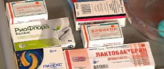
- It is strongly recommended not to take antibiotics unless absolutely necessary. If taking pills cannot be avoided, there are ways to protect your stomach while taking medications. It is necessary to maintain the natural intestinal microflora with the help of supplements with bifidobacteria.
- If the condition worsens, you should immediately stop all therapeutic manipulations and consult a doctor again.
Strict compliance with all the rules will not only improve your well-being, but also avoid possible complications.
Taking medications
Before developing a therapeutic course, the doctor must conduct all the necessary examinations. Regimen for taking and dosage of medications for the regeneration of the mucous membranes of the stomach.
There are many drugs that restore the gastric mucosa, each of which has certain pharmacological features, indications and contraindications for use. Let's look at the most popular ones:
- Venter is made from one active ingredient - sucralfate. It is designed to protect the gastric mucosa, which it provides thanks to the protective film that forms on it. Significantly accelerates recovery processes, heals and relieves inflammatory processes.
- Cimed is a complex medicinal product that contains two active ingredients - zinc and copper. The preparation contains an extract from sea buckthorn berries. The main task of the medicine is to activate the production of enzymes.
- Regesol is a herbal medicine that contains licorice, calendula, plantain and sea buckthorn. Accelerates the healing process of the gastric mucosa, increases its resistance to the harmful effects of radiation therapy and chemistry. Reduces inflammatory processes in the digestive tract, destroys pathogenic microflora and pathogenic bacteria.
Source: https://bdc03.ru/pomoshh-pri-otravlenii/kak-vosstanovit-zheludok-preparaty-vosstanavlivayushhie-slizistuyu-zheludka.html
Symptoms
Any type of hyperemia leads to a decrease in barrier function, inflammation, and corresponding symptoms. Patients complain of pain, burning in the epigastric region, and indigestion. As the disease progresses, heartburn, nausea, and vomiting may appear.
If hyperemia of the inner layer of the stomach is caused by somatic diseases (heart disease, kidney disease), the patient may experience:
- swelling;
- high blood pressure;
- difficulty urinating;
- drowsiness, etc.
In such situations, additional examination is required. Hyperemia is often observed in patients with unstable mental health and under stressful conditions.
Thrombosis of the collar vein
Ascites that appears against the background of portal vein thrombosis is persistent and is also accompanied by obvious pain, mild hepatomegaly, and splenomegaly. Due to the occurrence of collateral circulation, extensive bleeding from hemorrhoids or esophageal varices often appears. Thrombocytopenia, leukopenia, and anemia are detected in peripheral blood.
Ascites is a disease that accompanies portal intrahepatic hypertension and is characterized by moderate hepatomegaly and muscular dystrophy. On the skin of the abdomen, the expansion of the network of veins in the shape of a “jellyfish head” is clearly visible. Persistent ascites in postrenal portal hypertension is accompanied by jaundice, vomiting, nausea and severe hepatomegaly.
Swelling of the abdomen is also observed in heart failure. In sedentary patients with heart disease, there is an accumulation of fluid in the abdomen, sacrum, sides, and pelvic organs. Swelling, although considered the most characteristic sign of heart failure, is not the only one. Patients experience shortness of breath and tachycardia, which indicates advanced pathology.
With protein deficiency, ascites is most often minor; pleural effusion and peripheral edema are noted. In rheumatic diseases, polyserositis is expressed by specific skin symptoms, the presence of fluid in the cavity of the pleura and pericardium, ascites, arthralgia and glomerulopathy. If there are disturbances in the outflow of lymph (chylous ascites), the size of the abdomen quickly increases. Ascitic fluid has a milky hue, pasty consistency; lipoids and fats are determined in it during laboratory testing. The volume of fluid in the peritoneal cavity with ascites can reach 5-10 or even 20 liters.
The abdomen swells in older people much more often than in young people.
Diseases due to gastric hyperemia
In gastroenterology, mucosal hyperemia is associated with stomach diseases such as gastritis and peptic ulcers. In various forms of gastritis, in addition to focal hyperemia, the following signs are observed:
- Spicy . It is characterized by severe hyperemia and swelling of the folds, petechiae, erosions, and copious amounts of thick mucus.
- Chronic . The mucous membrane is pale, dull, grayish in color. Sometimes there are thinned areas (atrophy) with translucent vessels. This is the so-called false hyperemia.
- Superficial gastritis is characterized by diffuse hyperemia, the formation of foamy white mucus, swelling of the folds that do not level out when inflated. Submucosal hemorrhages are sometimes observed.
- Hypertrophic gastritis is characterized by thickening and pronounced diffuse hyperemia of the folds; they acquire a cherry color. Proliferative processes (nodules, warts) are revealed on the surface.
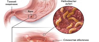
Hyperemia is also present in other forms of gastritis (phlegmonous, necrotic), as well as ulcers. It indicates an inflammatory process. When infected with Helicobacter Pylori, hyperemic manifestations are more pronounced.
Diagnostic methods
Hyperemic changes can only be diagnosed using endoscopy. For diagnosis, fibrogastroduodenoscopy or endoscopic video capsule is used. ultrasound , radiography , CT , MRI visually determine the appearance of the inner layer only indirectly, by identifying swelling of the mucous membrane.
Manifestation of the disease
In gastroenterology, mucosal hyperemia is associated with stomach diseases such as gastritis and peptic ulcers. In various forms of gastritis, in addition to focal hyperemia, the following signs are observed:
- Spicy. It is characterized by severe hyperemia and swelling of the folds, petechiae, erosions, and copious amounts of thick mucus.
- Chronic. The mucous membrane is pale, dull, grayish in color. Sometimes there are thinned areas (atrophy) with translucent vessels. This is the so-called false hyperemia.
- Superficial gastritis is characterized by diffuse hyperemia, the formation of foamy white mucus, swelling of the folds that do not level out when inflated. Submucosal hemorrhages are sometimes observed.
- Hypertrophic gastritis is characterized by thickening and pronounced diffuse hyperemia of the folds; they acquire a cherry color. Proliferative processes (nodules, warts) are revealed on the surface.
Hyperemia is also present in other forms of gastritis (phlegmonous, necrotic), as well as ulcers. It indicates an inflammatory process. When infected with Helicobacter Pylori, hyperemic manifestations are more pronounced.
Diagnostic methods
Hyperemic changes can only be diagnosed using endoscopy. For diagnosis, fibrogastroduodenoscopy or endoscopic video capsule is used. Other studies (ultrasound, radiography, CT, MRI) can visually determine the appearance of the inner layer only indirectly, by identifying swelling of the mucous membrane.
Pathology manifests itself for two reasons:
- Poor blood removal from the stomach (venous or passive).
- Large blood flow (arterial or active).
In the first case, further recovery is possible, but the passive form affects tissues due to a lack of oxygen.
Gastric hyperemia is characterized by a high blood content in each vessel of all linings of the stomach. In its normal state, it has a pinkish color and a reflective layer of epithelium, which can be seen during endoscopy. The thickness of the folds is 0.5-0.8 cm. When air is supplied to the stomach, they are smoothed out.
Arterial hyperemia develops when in each artery and even in small arterioles that carry blood to the stomach, an increased flow of blood is detected in each banded part of the organ. Various injuries to the smooth muscle of the vessel are characterized by strong blood filling, so the gastric mucosa turns red.
Based on the surface of the gastric mucosa, an experienced specialist can determine the type of disease:
- Superficial gastritis indicates a moderately hyperemic pathology. In this case, there is a white mucous foam on the surface of the stomach, and the folds have tortuous thickenings and do not straighten when air enters.
- In an atrophic state, the mucous membrane seems to become thinner, and upon examination a reddish vascular pattern is noticeable.
- With fibrous gastritis, purulent lesions are visible, since the causative agents of the disease are infections (scarlet fever, measles). Possible vomiting of blood.
- The phlegmous type of pathology indicates only focal areas that have been affected, for example, a fish bone.
The stomach lining becomes red and swollen because the blood vessels in the walls of the organ become overfilled with blood. It is not for nothing that in the old days this condition was called “plethora.”
The first type is called venous or passive hyperemia, the second - arterial or active. There is a significant difference between active and passive hyperemia.
Only active action leads to tissue healing, while passive action, on the contrary, contributes to further damage to the organ due to lack of oxygen in the tissues.
The gastric mucosa becomes hyperemic in most diseases of the gastrointestinal tract.
Based on the condition of the mucous membrane and the location of redness and swelling, the type of disease can be determined.
Most often, hyperemia is diagnosed as one of the types of gastritis, but it can be a symptom of duodenitis, stomach ulcers, or diseases of organs that are not related to the gastrointestinal tract at all.
Normally, the gastric mucosa should be pink, shiny, and well reflect the light of the endoscope.
The folds of healthy mucosa are 5–8 mm thick; when air is blown in, they straighten well, allowing the doctor to view all parts of the organ through an endoscope.
Hyperemic gastric lining occurs in several varieties. The type of hyperemia determines the diagnosis of the disease.
With superficial gastritis, hyperemia reaches a moderate degree. The inflammatory process can cover a separate area or become widespread. During the acute course of the disease, the endoscope reveals white foam, the folds of the organ look thicker than normal. When gas is injected, it is not possible to completely achieve a smooth inner wall.
Atrophic gastritis is characterized by focal thinning of the membrane. The vascular pattern in this place is clearly visible, the areas of the mucosa around the atrophic zone look paler.
If the hyperemic gastric mucosa is accompanied by the release of purulent masses, such gastritis has a fibrous form. The disease rarely has independent factors of genesis; in most cases, scarlet fever or measles have consequences in the form of hyperemia of the mucous membrane, followed by vomiting with blood.
This is how areas of dead mucous membrane with pus are rejected and is accompanied by intense pain.
Phlegmous gastritis is usually called hyperemia of an area of the mucous membrane that has been subjected to injury or sexually transmitted infection.
Alkali or acid in the stomach causes deep damage to many layers of the digestive organ. Necrotic areas are not the worst option for the development of necrotic gastritis. It is worse if provoking factors cause perforation of the walls of the organ, pouring its contents into the abdominal space and causing peritonitis.
Diagnostics
Having looked at the statistics, we can conclude that almost 90% of people need consultation with a gastroenterologist. To make a correct diagnosis, a specialist prescribes an examination, which is divided into laboratory and instrumental diagnostics.
Laboratory methods include: studies of gastric juice, blood, urine and feces. With their help, you can determine the secretory function, bacterial composition of the gastrointestinal tract, enzyme activity and other important functions. But without instrumental methods, the analysis results are uninformative.
Laboratory methods include: studies of gastric juice, blood, urine and feces. With their help, you can determine the secretory function, bacterial composition of the gastrointestinal tract, enzyme activity and other important functions. But without instrumental methods, the analysis results are uninformative.
- gastroscopy or esophagogastroduodenoscopy (EGDS) is a type of examination that is carried out using special equipment (gastroscope) with a flexible hose, equipped with viewing optics and a camera. Contraindications for manipulation are: heart disease, hypertension, mental disorders, severe respiratory failure. Before performing the procedure, the patient must refuse to eat food no earlier than 8 hours, and water 3 hours before, not take medications, smoke, or even brush their teeth;
- Symptoms of hyperemia of the gastric mucosa The gastric mucosa is focally hyperemic, there is a coating with whitish foamy mucus on the surfaces of the organ in “mucus lakes”, the folds are compacted and are not completely smoothed out with the help of air.
- As cells die, the surface becomes thinner and turns pale. In this case, the foci of the disease do not become hyperemic, and the vascular web is clearly visible.
- In the superficial form of gastritis, the mucous surface of the stomach is hyperemic throughout or only in the body and antrum of the stomach. Sometimes hyperemia is focal in nature or can be diffuse.
- If there is fibrous gastritis, hyperemia is most pronounced, while it is focal and characterized by the presence of pus. This inflammation can be caused by measles or scarlet fever infection. The patient may vomit blood frequently.
- The phlegmous form of the disease can be triggered by trauma to the stomach with sharp objects, such as fish bones. In such cases, this indicates possible hyperemic foci.
- Bulbit is characterized by swelling and redness, thickening of folds in the antrum. Among the reasons are Helicobacter pylori infection of the antrum of the stomach and unhealthy diet.
- Renal dysfunction (various degrees of swelling).
- Depression and permanent stress also provoke hyperemia.
Diagnosis and treatment methods
In the case of swelling and hyperemia of the gastric mucosa, treatment is prescribed only after identifying the cause and a complete examination of the body.
Survey
Diagnosis is carried out by a gastroenterologist. First, he examines and interviews the patient, finding out how severe the symptoms are. Afterwards, a series of diagnostic procedures are prescribed. What exactly these procedures will be depends on the cause of the disease. It can be:
- gastroscopy;
- X-ray of the pancreas;
- Ultrasound of the abdominal organs;
- tissue biopsy;
- CT scan.
Based on the results of the procedures described above, the doctor draws up a plan for further action.
Medications
Like diagnosis, the choice of medications depends on the cause of the disease and its severity:
- Antibacterial drugs. Helps fight infections.
- Antacids. Neutralizes hydrochloric acid, which causes irritation.
- Histamine receptor blockers.
- Drugs that stimulate the stomach.
- Proton pump inhibitors. Reduces the production of hydrochloric acid.
It is also recommended to take enzymes that have a beneficial effect on the functioning of the gastrointestinal tract as a whole.
Surgical intervention
The operation is performed if the cause of the swelling is a malignant tumor. In such cases, complete or partial resection (removal) of the stomach is performed. If the tumor spreads to neighboring organs, they will also have to be removed.
After surgery, chemotherapy and radiation therapy are indicated to prevent the occurrence of metastases. To restore the body, after all procedures are completed, the patient is prescribed vitamin complexes.
Traditional methods
Traditional medicine can only be used after consultation with a doctor. Like conventional medications, they have their side effects and contraindications.
There are several effective recipes:
- 3 tbsp. pharmaceutical chamomile pour 750 ml of hot water. Infuse in a thermos. Drink 1 glass 3 times a day. This remedy helps relieve pain.
- You can reduce inflammation with calendula alcohol tincture. A single dose is 50 drops (diluted in 250 ml of warm water).
- Sea buckthorn decoction will help eliminate nausea. To prepare it, 6 tbsp. berries (fresh or dry) need to be filled with 1 liter of hot water. Bring to a boil and cook for 5 minutes. Strain using cheesecloth. Take throughout the day. Add honey if desired.
Treatment methods
Since the gastric mucosa can become hyperemic for various reasons, this means that drug treatment is not always required. Sometimes it is enough to eliminate the impact of adverse factors on the body.
Treatment is carried out in accordance with the diagnosis. Appointed:
The patient must be prescribed dietary nutrition.
Clinical manifestations
Patients whose gastric mucosa is severely hyperemic rarely realize this. Some symptoms may not be related to the gastrointestinal tract at all. At the initial stage, the heart rate increases. Drowsiness or problems with urination appear. Depending on where the foci of severe hyperemia are localized, the patient experiences:
- fatigue;
- obesity;
- swelling of the face.
Causes and signs of the disease
Most often, swelling of the gastric mucosa is a consequence of the development of internal diseases. This is usually a form of gastritis. But there are others: peptic ulcer, duodenitis, bulbitis.
There are also a number of factors that can cause redness and swelling:
- damage to organ surfaces with sharp objects;
- unbalanced diet;
- infectious diseases such as measles and scarlet fever;
- the presence of Helicobacter pylori bacteria in the gastric microflora;
- disorders of the kidneys;
- prolonged depression;
- frequent stressful situations.
Another cause of stomach edema is the appearance of tumors. It is noteworthy that this applies to both benign and malignant neoplasms.
The clinical picture of the pathology directly depends on the cause that caused it. If, for example, it is a stomach tumor, the symptoms may be:
- constant fatigue;
- lack of desire to do anything;
- lack of appetite;
- bad mood and even depression;
- pale skin;
- weight loss;
- anemia.
Swelling due to gastritis manifests itself in several conditions:
- pain in the gastrointestinal tract in the morning after waking up and a couple of hours after eating;
- sour belching and heartburn caused by an excess of hydrochloric acid;
- poor appetite;
- bowel dysfunction, in particular constipation and diarrhea.
Common symptoms of stomach edema include:
- nausea;
- vomiting;
- problems with urination;
- severe drowsiness;
- swelling of the face, arms and legs;
- cardiopalmus;
- violations of movement coordination.
Recommendations for nutrition in pathology
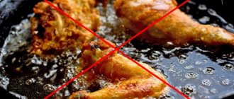
If hyperemic mucosa is detected, the patient is advised to follow a diet. Substances that irritate the mucous membrane should be excluded from the diet: spicy, salty, sour, fatty foods, smoked meats and marinades, alcohol, strong coffee. Fried foods are not recommended. For heat treatment, food should be boiled or steamed.
In the diet you can use:
- chicken, rabbit, turkey, veal, low-fat sea fish;
- a small amount of vegetable fats;
- skim milk, cottage cheese;
- non-acidic fruits and vegetables;
- cereals (rice, oatmeal, buckwheat);
- eggs, no more than 1 per day;
- wheat bread 1-2 grades, crackers.
Meals should be fractional (5-6 times a day), small portions. It is also necessary to observe the temperature regime; the dishes should not be too hot or cold. The optimal temperature is between 15 and 60 degrees Celsius.
How to treat
Arterial hyperemia is not treated in 85% of cases. In such a situation, intensive blood supply to tissues begins as a consequence of independent regeneration of damage. The cells of the stomach receive the necessary amount of oxygen and nutritional components, due to which metabolic processes are accelerated. As a result, cells begin to divide rapidly, and healthy tissues replace damaged structures. In the arterial form of the pathology, doctors only prescribe proper nutrition and help balance the diet.
In the remaining 25% of cases, active hyperemia, as well as venous blood filling, indicate the presence of gastritis. In case of inflammation of the mucous membrane, complex treatment is carried out. The patient must follow a strict diet, take antibiotics to suppress the growth of Helicobacter pylori and other medications to accelerate tissue regeneration. You are allowed to drink herbal infusions and eat honey.
Possible complications and prognosis
Prolonged irritation and damage to the mucosa are harbingers of gastritis. If you do not pay attention to this in time, plethora leads to the development of microthrombosis, hypoxia and the progression of damage to epithelial cells.
If hyperemia is detected, the patient should be observed by a doctor to avoid the development of chronic gastritis and peptic ulcer. With adequate treatment, the prognosis is favorable, since normally the surface epithelial layer is renewed every 7–10 days.
What is gastric edema
Inflammation of the stomach or swelling of the stomach is said to occur if the area of damage is limited to the superficial layer of the mucosa and the healing of the gastric mucosa subsequently takes place without a trace. Stomach edema occurs in acute gastritis. When the stomach swells, the abdomen becomes hard.
The causes of acute inflammation of the stomach or swelling of the stomach are dietary factors. It is enough to overeat, chew food poorly, eat very hot, spicy, fatty foods, drink an alcoholic drink or take a medicine that irritates the mucous membrane, and redness, local swelling, and painful irritation appear on its surface.
Systematic swelling of the stomach can lead to the development of chronic gastritis. Symptoms of gastric edema are pain in the epigastric region, a feeling of fullness in the stomach, decreased appetite, and moderate nausea.
Forecast
A favorable prognosis is possible only with timely treatment of the pathology. Treatment of the disease is complicated by the characteristics of its causative agent, the bacterium Helicobacter pylori. This microorganism is capable of temporarily stopping its vital activity and going into “sleep mode”. When a provoking factor occurs, activity resumes.
Therapy must be carried out fully. If you stop treatment early, the risk of complications will increase. Hypertension can cause peptic ulcers, large-scale atrophy of the mucous membranes of the digestive tract, and the formation of malignant tumors.
Sources
https://gastrosapiens.ru/zheludok/zabolevaniya-zh/ochagovyj-antralnyj-gastrit.html https://gastroved.com/ochagovyy-antralnyy-gastrit-lechenie.html https://ogastritah.ru/vidy/chto- takoe-ochagovyj-antralnyj-gastrit-i-kak-lechit-eto-zabolevanie.html https://gastrocure.net/bolezni/gastrit/antralnyj-gastrit.html https://medicalok.ru/zheludok/gastrit/antralnyj. html https://gastrotract.ru/bolezn/gastrit/antralnyj-gastrit.html https://zheludokok.ru/gastrit/antralnyj-gastrit-poverhnostnyj-i-ochagovyj.html https://zhkt.expert/gastrit/kak -raspoznat-ochagovyj-antralnyj-gastrit.html https://ogastrite.ru/lechenie-gastrita/ochagovyj-antralnyj-gastrit-prichiny-simptomy-diagnostika-lechenie/ https://gastrit.guru/lechenie/antralnyj-gastrit. html https://vseprogastrit.ru/gastrit/vidy-gastritov/antralnyjj-gastrit/
Prevention measures
To avoid irritation and inflammatory plethora of the mucous membrane, doctors recommend adhering to the following preventive measures:
- stop smoking;
- limit the intake of alcohol-containing drinks and strong coffee;
- Healthy food;
- avoid stress;
- See a doctor on time and treat chronic somatic diseases.
It is also necessary to exercise and not overwork. This will help boost immunity and ensure protection and rapid restoration of the mucous membrane.