When is a fluoroscopy of the stomach prescribed?
X-ray diagnostics have been helping to identify serious gastrointestinal diseases for more than 100 years.
X-ray of the stomach, performed using the traditional method (or using double contrast), is recommended in the following situations:
- suspected ulcer or cancer of the stomach and duodenum;
- suspicion of protrusion of the wall (diverticulum) of the esophagus;
- black color of stool;
- frequent pain or tightness in the stomach and intestines;
- difficulty swallowing;
- anemia or sudden weight loss for no obvious reason;
- after operations on the stomach and digestive organs.
X-ray of the stomach is considered a harmless procedure, but has several relative contraindications, including:
- pregnancy;
- general serious condition;
- massive bleeding from the gastrointestinal tract.
Indications and contraindications
Ulcers
X-ray examination of the abdominal cavity is an inexpensive and accessible diagnostic method. Radiation exposure is minimal. The procedure helps to quickly exclude (or confirm) serious diseases. Recommended in the following cases:
- suspicion of the presence of ulcerative pathology in the patient;
- for diseases of an inflammatory nature;
- the probable presence of tumors of benign or atypical etiology;
- during possible deformation processes.
X-ray of the stomach is prescribed when a certain clinical picture develops: sudden weight gain or loss, constant pain in the epigastric region, frequent occurrence of heartburn, blood in the stool.
Diagnosis is carried out according to the doctor’s recommendations; you will first need to visit a gastroenterologist. The procedure has some contraindications, they are characterized as absolute and relative.
Pregnancy is a relative contraindication; diagnosis is carried out with the permission of an obstetrician-gynecologist, but not earlier than in the third trimester. Women who are in the lactation period need to temporarily stop feeding.
Internal bleeding is also a contraindication for the study. This condition distorts diagnostic results. After eliminating the bleeding, an x-ray is available and will indicate the reasons that provoked this process.
X-rays are not prescribed for patients who are in severe physical condition to avoid deterioration. Also, a stomach image with barium is not prescribed if there is an allergic reaction to the contrast agent.
Preparation for barium fluoroscopy of the stomach
An empty stomach is required for the study: the last meal should be 6-8 hours before diagnosis.
Attention!
Immediately before contrast fluoroscopy, you should not smoke, take medications, alcohol or other drinks.
Other, special preparation for fluoroscopy of the stomach with barium is not required for most people.
For patients suffering from increased gas formation in the intestines, following a diet 2 or 3 days before the study will help them prepare for the examination of the stomach and duodenum.
It is recommended to exclude from the diet:
- dairy products;
- confectionery;
- fresh and rich bread;
- carbonated drinks;
- cabbage and pickles.
In rare cases, before the study, the doctor prescribes gastric lavage or cleansing enemas.
How to prepare for a stomach x-ray
Normally, preparation is not something special. The requirements for radiography are the same as for a number of many procedures:
- do not eat for 8-9 hours;
- do not smoke or drink for an hour.
However, if you have stomach problems, you need to switch to a certain diet in a few days. You need to completely remove from your diet everything that causes gas formation. Even if you take enzymes, all dairy and fermented milk is removed from the diet. Cabbage and any fiber are prohibited. The table should be strictly dietary, as if the patient had just undergone gastric surgery.
For elderly people or those with severe organic lesions in any part of the gastrointestinal tract, preliminary cleansing with the drug Fortrans is required. Cleanse according to instructions. It is not advisable to use Duphalac, as it cleanses only part of the intestines.
This type of X-ray irradiation is strictly contraindicated for pregnant women.
Cleansing is required for everyone if a double contrast is planned. Before preparing, you must abstain from alcohol in any quantity. The main point in preparation: minimizing gas formation. Gas formation seriously interferes with the procedure and affects accuracy. If there is excess gas, the patient may have the study canceled or rescheduled.
You need to understand if radiography, like fluoroscopy, is not an alternative to gastroscopy. If the patient is unable to clear the gastrointestinal tract and comply with the requirements, he will have to undergo an FGDS for diagnosis. FGDS is based on the gag reflex and is based on swallowing a special probe. Gastroscopy is a much more unpleasant procedure, especially difficult for patients with impaired gag reflex.
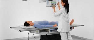
How to do a fluoroscopy of the stomach
The examination is carried out in a specially equipped (darkened) X-ray room using a monitor.
Fluoroscopy of the stomach is performed in several stages according to the following algorithm:
- First, a plain X-ray examination of the abdominal cavity is done in a standing position. It allows you to assess the general anatomical picture and functional features of the gastrointestinal tract. For seriously ill patients, fluoroscopy is provided in the supine position on a special lowering table - trochoscope.
- Next, the patient takes barium, which consistently fills the sections of his gastrointestinal tract.
- During the process of advancing the contrast agent, the doctor assesses the condition of the organ, for which he takes a series of photographs in different positions of the patient (on the back, on the stomach, on the side).
Typically, a stomach fluoroscopy takes 15 to 30 minutes. At the end of the procedure, the doctor analyzes the images and draws up a medical report.
On a note:
Immediately after diagnosis, you need to drink plenty of clean water. This will help remove the contrast agent from the intestines faster.
Features of fluoroscopy of the resected stomach
After resection, gastroscopy is performed to assess the size of the gastric stump, the function of the connection (anastomosis) and the speed of passage (passage) of contrast through the small intestine.
The study is carried out by:
- in the early postoperative period (on days 10-12);
- in three months;
- in two or three years.
X-ray of the stomach after resection allows us to identify early and late complications, as well as disturbances in motor-evacuation function caused by dynamic and mechanical reasons after surgery.
What does the examination show?
Without the introduction of a contrast agent into the body, X-rays easily pass through the soft tissues and cavities of the digestive system, forming a blurry image.
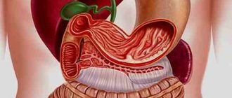
A high-molecular suspension of barium absorbs the flow of X-rays, and, filling the entire lumen of the digestive tube, like a cast, clearly depicts a “portrait” of the gastrointestinal tract on the screen. This “professional photo session” allows you to accurately examine the position, shape, structure, tone and contraction of the walls (peristalsis) of the hollow organs.
For example, during the first sips of barium suspension, with the patient standing, the movement of the contrast agent through the esophagus is monitored: its shape, size and motor-evacuation function are assessed.
And, a hiatal hernia (HH) can be detected in the Trendelenburg position - the patient’s pelvis is lifted up at an angle of 35-45 degrees.
Contrast studies can detect most severe gastrointestinal diseases in the early stages of pathological changes.
Barium X-ray of the stomach shows:
- foreign bodies in the gastrointestinal tract;
- diseases of the esophagus (constrictions, protrusion of the wall (diverticulum), neoplasms, varicose veins, etc.);
- diseases of the stomach and duodenum (inflammatory processes, ulcers, cancer, malabsorption and peristalsis, etc.);
- pathologies of the intestines and other organs.
To increase the information content of the study, images are taken in the patient’s vertical, lateral and horizontal positions.
How is diagnostics carried out?
The first images are taken in a standing position without prior administration of contrast. This will allow us to identify gross pathological processes.
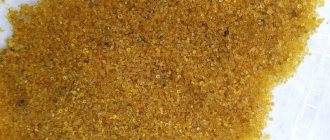
Next, the patient drinks barium sulfate diluted in water. The solution tastes like calcium. First, the patient takes a few sips of barium, after which the examination continues. The wall of the esophagus is examined.
The patient finishes the remainder of the solution, a little less than a glass, after a few minutes. After which the radiography continues, the stomach and other organs of the digestive tract are examined.
A contrast agent is necessary for a detailed study of the condition of the stomach. Barium prevents rays from passing through the organ, thus allowing its pathology to be detailed.
After taking barium, the patient is asked to lie on the table; during the manipulation process, the position will have to be changed several times for an informative examination. It is believed that a qualitative assessment of the condition of the stomach can be obtained with the patient in a supine position at an angle of 45˚ (Trendelenburg position).
X-ray of the stomach with barium is a non-invasive diagnostic method, painless, and not accompanied by discomfort. It is highly informative and helps to quickly diagnose serious problems.
X-ray of the stomach and duodenum
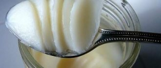
Typically, X-rays of the stomach and intestines are performed with barium. This substance is safe for humans; its consistency resembles a milkshake and tastes like chalk; taken orally (but when examining the rectum, a barium suspension is administered using an enema). After passing through the esophagus and then the stomach, the barium suspension enters the duodenum, which makes it possible to examine the initial section of the small intestine as well.
Contrast fluoroscopy of the stomach and duodenum is done in two stages:
- weak filling stage (the contrast envelops the mucous membrane, it is possible to study all its folds);
- stage of tight filling (the organ is completely filled with “contrast”; the shape, size, location, elasticity and other characteristics can be assessed).
There are two types of research:
- fluoroscopy of the stomach and duodenum combined with radiography (taking a series of radiographs during the examination);
- double contrast method (both barium and air are contrast).
Attention!
This type of diagnosis cannot be combined on the same day with fibrogastroduodenoscopy or colonoscopy.
What does an x-ray of the stomach and esophagus show?
The procedure will make it possible to track changes in the walls of the stomach and additional shadows in nearby tissues.
- Change in lumen. Narrowing occurs with cancer of the stomach or another organ that compresses the stomach from the outside. Enlargement is a sign of a diverticulum - acquired or congenital.
- Displacement of the stomach. May occur with injury or hernia.
- Violation of integrity. The changes are characteristic of peptic ulcer disease. The location of the defect is visualized as a dark area on the x-ray.
- Filling defects. The place where the contrast did not penetrate may be a tumor, polyp or neoplasm.
- Thinning of the folds is possible with atrophic gastritis.
Pathologies of the esophagus:
- Narrowing or protrusion of diverticula;
- Dilatation of veins;
- Foreign bodies.
Stomach diseases:
- Malabsorption;
- Duodenal ulcer.
- Cancer.
Digital fluoroscopy of the stomach
Modern methods of radiation diagnostics with contrast have been enriched by the use of computer technology and new highly sensitive sensors.
Digital fluoroscopy of the stomach has the following advantages:
- significant reduction in radiation dose;
- high quality x-ray images;
- high speed and ease of research;
- X-ray images can be conveniently enlarged, printed, saved on digital media, or sent via the Internet.
The digital technique does not use laboratories and chemical reagents to develop X-ray films, making it cheaper and more environmentally friendly.