Most diseases associated with the digestive system are accompanied by swelling and redness of the mucous membrane. Hyperemic gastric mucosa very often appears during gastroduodenoscopy. Typically, this type of examination is recommended by a doctor to confirm diseases such as gastritis, pancreatitis, and ulcers. These are those diseases that have accompanying symptoms: pain in the epigastric region, belching, nausea, flatulence.
Manifestations of violation
If the gastroscopy conclusion indicates that hyperemic gastric mucosa is observed only in some places, that is, focally, then this indicates the initial stage of inflammation in the walls of the stomach. It is important to understand that this is not an independent disease. The cause of hyperemia of the gastric mucosa is the development of one or another pathology in the epigastric region.
Under no circumstances should you leave your well-being to chance if you begin to be bothered by painful sensations in the upper stomach, nausea and heartburn. Contact your doctor immediately. Hyperemic focal gastric mucosa is one of the symptoms of many diseases of this organ, but they can only be identified with a thorough examination.
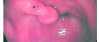
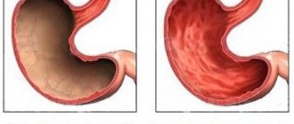
In a normal state, the gastric mucosa has a pink tint, a smooth surface, and reflects the shine of the corresponding equipment. The thickness of the folds of the mucosa is 5-8 mm. When straightened with air, the folds should completely disappear and expand.
Reasons for the development of the pathological condition
Hyperemia of the gastric mucosa occurs in the following diseases.
Reflux esophagitis
A chronic disease of the esophagus, which is characterized by inflammation of its mucous membrane due to the constant reflux of stomach contents into it. Sometimes, when the disease occurs, the pain radiates to the sternum and resembles symptoms of heart disease.
Often, patients mistake pain for angina pectoris, without even thinking about digestive problems. The main signs of pathology include: belching of air or food, nausea, severe heartburn, sour taste in the mouth, regurgitation, prolonged hiccups. The chronic form of esophagitis is characterized by alternating periods of exacerbation and remission.
Gastritis
Inflammation of the gastric mucosa and its dystrophic changes. The form of the disease is determined by the location and nature of the redness and swelling: if the gastric mucosa is moderately hyperemic and there is a slight whitish coating, then we can talk about minor inflammation.
If the redness is severe, the mucous membrane is thinned and blood vessels are visible, then atrophic gastritis is diagnosed. Focal hyperemia is observed during purulent-inflammatory processes, characterizing the fibrous form. If the gastric mucosa is diffusely hyperemic, then perhaps we are talking about superficial gastritis.
The clinical picture of the disease includes the following symptoms: pain and a feeling of fullness in the epigastric region, nausea and vomiting, increased salivation, decreased or loss of appetite, frequent belching, bloating, weight loss. The chronic form of gastritis does not have pronounced symptoms, but is characterized by periodic exacerbations with disruption of the gastrointestinal tract.
Peptic ulcer
A pathology characterized by damage to the gastric mucosa and the formation of ulcers in it. Signs of the disease can be different and are related to the size and location of the defects, pain threshold, stage of the disease, age of the patient, etc.: pain that can occur on an empty stomach and go away after eating, and vice versa, heartburn, belching sour or bitter, a feeling of heaviness in the stomach, rapid satiety, flatulence, decreased or loss of appetite.
Of all the pathologies of the stomach, peptic ulcer is the most insidious and can be accompanied by a number of complications. These include penetration, perforation, malignancy, pyloric stenosis and bleeding.
Bulbit
A disease in which there is redness and swelling of the mucous membrane of the bulbar part of the duodenum. The disease can be asymptomatic or have a pronounced acute period. The main signs of bulbitis are:
Stomach problems
- bitter taste in the mouth;
- minor pain in the upper left abdomen;
- attacks of nausea and vomiting;
- often constipation.
In addition, other unpleasant symptoms may appear, such as a whitish coating on the tongue, increased gas formation, cramping abdominal pain on an empty stomach or after eating. If the pathology is not treated in any way, then there is a likely risk of developing gastrointestinal bleeding.
Duodenitis
An inflammatory disease characterized by an inflammatory process in the duodenum. Often the disease is combined with gastritis, which most often affects the antrum of the stomach.
Characteristic signs of pathology are:
- epigastric pain that increases with palpation of the abdomen;
- constant nausea;
- rarely vomiting mixed with bile;
- rumbling in the stomach;
- flatulence;
- loss of appetite and weight loss.
When bile stagnates, yellowness of the skin and sclera of the eyes may appear. In elderly people, duodenitis is often asymptomatic and is diagnosed accidentally during an FGDS. But there are also factors due to which the gastric mucosa is hyperemic:
- mechanical damage to the digestive organ by any object;
- irrational and unhealthy diet;
- infectious diseases (measles, scarlet fever);
- bacterial infection (Helicobacter pylori);
- renal failure;
- prolonged exposure to stress and depression.
Remember! If you experience any discomfort behind the sternum or in the upper abdomen, as well as nausea and vomiting, you should seek help from a specialist as soon as possible.
Symptoms
If pathology begins to develop, the following symptoms appear:
- the mucous membrane becomes thinner or, on the contrary, thickened;
- redness appears;
- it swells;
- ulcers appear on the surface of the mucosa.
If the inflammatory process begins, the gastric mucosa becomes hyperemic either in one place or diffusely. Visually, upon examination, you can see that the mucous membrane is red, swollen, and blood fluid appears in the vessels.
Excessive filling of blood vessels can be a consequence of such problems:
- dysfunction of blood outflow from the walls of the stomach;
- excessive filling of the walls with blood.
By the way, active hyperemia can be considered a completely positive phenomenon, because it is a signal of recovery, and if there is a lack of blood supply and the regeneration function is inhibited, then the pathology of the stomach walls worsens. These negative phenomena are accompanied by oxygen starvation of tissues. Only with the help of an examination can a specialist determine the severity of the pathology and prescribe a competent course of treatment.
Diagnostics
Having looked at the statistics, we can conclude that almost 90% of people need consultation with a gastroenterologist. To make a correct diagnosis, a specialist prescribes an examination, which is divided into laboratory and instrumental diagnostics.
Laboratory methods include: studies of gastric juice, blood, urine and feces. With their help, you can determine the secretory function, bacterial composition of the gastrointestinal tract, enzyme activity and other important functions. But without instrumental methods, the analysis results are uninformative.
Instrumental methods include:
- gastroscopy or esophagogastroduodenoscopy (EGDS) is a type of examination that is carried out using special equipment (gastroscope) with a flexible hose, equipped with viewing optics and a camera. Contraindications for manipulation are: heart disease, hypertension, mental disorders, severe respiratory failure. Before performing the procedure, the patient must refuse to eat food no earlier than 8 hours, and water 3 hours before, not take medications, smoke, or even brush their teeth;
- X-ray of the stomach with contrast agent. With its help, you can identify the condition of the gastric mucosa and diagnose improper functioning of the gastrointestinal tract. The procedure is contraindicated during pregnancy and lactation, intestinal obstruction, perforation of the stomach wall, or allergies to barium preparations. Before the procedure begins, the patient must take a contrast agent. A few days before the x-ray, completely abstain from legumes and dairy products; on the evening before the procedure, refrain from sweet products, raw vegetables and fruits;
- Ultrasound diagnostics or echography is a method that is based on the ability to reflect sound waves. This method is not very informative and is most often prescribed to young children. Using echography and ultrasound, you can determine the presence of tumors, ulcers, thickening of organ walls, etc.
Gastritis and the consequences of hyperemia of the gastric mucosa
Symptoms of gastritis in adults are as follows:
- A mild inflammatory process causes mild hyperemia and focal damage to the gastric mucosa. The mucous membrane on the surface looks swollen, a whitish coating is visible on it, thickening of the folds can be diagnosed, stretching with air does not lead to their complete smoothing.
- During the atrophic process, serious depletion of the mucous membrane occurs, it becomes pale, and red vessels are clearly visible. Hyperemia develops locally.
- Gastritis of the fibrous form causes pronounced hyperemia, which is accompanied by purulent processes and is located focally. Pathology develops as a result of infection with measles and scarlet fever. An accompanying symptom may be bloody vomiting. This may indicate that the festering film is being rejected.
- In the phlegmous form, focal lesions become noticeable, which are the result of trauma to the stomach with something pointed. Such an object could be, for example, a fish bone.
- If a patient is diagnosed with bulbitis, swelling accompanied by redness can be detected, and in the antrum the folds are thickened. The mucous membrane is swollen and red. The disease is a consequence of an unbalanced diet or the influence of the Helicobacter pylori bacterium.
- If kidney function is impaired, many patients can be diagnosed with swelling and hyperemia of the gastric mucosa, which is expressed in varying degrees of severity, depending on the type of pathological process.
- Hyperemia can be a consequence of factors such as prolonged depression, chronic stress, and constant emotional outbursts. With such negative psychological factors, the vascular walls of the stomach become overfilled with blood fluid.
Ignoring the symptoms of this disease can lead to atrophy of the gastric mucosa.
What is hyperemia of the gastric mucosa
In medicine, the term “hyperemia” means redness and swelling, in particular of the mucous membranes and skin. This phenomenon occurs as a result of the vessels in the affected area overflowing with blood.
If during gastroscopy it is discovered that the gastric mucosa is swollen and hyperemic, then this condition indicates that the inflammatory process of the organ wall has begun. Hyperemia can be localized diffusely or focally.
This pathology is a symptom of many stomach diseases. Normally, when the mucous membrane has a pink tint, it reflects the glare of the endoscope, and its thickness ranges from five to eight millimeters.
When wrinkles expand under the influence of air, they quickly smooth out. It is considered normal when the epithelium in the antrum is pale pink.
Diagnosis of pathology
If you are bothered by unpleasant symptoms, such as nausea, heartburn, pain in the stomach, belching, bloating, you need to make an appointment with a gastroenterologist as soon as possible. He will prescribe tests and carry out appropriate diagnostic measures.
To diagnose pathology, the basic methods of examining the gastrointestinal tract are used. Such diagnostic methods can provide a complete picture of the disease. In the case of hyperemic gastric mucosa, the best method is esophagogastroduodenoscopy. This procedure is performed using an endoscope - a special probe, at one end of which there is a camera that records internal changes.
Using this type of examination, it is possible to accurately assess the condition of the gastric walls, take a biopsy, see the pathology and develop a competent course of treatment.
An experienced doctor can easily diagnose the development of pathology if, during examination, the mucous membrane turns out to be hyperemic, because healthy tissue has a special shine and produces mucus normally.
Types and symptoms of pathology
Hyperemia of the gastric mucosa is divided into several types, each of which is characterized by a special clinical picture. With the passive type, there is an excessive flow of blood. The stomach stops working and becomes further damaged due to lack of oxygen. The second type is arterial hyperemia in the stomach, characterized by impaired blood flow from the walls of the internal organ.
The mucous membrane of a healthy digestive organ has a pale pink color.
In a healthy patient, the gastric mucosa has a pale pinkish tint. When the organ is swollen and reddened moderately, the clinical picture may take a long time to manifest. If hyperemia occurs against the background of bulbitis, then thickening occurs in the antrum of the stomach and the duodenal bulb of the intestine. In this area, swelling occurs and the mucous membrane becomes motley. Hyperemia occurs with general symptoms:
- severe epigastric pain;
- heartburn;
- attacks of nausea accompanied by vomiting;
- problems emptying the bladder;
- constant desire to sleep;
- swelling of the legs and face;
- tachycardia;
- loss or gain of body weight;
- impaired coordination.
A common cause of gastric hyperemia is an inflammatory reaction that occurs in several forms:
- Moderate. Hyperemic mucosa is characterized by swelling, which looks like a foam-like coating on the top layer. Hyperemia may be accompanied by one focus or the mucous membrane is damaged unevenly. Such signs indicate mild inflammation of the stomach.
- Local. The folds of the mucous membrane turn pale and become thin, and blood vessels are visible. Such manifestations signal atrophic gastritis.
- Phlegmous. The mucous membrane is significantly swollen, which is associated with mechanical trauma to the stomach with a sharp object.
- Fibrous. Hyperemia covers several foci that turn red and fester. A dangerous symptom of this form is vomiting blood.
Therapeutic measures
For people interested in how to restore the gastric mucosa, it is important to know that when this disease is diagnosed, in most cases no treatment is prescribed, since it is believed that the body independently fights the problem through self-regeneration. During this process, accelerated metabolism occurs, which causes the process of tissue self-healing to enter an active stage.
This condition is a positive phenomenon in the development of arterial hyperemia. In some cases, doctors create an additional flow of blood fluid to stimulate regenerative processes and speed up recovery. If you are interested in how to restore the gastric mucosa in case of other pathologies, you need to contact your doctor with this question.
How to treat
Arterial hyperemia is not treated in 85% of cases. In such a situation, intensive blood supply to tissues begins as a consequence of independent regeneration of damage. The cells of the stomach receive the necessary amount of oxygen and nutritional components, due to which metabolic processes are accelerated. As a result, cells begin to divide rapidly, and healthy tissues replace damaged structures. In the arterial form of the pathology, doctors only prescribe proper nutrition and help balance the diet.
In the remaining 25% of cases, active hyperemia, as well as venous blood filling, indicate the presence of gastritis. In case of inflammation of the mucous membrane, complex treatment is carried out. The patient must follow a strict diet, take antibiotics to suppress the growth of Helicobacter pylori and other medications to accelerate tissue regeneration. You are allowed to drink herbal infusions and eat honey.
Important information: How to treat chronic disseminated intravascular coagulation (DIC) during pregnancy and what are the symptoms of thrombohemorrhagic
Treatment of hyperemia with gastritis
Quite often, redness of the epithelium is one of the symptoms of gastritis. The course of therapy in this case is quite long and complex:
- a special diet must be prescribed;
- medications are prescribed, including antibacterial and anti-inflammatory drugs, sorbents, enzymes and painkillers.
Enveloping drugs can be prescribed as auxiliary agents, which help eliminate inflammation, eliminate redness of the epithelial surface, and reduce the swelling process. In some cases, non-traditional treatment methods help. Thus, decoctions of medicinal plants and honey are actively used. And thanks to a special therapeutic diet, it is possible to prolong the remission of the disease for a long time.
Even when the patient has recovered, he is strongly recommended to make an appointment with a gastroenterologist for a preventive examination. You should visit a doctor at least twice a year so that if the inflammatory process recurs, you can notice it in the initial stages and begin treatment immediately.
Preventive actions
To avoid atrophy of the gastric mucosa and other complications, as well as to completely get rid of unpleasant symptoms, it is necessary to consult a doctor at the first signs of a clinical picture to make an accurate diagnosis and determine the causes of the development of the pathology.
Twice a year it is recommended to undergo diagnostics using gastroscopy to avoid recurrence of the disease. Take preventive measures prescribed by your doctor in a timely manner. Do not neglect your health, follow all the recommendations of your doctor and do not ignore unpleasant symptoms. If the disease is recognized in time, serious consequences and long-term treatment for advanced pathology can be avoided.
Hyperemia and other diseases
Most often, arterial hyperemia on the skin is accompanied by an increase in temperature in the problem area, redness, tingling and even swelling. In addition, the patient may experience other symptoms, but they will be associated with the underlying disease, which caused the hyperemia.
As an example, consider conjunctive hyperemia. If a patient has a similar problem, this indicates the development of pathology, and, most likely, an inflammatory process. In this case, lacrimation, pain in the eyes, purulent and mucous discharge will be noted. Conjunctival hyperemia is a common symptom of conjunctivitis, allergic reactions and mechanical effects, for example, friction from dirt and sand.
In representatives of the fair sex, the appearance of hyperemia on the genitals is not excluded. The mucous membrane often suffers from various irritants, which causes increased blood flow, redness and an increase in temperature. This is often accompanied by itching, an unpleasant odor, discharge and other signs of problems with the genitourinary system. In this case, hyperemia is a clear symptom of health problems.
Most often, if a woman experiences redness of the genitals, which is accompanied by other unpleasant symptoms, doctors make diagnoses related to a bacterial infection or sexually transmitted diseases. Diagnosis is carried out using the results of a blood test and a smear of the vaginal mucosa. But vaginal hyperemia is not always a sign of a serious illness. Often this is how an allergic reaction to taking medications and using condoms manifests itself. But in this case, redness and increased blood flow will occur only after contact with the allergy pathogen.
Representatives of the fair sex may encounter the fact that they will experience hyperemia not only on the external, but also on the internal genital organs. For example, a doctor may diagnose redness of the cervix. This often happens after sexual intercourse if it was too active. Treatment in such a situation is not required, since it is enough to simply abstain from sexual relations for a while and wait until everything returns to normal.
With the development of a bacterial and viral infection in the human body, hyperemia of the throat may occur. Inflammatory processes in the nasopharynx can be involved here, which will cause very unpleasant processes. Additional symptoms that will accompany hyperemia due to inflammation or infection are increased body temperature, pain during swallowing and severe swelling. This is often accompanied by nasal congestion and even the formation of pus.
In internal organs, the clinical picture of hyperemia can most often be observed in the stomach. This is a clear symptom that an inflammatory process is planned or is already occurring. Gastric hyperemia is a symptom of ulcers and gastritis. Most often, redness on the mucous membrane of the organ is caused by the bacterium Helicobacter pylori. It is this that initially causes gastritis, and then peptic ulcer disease.
Hyperemia does not always need to be treated. However, it is necessary to determine its cause. It is possible that this will be a sign of a serious illness or inflammatory process that requires active treatment.
