Why is gastric erosion dangerous?
Lack of timely treatment threatens exacerbation of the disease, leading to the following consequences:
- Ulcer . With ulcerative lesions, the deeper layers of the mucous membrane affecting the muscle tissue are affected. The disease is characterized by frequent relapses, accompanied by temporary improvement and a sharp exacerbation of symptoms. The presence of an ulcer leads to nausea after meals and causes sudden weight loss.
- Chronic form of erosion . The chronic course is dangerous due to the formation of polyps that can provoke bleeding. This disease is rare and with complete removal of the formations has a favorable prognosis.
- Gastric resection . Resection, that is, a surgical method of removal, is used in case of extensive damage (ulcers, malignant tumors). The painful recovery period takes about 1.5 months.
- Gastrointestinal bleeding . Vomit that is dark brown or black in color is an important symptom, indicating deep damage to the walls of the stomach. Internal bleeding can also be recognized by the graininess of the vomit. In this case, the patient experiences anemia, which affects the normal functioning of the kidneys.
- Stomach cancer . Gastric oncology has a high mortality rate and is more common in the male half of the population. Fatty and fried foods, flavored with copious amounts of hot spices, are the main factors that irritate the delicate mucous membrane. With constant contact with junk food, erosive formations can develop into cancer.
How is an ulcer different from an erosion?
Even though both diseases have a number of common features, each of them is characterized by the presence of its own characteristics in pathogenesis and clinical picture, which makes it possible to differentiate them from each other and make the correct diagnosis. The difference between erosion and gastric ulcer lies in a number of factors, which are presented in the table:
The difference between erosion and ulcer Factor Erosion pathology Peptic ulcer
| Causes | Psychogenic factor | Circulatory disorders in the stomach wall |
| Long-term use of non-steroidal anti-inflammatory drugs | ||
| The presence of pathogenic microflora (bacteria Helicobacter pylori) | ||
| Localization of the defect | Multiple point lesions of the mucous membrane | The defect extends to the muscle layer of the gastric wall |
| Symptoms | Pain while eating | Pain before and after eating |
| Nausea | ||
| Stool instability | Nausea | |
| Loose stools | ||
| Exacerbation regardless of season | Heartburn | |
| Seasonal exacerbations | ||
| Consequences of the disease in case of recovery | The mucous layer completely regenerates without scar formation | A scar forms |
The identity of the causes of occurrence, the mechanism of development and the clinical picture of the disease creates a problem in making the correct diagnosis. An ulcer can be considered a complication of erosion. The disease is more difficult to tolerate, leads to more serious complications and requires immediate medical or surgical intervention by a specialist when the first symptoms appear.
Comparison
The two phenomena in some cases represent stages of a single destructive process. Moreover, the difference between erosion and ulcer is that the first of them forms at an early stage, and the second - after some more time.
Initially, there is a negative impact of one or more factors. Disturbances in the stomach can occur, for example, due to irregular eating, constant consumption of hot liquids or taking irritating medications. All this, and much more, can lead to the destruction of mucosal cells and the occurrence of erosion.
It is a milder form of the disease because it affects only the superficial layer. The damaged area has a round or jagged shape and differs in color from the surrounding healthy tissue. The integrity of the mucous membrane during erosion can be impaired in several places at the same time, which aggravates the situation.
The development of erosion is indicated by spasms, discomfort when food is ingested, as well as bloody inclusions in stool or vomit. Fortunately, such a defect does not always degenerate into an ulcer. The pathogenic process may stop at this stage, especially if the necessary treatment has been carried out. With a favorable outcome, the tissues are completely restored, not even a scar remains.
But if provoking factors continue to act and a person is in no hurry to see a doctor, there is a risk of developing a more dangerous defect - an ulcer. With it, in addition to the mucous membrane, the deeper layers of the organ are also corroded. Unlike erosion, this damage is detected not only by endoscopic examination, but also by x-ray examination.
What is the difference between erosion and ulcer regarding symptoms? The fact is that the latter, for obvious reasons, is felt stronger. Pain occurs here both during and after eating. The stomach may not accept the food consumed, and vomiting occurs. The ulcer is often accompanied by severe heartburn and periodic bowel movements.
The disease takes a long time to treat and tends to periodically worsen. The diet is prescribed in both cases, but with an ulcer it is more strict. In case of successful healing, a scar remains at the site of such a deep defect.
Minor differences
How is erosion different from a stomach ulcer? Damage to the upper, mucous layer in the first disease, and damage to the mucous layer, including the submucosal plate, in the second.
In connection with the above, the second disease is characterized by regularly recurring relapse, long treatment times and scarring of the place where the ulcer was located.
The process is explained by the presence of a transition from the acute to the chronic type, when after healing the ulcer leaves a scar.
Visual differences between lesions
Depending on quantity
- single;
- multiple (point).
Erosion is classified by form into:
Acute erosion
In the acute form of the disease, oval or round defects appear on the mucous membrane. Mature erosion ranges in size from 2 to 4 mm. Most often they form on the walls of the organ body or on the bottom. In this case, the surface epithelium is absent, the epithelial cells become denser, and the amount of DNA in the cell nucleus increases. Healing of erosion of the stomach and duodenum takes 10 days, in rare cases up to 2 months.
Erosive and ulcerative skin lesions
V.N. Mordovtsev, V.V. Mordovtseva, L.V. Alchangyan Central Research Institute of Dermatovenerology, Ministry of Health of the Russian Federation, Moscow
Erosive-ulcerative skin lesions are a heterogeneous group of diseases, for which a common feature is a violation of the integrity of the skin and the formation of a defect within the epidermis (erosion) or reaching the dermis itself (ulcer). The formation of erosions and ulcers can be caused by various reasons: they can form at the site of the primary cystic elements as a result of insufficient local blood circulation (ischemia), and also arise as a consequence of an infectious inflammatory process or injury. In the case of a chronic, long-term non-healing ulcer, especially in an unusual location, a histological examination is necessary to exclude a malignant process (basal cell carcinoma, squamous cell carcinoma, lymphoma, metastatic cancer). Thus, we can propose the following pathogenetic classification of the main erosive-ulcerative skin lesions, including mainly those dermatoses where the formation of ulcers is a consequence of the natural evolution of the pathological process, and not other changes (for example, infection of erosions).
Pemphigus Epidermolysis bullosa
Circulatory failure Trophic ulcers of arterial origin Trophic ulcers of venous origin Neurotrophic ulcers Martorella's ulcer
Inflammatory vascular diseases Vasculitis (Wegener's granulomatosis, periarteritis nodosa, etc.) Pyoderma gangrenosum
Traumatic Pathomimia
Infectious inflammatory processes Tuberculosis (scrofuloderma, indurated erythema of Bazin) and other mycobacterioses Leishmaniasis Pyoderma (ecthyma, chronic ulcerative-vegetative pyoderma, chancriform pyoderma)
Pemphigus
The development of the disease is based on an autoimmune process in which antibodies are produced to various antigens of intercellular bridges - desmosomes, as a result of which epidermal cells lose contact with each other (acantholysis) and blisters form. Pemphigus is characterized by the development of blisters with a flaccid operculum, transparent contents, on the skin of the face, torso, in folds and on the mucous membrane of the oral cavity. Often blisters and erosions in the oral cavity are the first manifestation of the disease. Under the weight of the exudate, large blisters can take on a pear shape. The blisters spontaneously burst with the formation of extensive eroded areas of the skin. When pulling on the covering of the bladder, the phenomenon of epidermal separation in the adjacent area of unaffected skin with an increase in the cavity of the bladder is observed - Nikolsky's symptom. The disease often becomes generalized and severe, threatening the life of the patient.
Epidermolysis bullosa
Epidermolysis bullosa/hereditary pemphigus (Fig. 1 on color insert, p. 198) is a genetically determined disease, including over 20 clinical variants, characterized by the tendency of the skin and mucous membranes to develop blisters, mainly at sites of minor mechanical trauma (friction, pressure, ingestion of solid food). This is one of the most severe hereditary skin diseases, often resulting in death in young children and causing disability in adults. It develops in the first days of life, can exist from birth, and also develop at a later age. The condition worsens in the summer months. According to the level of formation of blisters in the epidermis, all forms of epidermolysis bullosa are divided into 3 groups: simple epidermolysis bullosa (intraepidermal blisters), borderline epidermolysis bullosa (blisters in the area of the basement membrane plate) and dystrophic epidermolysis bullosa (bubbles between the epidermis and dermis). Recessively inherited forms are the most severe. They are characterized by a generalized eruption of blisters that heal slowly to form scars. The repeated appearance of blisters on the skin of the hands, feet, in the area of the knee, elbow, and wrist joints leads to the development of scar contractures and fusion of the fingers. Scarring of blisters on the mucous membranes of the digestive tract also ends with the development of strictures and obstruction. The course and prognosis are worsened by secondary infection of bullous elements and tumors developing at the site of long-existing erosive and ulcerative skin lesions.
Familial benign chronic pemphigus
Familial benign chronic pemphigus is manifested by grouped vesicular and cystic rashes, prone to recurrence, with a predominant localization in the folds. It is inherited in an autosomal dominant manner, most cases are familial. The disease usually develops during puberty, but often at the age of 20-40 years. Clinically, multiple blisters or small blisters are detected. Favorite localization is the neck, axillary, inguinal folds, navel area, under the mammary glands. Rashes can appear on the mucous membranes and become generalized. The elements quickly open up, and when they merge, lesions are formed with a weeping surface, winding erosions - cracks, between which there are vegetations in the form of low ridges, bordered by an edematous corolla growing along the periphery. Nearby, Nikolsky's sign may be positive. A secondary infection is often observed.
Pyoderma
Pyoderma most often develops in children and adolescents. Caused by staphylococcal or streptococcal flora. Ecthyma begins with a superficial pustule, flaccid, with turbid contents, prone to peripheral growth. Gradually, the process becomes deep, acquires an infiltrative character, and a round ulcer is formed, covered with a dense crust. Ulcerative-vegetative pyoderma. Ulcerative-vegetative pyoderma is characterized by the development in place of pustules of ulcerated lesions of a purplish-red color with uneven outlines. The surface is covered with papillomatous growths, and there is purulent discharge in the area of ulceration. Chancriform pyoderma (Fig. 2 on color insert, p. 198). The ulcer in chancriform pyoderma resembles syphilitic chancre. The disease begins with the appearance of a vesicle, in its place a painless erosion or ulcer with a compacted pinkish-red bottom and raised edges forms. Staphylococci and streptococci are usually found in scanty serous-purulent discharge. Regional nodes are dense, painless, and not fused to the underlying tissues.
Trophic ulcers
The most common cause of trophic ulcers is diseases of the venous vessels of the lower extremities. As a result of valve insufficiency, blood is redistributed, pressure in the vessels increases, and blood flows back into the capillaries. Venous ulcers are usually located on the lateral surfaces of the legs, as a rule, they are superficial and painless, with jagged edges. There are other signs of varicose veins - swelling of the limbs, varicose nodes, hemorrhages (purpura) or hyperpigmentation of the skin as a consequence, eczema, white skin atrophy (white scar covered with a network of dilated vessels) at the site of the previous ulcer. Trophic ulcers of arterial origin are a consequence of atherosclerosis. They usually form in areas of poor blood supply - on the tips of the toes, the back of the feet, and on the legs. Arterial ulcers are deep and painful, with smooth edges. The affected limb is pale, cold, and the peripheral pulse is not palpable. A characteristic sign of chronic limb ischemia is cessation of hair growth. If left untreated, gangrene may develop. Neurotrophic ulcers occur at the site of injury due to loss of sensitivity in the limb (for example, with diabetes). Most often, such ulcers develop over bony prominences (for example, in the heel bone). Such ulcers are deep, painless and often covered with thick horny layers. Ulcers in diabetes can have another origin, namely, as a result of diabetic angiopathy. In these cases, as a rule, the ulcers quickly progress to wet gangrene of the limb. Ulcerations can also be observed in necrobiosis lipoidica, which is often detected in patients with diabetes mellitus. Martorella's ulcer. Develops in patients with severe arterial hypertension on the skin of the legs as a result of spasm of small arteries. The ulcers are very painful, with smooth edges, surrounded by a halo of hyperemia.
Tuberculosis
Scrofuloderma. It is a secondary skin lesion due to abscess formation in lymph nodes, bones or joints affected by tuberculosis. It is characterized by the appearance of round-shaped nodes in the subcutaneous tissue that are dense to the touch. At first, the nodes are mobile, but as they increase in size, they become fused with the surrounding tissues. The skin over the nodes gradually acquires a bluish-red color. The nodes are opened with the formation of sluggish granulating ulcers with irregular, star-shaped outlines and deep undermined edges. The discharge from the ulcer is purulent-hemorrhagic or crumbly due to necrotic masses. Indurated erythema of Bazin. The disease is based on deep allergic vasculitis in combination with panniculitis, caused by increased sensitivity to mycobacteria, which enter the skin mainly through the hematogenous route. The clinical picture is characterized by the appearance on the legs of symmetrical, deeply located nodes of a doughy and densely elastic consistency. The nodes are usually slightly painful and isolated from each other. As they grow, the skin over the nodes becomes hyperemic, bluish, and fused with them. Some of the nodes in the center soften and ulcerate. The resulting ulcers are often shallow, have a yellow-red bottom, covered with flaccid granulations and serous-purulent discharge. The edges of the ulcers are steep and dense due to the rim of undissolved infiltrate. Other mycobacterioses (Fig. 3 on color insert, page 198). Infection with Mycobacterium marinum usually occurs in an aquatic environment (swimming pool, fish tank, etc.) at the site of injury, usually on the extremities. An inflammatory nodule develops with a verrucous or hyperkeratotic surface, which can reach 3-4 cm in diameter. Subjectively, itching and sometimes pain are noted. The nodes often ulcerate. The ulcers are covered with crusts, when removed, serous or purulent discharge is visible. The formation of daughter nodes, draining sinuses and fistulas is possible. When localized on the shoulder or forearm, the development of lymphangitis and inflammation of regional lymph nodes are characteristic.
Leishmaniasis
Cutaneous leishmaniasis is an endemic infectious disease caused by protozoa of the genus Leishmania. In Russia, there are two varieties - the anthroponotic type (caused by Leishmania tropica minor) and the zoonotic type (caused by Leishmania tropica major). Carriers are various types of mosquitoes. Anthroponotic type of cutaneous leishmaniasis. At the site of the bite, a small dense tubercle of flesh-colored or reddish color with a shiny surface forms. It grows slowly and a depression forms in the center. Then the tubercle disintegrates and ulcerates. The ulcer is usually shallow, with uneven, abrupt edges and scanty serous-purulent discharge or without it. Heals within a year or more with the formation of a scar. Zoonotic type of cutaneous leishmaniasis. At the site of the bites, multiple acutely inflammatory painful bumps form, which quickly increase in size against the background of inflammatory swelling of the skin. Quite quickly, ulcers form with abrupt edges and a necrotic bottom, copious serous-purulent discharge, which sometimes shrinks into crusts. Along the periphery of the ulcers there may be a significant inflammatory infiltrate, as well as small tubercles of contamination. From the process of tubercle formation to scarring of the ulcer, no more than 4-6 months pass.
Patomimia
Patomimia (dermatitis artefacta) (Fig. 4 on color insert, p. 198)). Pathomimia is often a manifestation of severe mental illness. In the presence of ulcers of bizarre shape (for example, triangular or linear) and unusual localization, first of all, injury to the patient himself should be excluded. In typical cases, patients colorfully describe how, upon waking up in the morning, they noticed suddenly formed red spots, in the place of which ulcers quickly developed. It is noteworthy that the ulcers are localized only in those areas of the skin that the patient can reach. When collecting an anamnesis, it is usually possible to establish that similar or even stranger cases have “happened” before.
Periarteritis nodosa
This is a multisystem necrotizing vasculitis affecting small and medium-sized arteries. In rare benign cases, there is isolated involvement of skin arteries, mainly of the lower extremities, in the pathological process. It is characterized by the formation of painful subcutaneous nodes along the affected arteries, prone to ulceration. The skin over the nodes is hyperemic. At the same time there is livedo reticularis. Patients complain of muscle pain, paresthesia, numbness of the limbs. Ulcerative-necrotizing vasculitis is one of the most common forms of allergic vasculitis.
Wegener's granulomatosis
It is a chronic systemic vasculitis with damage to the arteries and veins and the formation of granulomas in the upper respiratory tract and lungs. Characterized by nosebleeds and the formation of ulcers in the nasal and oral cavity. One of the main manifestations is glomerulitis. More than half of the patients have skin rashes with a predominant localization on the lower extremities. They can be papular, vesicular, hemorrhagic. However, subcutaneous nodes prone to ulceration or ulcers resembling pyoderma gangrenosum are more often observed.
Pyoderma gangrenosum
This is a chronic condition of unknown etiology, most often observed in combination with systemic diseases such as chronic ulcerative colitis, rheumatoid arthritis, and Crohn's disease. It is characterized by an acute onset with the appearance of a painful node or bubble with hemorrhagic contents, which is opened and a painful ulcer is formed with uneven, overhanging edges of a purple color and a bottom covered with purulent exudate.
Treatment of erosive and ulcerative skin lesions
In addition to special (pathogenetic) drugs for the treatment of the listed diseases (for example, corticosteroids and immunosuppressants for pemphigus, systemic vasculitis, drugs that improve peripheral circulation (arterial and venous) for trophic ulcers; antibiotics for pyoderma, etc.) common to this group of diseases is a therapy aimed at stimulating the healing of erosions and ulcers. Zinc hyaluronate has proven itself well in the treatment of erosive and ulcerative skin defects. Thanks to the hyaluronic acid included in the drug, rapid epithelization of the lesions occurs, and zinc provides an antimicrobial and anti-inflammatory effect, which eliminates the need to use local anti-inflammatory and antibacterial drugs, which usually inhibit the healing process.
Illustrations
(Visited 644 times, 1 visits today)
Causes
Erosive gastritis is the reason that the protective layer is not able to perform its functions; it is destroyed from the negative effects of gastric juice. The intercellular space in the epithelial tissue expands, which makes the mucous membrane more prone to lesions.
Factors that can provoke an imbalance in the protective layer of the stomach:
- frequent stress;
- injuries;
- burns;
- sepsis;
- long-term therapy with non-steroidal drugs for inflammation;
- surgical intervention on the digestive organs;
- shock;
- smoking;
- diabetes;
- poisoning with heavy metals or other dangerous compounds;
- burn of the mucous membrane caused by alcoholic drinks or irritating foods;
- violation of metabolic processes;
- alcoholism;
- chronic diseases of the respiratory system, which causes the body to receive little oxygen;
- heart failure;
- diseases of the digestive system;
- Helicobacter;
- bowel or stomach cancer.
Erosive gastritis is characterized by vivid symptoms:
- strong cutting pain;
- bleeding;
- blood in stool;
- vomiting with blood;
- dark stool color;
- bad and brittle nails;
- hair condition worsens;
- dry skin;
- violation of taste preferences;
- signs of anemia;
- problems with urination;
- nausea;
- heartburn;
- hunger pains;
- digestive disorders.
As you can see, the symptoms of erosion are similar to the symptoms of ulcers.
Signs of erosive damage to the mucous membranes of the stomach
Very often, in young patients with an unchanged mucous membrane, acute gastric erosions do not manifest themselves in any way, but in most cases their symptoms are difficult to confuse with any other ailment.
The difference between erosion and damage to the stomach wall in acute and chronic gastritis
Thus, patients exhibit the following characteristic signs of gastric erosion:
- severe pain in the epigastric region, appearing not only during the day, but also at night;
- nausea, vomiting;
- the presence of blood impurities in stool and vomit;
- the appearance of brittle nails and hair;
- impaired sense of smell;
- decreased hemoglobin levels;
- biliary dyskinesia;
- frequent belching;
- dry skin;
- distortion of taste, and some patients may experience an irresistible desire to try foods not intended for food;
- anemia;
- heartburn;
- disruption of the digestive process;
- presence of internal bleeding.
Important: only in exceptional cases do people experience all of the above symptoms. Typically, patients complain of only a few different manifestations of the disease that cause them discomfort, and their severity depends on the degree of damage to the organ.
Distinctive signs of various forms of the disease
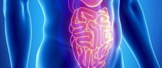
Layout of the gastric sections
Also, different forms of pathology are characterized by different signs. Thus, erosion of the esophagus and stomach is more often manifested by bloody vomiting than all other forms.
In addition, patients suffer from:
- frequent hiccups;
- regurgitation;
- belching;
- pain when swallowing;
- increased salivation;
- bad breath.
Erosion of the prepyloric part of the stomach is quite often diagnosed. In this form of the disease, the pathological process can spread to the antrum and, consequently, to the pylorus and duodenum, which is usually accompanied by severe bleeding, anemia and hungry abdominal pain, causing patients particular discomfort at night. Erosion of the pylorus of the stomach due to hyperemia of the surrounding tissues can in rare cases cause the development of its stenosis with all the ensuing consequences. The narrowing of the pyloric lumen is accompanied by food retention and, consequently, the occurrence of rotting processes in it. These processes manifest themselves in the appearance of bad breath, nausea, vomiting, etc.
Attention! Multiple erosions of the stomach pose a particular danger to the health and even the life of the patient. Since the onset of massive bleeding from them can lead to a threat to the patient’s life.
Chronic gastric erosion generally has no characteristic symptoms. Most often, patients experience symptoms of diseases against which ulcerations have formed. Patients do not suffer much or too often from pain. But since erosive lesions of the mucous membranes often transform over time into hemorrhagic erosions of the stomach, their symptoms become more pronounced.
Article on the topic: Colon cleansing drugs - what they are and when they are used
Endoscopic picture
On gastroduodenoscopy, in some cases, as, for example, in the photo, gastric erosions look like flat, small (no more than 1–2 mm) epithelial defects of various shapes. Sometimes they are covered with hemorrhagic or fibrous plaque, and the surrounding tissues may be hyperemic. In other cases, endoscopic examination reveals complete erosion of the stomach. This defect takes the form of a conical polyp-like formation located on the mucous membrane. There is a depression or ulceration in its central part. Such formations are covered with dark-colored fibrin.
Diagnosis and treatment
Modern diagnostic measures make it possible to most accurately determine the form of pathology and the severity of damage to the mucous membrane. In order to identify the disease at the initial stage of development, it is necessary to carry out an examination consisting of several stages.
First of all, a laboratory analysis of stool and urine is performed. General and biochemical blood tests are mandatory. This allows you to identify the presence of an infectious pathogen in the body and determine acidity.
Gastroscopic examination helps to visually examine the inner lining of the stomach. For this, a special probe is used, which is inserted through the oral cavity using a long hose. In some cases, it may be necessary to collect tissue cells from the inner surface of the stomach. This method of examination is used to identify pathogenic microorganisms.
An ultrasound examination is carried out to determine the condition of other internal organs. This allows you to confirm or exclude the development of concomitant pathologies.
In order to reduce the secretion of gastric juice, proton pump drugs (Omez, Omeprazole, Controloc, Lansoprazole) or drugs whose action is based on blocking histamine receptors (Quamatelom, Famotidine, Ranitidine) are used. In order to reduce the negative effect of hydrochloric acid on the walls of the stomach, drugs such as Maalox, Phosphalugel, Almagel are prescribed. These medications form a protective film on the inflamed area.
If during the examination the presence of Helicobacter pylori microorganisms was revealed, the use of antibiotics, for example Amoxicillin, Metronidazole, Clarithromycin, is necessary. To restore the functioning of the duodenum and stomach, the doctor prescribes Motilium, Cerucal or Metoclopramide. These drugs help eliminate nausea and bloating. With erosive changes in stomach tissue, accompanied by ulcerative lesions, bleeding is often observed, which can be stopped with the help of Vikasol or Dicinon. Most of the drugs listed are dispensed from pharmacies without a doctor's prescription.
In order to eliminate gastritis, the treatment of which should be as effective as possible, it is first necessary to get rid of the causes that caused the pathology. For erosive gastritis caused by the bacteria Helicobacter pylori, antibiotics are mandatory. The course of treatment with such medications is quite long. In no case should you interrupt taking medications until complete recovery, as this will provoke the re-production of bacteria, which will greatly worsen the person’s condition.
The next very important step in the treatment of gastritis accompanied by erosions is normalizing the acidity level. The mucous membrane of gastritis must be strongly protected from the negative effects of gastric juice. The doctor prescribes medications that have an antacid effect, such as Maalox and Rennie. It should be remembered that you can take certain medications only after consulting a doctor.
Medication
Most often, treatment for diseases of the stomach and duodenum is the same as for peptic ulcers; the patient is prescribed proton pump inhibitors and H2 blockers. An exacerbation cannot occur without gastroprotectors, for example, bismuth preparations. Effective gastroprotectors include:
- film-forming agents;
- cell regeneration stimulators;
- enveloping drugs;
- mucus-forming agents
If the stomach is damaged by Helicobacter bacteria, antibiotics are prescribed
If the eroded stomach is the result of exposure to Helicobacter, the patient needs antibacterial agents. If the disease is secondary, the patient needs synthetic prostaglandins and cytoprotectors. These medications accelerate tissue regeneration. Epithelization in severe cases can take up to 2 months. If this does not happen, the possibility of surgery cannot be ruled out.
Physiotherapy
If severe pain is present, the patient may be advised to undergo a course of electrophoresis with novocaine and magnesium. This will help relieve pain.
Surgery
An additional method of treating erosion (especially hemorrhagic) is exposure to a low-intensity laser, which improves blood flow in the affected area. This procedure is performed surgically. Sometimes, if the disease is advanced and the eroded area does not stop bleeding, the patient needs surgical intervention, which involves resection of the diseased part of the stomach. If the disease is chronic and drug treatment is ineffective or if polyps have formed, the patient is sent for cauterization:
- diathermocoagulation (focal cauterization with current);
- laser coagulation (laser cauterization).
Sometimes there is a need for radio wave cauterization.
Treatment with folk remedies
The use of herbs and other non-drug treatments is prohibited without first consulting your doctor. This is not therapy, but only auxiliary treatment methods. Celandine is considered the most effective traditional medicine. A decoction is made from it. To do this, you need to steam 10 g of the plant in a thermos with 200 g of boiled water. Take the medicine 1 tsp. three times a day half an hour before meals for 30 days. After this, take a break for 1.5 weeks. Celandine is a poisonous plant, so you must strictly adhere to the dosage.
In addition to treatment, herbal infusions are prepared
The following infusion will help cope with erosion, ulcers and gastritis. Compound:
- St. John's wort - 1 tbsp. l.;
- celandine - 1 tsp;
- yarrow - 1 tbsp. l.;
- chamomile (flowers) - 1 tbsp. l.
Steam 2 tablespoons of the mixture with 200 g of boiling water and take half a glass three times a day before meals. It is useful to eat a spoonful of propolis or natural honey on an empty stomach.
Diet during therapy
When treating any diseases of the gastrointestinal tract, it is necessary to adhere to dietary nutrition. Diet for erosive and ulcerative disorders is a necessary factor contributing to the effective treatment of the disease. When the inflammatory process develops, doctors prescribe diet food No. 1. After the exacerbation of the pathology has been eliminated, the patient should adhere to diet No. 5.
The diet for erosive-ulcerative disease includes regular meals (every 5-6 hours). It is necessary to ensure that the food is at a normal temperature, not too hot, but not too cold.
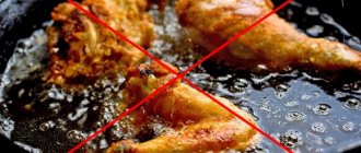
During therapy, it is necessary to completely exclude junk food from the diet. This includes carbonated drinks, canned foods, fried, smoked and spicy foods. It is necessary to reduce the amount of salt consumed as much as possible. It is not recommended to drink coffee and strong black tea with this disease. In addition, you should stop smoking and drinking alcoholic beverages.
It is recommended to eat dishes such as various broths and soups, cereals, vegetable and fruit purees. Raw vegetables and fruits are contraindicated as they can increase acidity in the stomach. As drinks, it is advisable to consume compotes, fruit drinks, clean water without gases, and green tea.
During treatment, you must adhere to the following nutritional rules:
- Consume boiled or steamed foods.
- Eliminate from the diet foods that can increase the amount of gastric juice produced (fried and fatty, smoked foods, canned food and salty foods).
- Eat often, in small portions.
- The food consumed should be at medium temperature. It is better to eat food warm.
Diet during therapy
When treating any diseases of the gastrointestinal tract, it is necessary to adhere to dietary nutrition. Diet for erosive and ulcerative disorders is a necessary factor contributing to the effective treatment of the disease. When the inflammatory process develops, doctors prescribe diet food No. 1. After the exacerbation of the pathology has been eliminated, the patient should adhere to diet No. 5.
The diet for erosive-ulcerative disease includes regular meals (every 5-6 hours). It is necessary to ensure that the food is at a normal temperature, not too hot, but not too cold.
During therapy, it is necessary to completely exclude junk food from the diet. This includes carbonated drinks, canned foods, fried, smoked and spicy foods. It is necessary to reduce the amount of salt consumed as much as possible. It is not recommended to drink coffee and strong black tea with this disease. In addition, you should stop smoking and drinking alcoholic beverages.
It is recommended to eat dishes such as various broths and soups, cereals, vegetable and fruit purees. Raw vegetables and fruits are contraindicated as they can increase acidity in the stomach. As drinks, it is advisable to consume compotes, fruit drinks, clean water without gases, and green tea.
During treatment, you must adhere to the following nutritional rules:
- Consume boiled or steamed foods.
- Eliminate from the diet foods that can increase the amount of gastric juice produced (fried and fatty, smoked foods, canned food and salty foods).
- Eat often, in small portions.
- The food consumed should be at medium temperature. It is better to eat food warm.
Consequences and complications
If erosive gastritis is not treated, it can develop into a chronic form, which is difficult to treat. Erosion of the antrum of the stomach (which is separated from the intestine only by the pyloric ring) is often accompanied by bleeding, so the risk of severe blood loss is high. Such bleeding is difficult to stop, even if you immediately seek help.
The most common complication of the disease is peptic ulcer of the stomach and duodenum. The chronic form of the disease can transform into hyperplasia and cause polyps. Focal hyperplasia is the formation of a neoplasm due to impaired cell reproduction.
Erosion of the pylorus can lead to stenosis. The pylorus is a sphincter that separates the stomach from the lower gastrointestinal tract. The narrowing of the lumen in the pylorus disrupts the transport of food, which leads to it rotting in the stomach. Pyloric stenosis can only be treated with surgery. If the narrowing of the pyloric lumen is not treated, a disruption of homeostasis may occur. Due to erosion in the pylorus, polyposis may develop.
If the disease occurs in an acute form, then if you consult a specialist in a timely manner, the prognosis for a full recovery is good.
Forecast and preventive measures
If, after identifying gastritis, treatment was started in a timely manner, the prognosis is quite favorable. It should be remembered that the lack of therapy can provoke the disease to acquire a chronic form.
If gastric bleeding is primary in nature, and its cause cannot be determined, the patient must be hospitalized in the surgical department. If the patient's condition is stable, in the absence of bleeding and the risk of relapse, the disease can be completely cured even on an outpatient basis.
Prevention of erosive type gastritis consists of implementing anti-epidemic measures, maintaining a healthy lifestyle and proper nutrition. If a person has a high risk of developing a secondary form of erosive gastritis, specific prevention is carried out, which involves the introduction into the stomach of H2-blockers of histamine receptors and medications that have an antacid effect.
It should be remembered that the earlier the diagnosis is made, the faster and more effective the treatment will be. Therefore, at the first signs of stomach disease, you should consult a specialist.
Symptoms
Erosive and ulcerative lesions of the stomach are not always characterized by severe symptoms. Such cases occur infrequently, common signs:
- Painful symptoms in the upper abdomen, and in some patients the intensity of pain is low, in others the pain is severe, intensified by drinking alcohol, spicy food, with long breaks between meals or playing sports.
- Heartburn is characterized by a burning sensation in the area just above the abdomen.
- Nausea, sometimes with vomiting, bringing relief.
- Decreased appetite.
- Belching with a bitter or acidic taste.
- Increased gas formation.
- Feeling of heaviness in the stomach after eating.
- Quick feeling of fullness.
- Various stool disorders.
Forms of the disease
Erosive and ulcerative lesions of the stomach have two forms: acute and chronic; when working with a patient, the doctor must take this into account. The acute form is characterized by localization of pain at the bottom of the gastric wall, epithelium is not layered on the area of erosion, there is no fibrin, pain is more pronounced and appears more often. Painful symptoms of acute erosion correspond to the severe pain described above. The acute form is treated in two weeks.
In the chronic form, pain is localized at the junction of the stomach and the beginning of the duodenum. The pain is milder, less frequent, and treatment continues for up to six months. Depends on the strength and scale of the lesion, the perception of drugs by the patient’s body.
- Hemorrhagic type (from Latin hema - blood). Such erosions reveal plaque in the form of blood and a pale, hyperemic, reddened membrane.
- Superficial flat - distinguished by a white rim around the erosion damage, there is a white coating, the inside is clean. There are exceptions.
- Full - similar in appearance to a polyp, most often formed in a fold of the gastric walls. Other names: hyperplastic, inflammatory.
Provoking factors
Unfortunately, doctors cannot determine exactly what causes erosion, but they know what factors contribute to the development of the disease. Risk factors include poor diet, smoking, inappropriate use of medications, especially anti-inflammatory drugs, alcohol abuse, and depression.
Stomach erosion in a patient
Identifying causes
Provided that gastric erosion is not an independent pathology, you will have to look for problems in other organs. Most often in the liver and blood vessels. The treatment is complex - to achieve the highest effect. During the process, endoscopy is required to monitor the results of therapy and correction.
What happens if erosion is not treated?
Untreated erosion is fraught with the development of the following pathologies:
- Stomach ulcer. This disease affects the deeper tissues of the stomach, which is quite dangerous to health. The main danger of an ulcer is that it can break through the organ, with all the contents entering the abdominal cavity, where over time purulent peritonitis (inflammation of the peritoneum) develops. There is no drug treatment, only surgery.
- Polyp formation. Initially they are benign formations, but can degenerate into malignant ones. The threat is that they practically do not manifest themselves until they reach large sizes. They are usually found during endoscopic examination.
- Anemia. With internal bleeding, hemoglobin decreases, which affects general well-being. Concentration decreases, appetite and quality of sleep are disrupted, and heart failure may even develop. It is often difficult to immediately determine the cause of anemia, because not all people are ready to undergo gastroscopy for this, because do not believe that the stomach and hemoglobin can be connected.
- Stomach cancer. In the early stages there may be no symptoms at all, or the patient mistakes it for other diseases; some take the signs for an exacerbation of peptic ulcer disease.
The first thing the patient needs to know
Before starting treatment, you need to find out the cause of the disease, then treat it. Otherwise, in addition to the existing erosive and ulcerative lesions of the stomach, you can get additional ones. Such a disorder appeared due to incorrect functioning of the liver or kidneys. Therapy to normalize the functioning of organs is mandatory, otherwise the effect of therapy will drop to zero.
Causes of ulcerations
Acute erosion of the gastric mucosa develops as a result of:
- Irrational use of medications, for example, NSAIDs, nitrofurans, digitalis preparations, corticosteroids, Veroshpiron, ethacrynic acid, etc.
- Alcohol intoxication.
- The presence of severe somatic pathology.
- Sepsis.
- Stress exposure.
Attention! Regardless of what causes the erosion of the stomach, after appropriate treatment, the erosion of the stomach walls disappears, and no connective tissue scars remain in their place.
What methods of treating this disease exist today? Most often, doctors use drug therapy in combination with a diet, but in complex cases surgery may be required. We discussed this issue in more detail in the article: Treatment of erosive lesions of the stomach using traditional medicine.
Basic diet rules
Diet in the treatment of erosion is necessary; without the right conditions for the functioning of the stomach, healing of the mucous membrane is impossible. The temperature of the food plays a significant role; only warm, not hot or cold food is suitable.
If there is a diagnosis of “stomach erosion,” too cold or, conversely, hot food injures the deep layers of the stomach lining, leading to aggravation of the diagnosis. In the absence of a painful condition, a healthy person feels only slight discomfort; the mucous membrane prevents injury to the stomach.
Is it possible to do without medications?
It is believed that by following a proper diet, it is possible to cure the disease without medication. According to the recommendations, meals should be taken frequently and regularly, every 3.5 or 4 hours, the portion should be 250 - 300 ml; with an increase in volume, aggravation of the diagnosis is quite expected.
Potato juice
Wash several potato tubers (preferably pink) thoroughly and grate them without peeling. Then squeeze it out and drink it on an empty stomach before breakfast. The course of treatment is 4–6 weeks. Drink only freshly prepared juice.
Cabbage juice
Dilute fresh cabbage juice with water in a 1:1 ratio to obtain 100 ml of liquid. This amount should be drunk every day before meals for a week. If there is no discomfort in the stomach, in the second week the amount of juice can be doubled, in the third week they drink 400 ml of diluted juice per day, and in the fourth - 800 ml. Salt and spices should not be added to juice to improve taste.
Rose hip decoction
If you have high acidity, it is not recommended to take rosehip decoction; it can cause complications. For erosive gastritis with low acidity, this folk remedy will be a source of vitamins and will help heal lesions on the gastric mucosa. The berries should be crushed, boiled for 3 minutes, allowed to cool and instead of tea, drink one glass a day.
Following a diet for erosive gastritis is not an easy task and requires patience and self-discipline. But this is the only way to eliminate factors that irritate the gastric mucosa and prevent erosion from turning into ulcers or cancerous formations.
What foods are forbidden to eat?
You will have to completely abandon:
- Alcoholic drinks.
- Coffee and coffee drinks.
- Cabbage, chocolate and products containing dyes and preservatives.
- Smoked, salted, bee products are not allowed, although you can leave propolis, it has restorative properties. There is no difference in diet in the last point. Suitable for one diagnosis, suitable for another.
Often the disease is caused by taking medications that harm the gastric mucosa. Antibiotics are rarely recommended by doctors. Only if an infection is detected in the patient’s body.
Treatment
Often the disease is caused by taking medications that harm the gastric mucosa. Antibiotics are rarely recommended by doctors. Only if an infection is detected in the patient’s body.
What can you eat if you have stomach erosion and what can’t you eat?
The diet must meet the requirements presented in Table 3.
Table 3. Nutrition for gastric erosion
| You can eat | Can't eat |
| I eat it boiled, steamed, stewed | Food with a rough texture |
| Lean meat and fish - chicken, beef, rabbit, pike perch | Fatty meat with elements of connective tissue and skin; offal |
| Stewed vegetables (pumpkin, beets), neutral-tasting fruits - apples, pears, peaches | Raw plant foods, sour vegetables and fruits, citrus fruits, walnuts, seeds |
| You can drink jelly, sweet fruit infusions, weak tea | Juices, fresh drinks, sparkling drinks, coffee, cocoa |
| Porridge - buckwheat, rice, oatmeal | Yeast baked goods, products made from low-grade flour |
| Baked goods made from premium flour, vegetable oil | Butter, margarine |
| Honey, marshmallow | Chocolate |
| Milk porridges, puddings, low-sour cottage cheese | Fermented milk products, kefir |
| Boiled eggs, omelet | Legumes, celery, turnips, radishes |
| Alcoholic drinks, including beer |
How to eat during remission
After overcoming the acute form of the disease, you can eat a little more varied. The weekly menu for erosive gastritis of the stomach includes dishes from meat and fish, vegetables and fruits, dairy products and eggs. An example of a weekly menu with recipes is given below: each meal has options for 7 days of the week.
First breakfast
- Soft-boiled egg, rice porridge, tea with biscuits.
- Oatmeal with honey, tea with crackers.
- Omelet, jelly.
- Buckwheat porridge, tea with jam.
- Steamed cheesecakes, compote.
- Cottage cheese casserole, jelly.
- Lazy dumplings, apple pudding, tea.
Is it possible to do without medications?
It is believed that by following a proper diet, it is possible to cure the disease without medication. According to the recommendations, meals should be taken frequently and regularly, every 3.5 or 4 hours, the portion should be 250 - 300 ml; with an increase in volume, aggravation of the diagnosis is quite expected.
Diagnostics
Making a correct diagnosis depends on a thorough history taking. A significant loss of body weight in a short period of time, uncontrolled medication use and alcohol consumption speaks volumes.
Where does it hurt
The chronic form of erosive gastritis can be painless. Acute is characterized by cutting pain in the stomach and sternum.
What hurts
Painful spasmodic attacks constrain the abdominal cavity. In some cases it can radiate to the back and under the left shoulder blade.
What needs to be examined
To diagnose erosive gastritis of the stomach, laboratory and instrumental studies are first performed. A blood sample is taken for a general analysis to confirm (refute) anemia. Feces for blood content.
Blood biochemistry reveals complications and concomitant diseases.
To identify the pathogen that provoked erosive gastritis of the stomach, a bacteriological analysis of the contents of the stomach, vomited and feces, is carried out.
- Of the instrumental methods, esophagogastrodudenoscopy with biopsy sampling is most often used. During the procedure, the degree of destruction of the mucosa, the depth and extent of erosions and the location of bleeding are assessed. In case of heavy blood loss, the manipulation should be carried out immediately, if the patient’s condition is stable, within 12-20 hours.
- If EGDS is not possible, the fluoroscopy method is used. The procedure can be carried out either through conventional gastrography or with the introduction of a dye into the organ. An x-ray can show swelling and an increase in the thickness of the folds of the gastric mucosa. Double contrasting of the gastric walls is more informative. With such a study, linear or extended defects of the shell can be seen and torn edges can be visualized.
- pH metering. Allows you to determine the content of hydrochloric acid in gastric juice.
Important! Examination of gastric juice is impossible after surgery and with stenosis.
Who to contact
When the first symptoms of the disease appear, you must contact a gastroenterologist at your local clinic to determine a diagnosis and prescribe adequate treatment.
Stomach erosion: treatment with folk remedies
Along with drug therapy, alternative methods are used. Many patients are interested in how to treat gastric erosion with folk remedies. The following are the most effective natural products that can be used at home. Recipes from them are presented in the table.
Table 4. The most effective folk remedies for treating gastric erosion
| Potato juice | The juice obtained by rubbing 2-3 potato tubers is diluted with a small amount of water. Drink up to 3 times a day half an hour before meals for 14 days |
| Sea buckthorn oil | Consume warm, 5 ml 2-3 times a day before meals. |
| Flax seed | Boil one tablespoon of seeds for 5 minutes. They insist for two hours. Take the mucous mass 3-4 times a day before meals |
| Aloe honey | Aloe juice is mixed with honey. Take daily in the morning on an empty stomach for a month |
| Calamus root | Pour one teaspoon of the mixture into a glass of boiling water and cook for another 15 minutes over low heat. Drink 50 ml of the decoction before meals for 14 days. |