In the article we will look at where the cardiac part of the stomach is located. We will also find out what diseases can develop in this department.
The coordinated work of all organs and systems ensures the normal functioning of the human body. The proper functioning of the digestive system is of considerable importance in this process. The stomach is the main organ of the gastrointestinal tract. It is composed of muscle fibers and has a high degree of elasticity, which allows it to stretch, increasing in size up to seven times. Each section of the stomach is responsible for performing a specific function. Their correct performance of their duties determines the correct digestive process.
Description
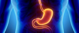
The stomach is a hollow space in the digestive system that resembles a bag in appearance. The organ serves to connect the upper part of the esophagus and the lower part of the duodenum. The stomach includes several sections, each of which performs certain functions, and in general the organ contributes to the normal functioning of the human body.
After entering the mouth, the food is thoroughly chewed by the person and then swallowed. Next, the food, roughly processed by the teeth and saliva, falls into the stomach. It accumulates food eaten, some of which is digested with the help of hydrochloric acid and special gastric enzymes. The latter allow you to break down fats and proteins. After the stomach, food enters the distant parts of the digestive system, namely the gastrointestinal tract.
Departments
The gastrointestinal tract consists of several parts. Each department performs a specific function and takes part in the process of digesting food.
Cardiac. The department is located next to the heart, which is why it got its name. This is the border between the esophagus and the stomach, where the cardiac sphincter is located. It consists of muscle fibers. The pulp prevents food from entering the esophagus area.
Fundus of the stomach. A section that is located at the level of the esophagus. Externally it looks like a dome. It is called the bottom (vault). This part collects air, which enters the digestive organ along with food. The mucous membrane of the fundus contains a large number of glands that secrete hydrochloric acid. It is necessary for digesting food.
Body. The main and largest part of the digestive organ. Its beginning lies in the cardiac region and ends in the pyloric part. The absorbed food collects in the body.
Pyloric department. The area, which is also called the gatekeeper. It is located below all departments. After the pyloric region, the small intestine begins. It includes a canal and a cave. These two areas also perform certain functions. The canal facilitates the movement of food from the stomach to the duodenum. The cave preserves partially digested food.
All parts of the stomach support the proper functioning of the digestive system. Each area requires a certain amount of time to function. Fruit juices or broths are digested for 20 minutes. Meat dishes require 6 hours.
Functions
The functional responsibilities of the stomach are quite numerous. The main ones among them are the following:
- Preservation of eaten food.
- Control of gastric juice production.
- Carrying out chemical processing of food.
- Promotion of food and timely cleansing of organ contents.
- Most of the absorption of various nutrients occurs in the stomach.
- Bactericidal effect.
- Protection from harmful effects.
During the digestive process, all residual metabolic products are eliminated. This also applies to substances that negatively affect the functioning of the endocrine glands.
Few people know where the cardiac part of the stomach is located.
Physiology[edit | edit code]
The glands of the gastric mucosa secrete gastric juice containing the digestive enzymes pepsin, chymosin and lipase, as well as hydrochloric acid and other substances. Gastric juice breaks down proteins and partially fats, and has a bactericidal effect.
Due to the muscular layer, the stomach mixes food and gastric juice, forming chyme - a liquid pulp, which is removed in separate portions from the stomach through the pyloric canal. Depending on the consistency of the incoming food, it lingers in the stomach from 20 minutes (fruit juices, as well as vegetable juices and broths) to 6 hours (pork).
In addition, the stomach wall absorbs carbohydrates, ethanol, water and some salts.
Sections of the stomach
The gastrointestinal tract includes several main sections. Each of them has a number of functions and, to one degree or another, is involved in the process of food processing. The following main sections of the stomach are distinguished:
- Cardiac section of the stomach. Located near the heart, which explains its name. This section is located between the esophagus and the stomach, in the area of the cardiac sphincter. The cardiac section consists of muscle fibers. The pulp prevents food from entering the esophagus area.
- Gastric fundus. This section is located directly at the level of the esophagus. In terms of external characteristics, it resembles a dome or vault. Air that is swallowed along with food accumulates here. The mucous membrane of the gastric fundus contains many glands that are responsible for the production of hydrochloric acid necessary for the digestion process.
- Body of the stomach. The largest part of the digestive organ. The body begins in the cardiac region and ends in the pyloric region. The food eaten accumulates in the gastric body.
- Pyloric. Another name for this department is the gatekeeper. This section is located lower than the others, and then the small intestine originates. The pyloric section includes a cave and a canal, which also perform certain functions. The canal carries food into the duodenum, and the cave stores the digested part of the food for further processing.
Together, all sections, including the cardiac and pyloric sections of the stomach, ensure the normal functioning of the digestive system. Each department processes food for a certain time, which also depends on the nature of the products consumed. Fruit juice is digested in a third of an hour, and a meat dish will remain in the stomach for at least 6 hours.
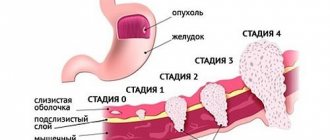
Causes of stomach cancer
The cause of stomach cancer in most cases is inflammatory diseases of the digestive system.
Let's look at some of them:
- Atrophic gastritis. It precedes gastric cancer in 70-80% of cases. Due to inflammatory processes, the gastric glands are destroyed, the ability to secrete acid decreases, and achlorhydria develops (sometimes pernicious anemia). This is followed by the colonization of the organ cavity by pathogenic microorganisms that are capable of synthesizing carcinogenic substances.
- Intestinal metaplasia. In this condition, the normal lining of the stomach is replaced by cells similar to the lining of the intestine. Often intestinal metaplasia develops against the background of atrophic gastritis, but how and why it progresses to cancer is not yet known.
- Adenomatous polyps (adenoma). With this disease, various dysplastic changes are observed in the mucous membrane around the polyps, and therefore the incidence of malignancy is 30-40%.
- Cardioesophageal cancer is possibly associated with a disease called Barrett's esophagus, where the cells in the lining of the esophagus undergo pathological changes and turn into columnar epithelial cells. Barrett's esophagus is closely associated with gastroesophageal reflux.
It is worth noting ,! The development of these diseases is closely related to the bacterium Helicobacter pylori. Some of its subtypes are capable of converting substances from food into chemical carcinogens that cause mutations in the DNA of cells in the gastric mucosa.
Scientists have identified several risk factors for gastric cancer with varying degrees of evidence:
- The first, most proven factor is nutrition. The consumption of semi-finished products, fats, smoked meats, and pickles has a negative effect. The disease is also associated with non-compliance with sanitary standards (for example, washing hands before eating, thoroughly washing vegetables and fruits) and improper storage of food.
- Smoking and alcohol. These factors most likely have a negative impact on the state of the digestive system and the entire body, although this has not been fully studied.
- In rare cases, hereditary stomach cancer has been observed.
- Operations. People with a resected stomach are several times more likely to develop cancer. This may be due to decreased stomach acid production and reflux of bile from the small intestine.
- Hereditary syndromes (Lynch syndrome, familial adenomatous polyposis, Li-Fraumeni syndrome, etc.). It has been noted that people with such abnormalities are more susceptible to DNA mutations.
- Overweight (not proven).
Read here: Luminal breast cancer
Risk factors sometimes contribute to the development of cancer, but many people who get it have no risk factors at all. However, some of them can be eliminated on your own and this will be the right choice.
Stomach diseases: ulcers
There are a number of diseases specific to the cardiac region of the stomach. Ulcer is one of the most common pathologies. This disease is seasonal and is accompanied by pain and other unpleasant symptoms. Ulcers in this section are slightly less common than in the body or bottom of the stomach, as well as in the intestines. However, due to the accelerated pace of life, constant stress and poor environmental conditions, the number of cases of ulcers in this particular department has been steadily increasing recently.
Hereditary character
The tendency to peptic ulcer of the gastric cardia can be hereditary. The factors listed above trigger a genetic program that leads to the occurrence of ulcers. Another genetically determined cause of ulcers is increased activity in the production of gastric secretions. As a result, the balance between negative effects and immune defense occurs.
An ulcer of the cardial part of the stomach is characterized by the appearance of pain after eating, when irritation of the mucous membrane occurs. The main tactics for treating peptic ulcer disease is following a special diet. The first thing to do after a diagnosis is made is to remove any foods that irritate the stomach from your diet. The diet involves the absence of rich broths, fried foods and stewed meat or fish.
In addition, you will need to exclude from your diet any sauces, smoked foods, pickled and salted vegetables, and fruits that contain too much acid. Fruits are best consumed in the form of jelly, since it is this substance that can coat the stomach walls and accelerate the healing of ulcers. The use of pepper and various seasonings is prohibited; salt should be in strictly limited quantities. Green tea is allowed as a drink.
Sometimes, in particularly advanced cases, surgery may be required in addition to diet. This may mean that conservative therapy is not effective, and relapses of exacerbations of peptic ulcer disease are becoming more frequent.
Polyp of the cardia of the stomach
Polyps are also very common in the gastrointestinal tract. They are neoplasms on the mucous membrane. Polyposis occurs in all parts of the stomach, but the most common variant is antral-pyloric. Less common is polyposis in the cardiac region of the stomach.
Polyps in this section are also called cardia, since they are located very close to the heart. Between the esophagus and the stomach, as mentioned above, there is a cardiac sphincter, which prevents food from passing back into the esophagus from the gastric cavity. When a disturbance in the functioning of the stomach occurs, acid enters the esophagus, which leads to inflammation and its further transformation into a malignant formation of the cardiac part of the stomach.
Symptoms of pathology
Gastroesophageal reflux develops at the third stage of the pathology.
Gastric cardia insufficiency is 95% an acquired disease and only 5% is a congenital defect. Valve non-closing has 3 degrees of violation:
- The sphincter of the stomach does not close completely. This occurs when the stomach is overloaded or when breathing deeply. A burp of air appears.
- The hole increases to half and means a displacement of the upper wall of the stomach towards the cardia.
- The rosette of the cardia does not close, and pronounced gastroesophageal reflux is observed.
Cardiac insufficiency of the 2nd degree can be cured comprehensively, but non-closure of the valve of the 3rd degree must be treated with surgery. Echosigns are poorly defined and resemble reflux esophagitis in nature. Valve deficiency is determined by the main symptoms:
- belching of air or acid;
- heartburn in the esophagus, throat and stomach;
- bitter taste in the mouth;
- multiple stomatitis;
- vomiting bile;
- pain, bloating, distension behind the sternum;
- lump in throat;
- heavy salivation;
- weakness.
Operation methods
Treatment of polyposis involves surgical removal. There are several methods of performing the operation:
- Laser or radio wave surgery. There are no contraindications for these methods. They are minimally invasive and do not require a long recovery period.
- Endoscopic surgery. It is done through a small incision using a flexible manipulator. In this way, it is possible to remove polyps from the cardiac region.
- Resection. It is a last resort and is used in advanced cases when minimally invasive treatment methods do not produce positive dynamics. After resection, a person has to go through a long rehabilitation period and completely reconsider their lifestyle.
Submucosal formations
These are pathological formations growing inside the stomach walls. Submucosal formations of the cardiac part of the stomach of a benign nature are distinguished, such as lioma, hemangioma, leimioma, fibroma, etc., as well as malignant tumors, such as fibrosarcoma or leiomyosarcoma. In the case of a benign course of the pathological process, there is no danger to human life.
The development of pathological formations occurs in a latent form. The sizes of tumors can vary. With a benign course, their size reaches an average of 3-4 centimeters. Their contours and location may also vary. Submucosal tumors are characterized by clear contours and a homogeneous nature. Uneven edges indicate malignancy of the neoplasm.
Indications for use
Because bariatric surgery is primarily a medical and not an aesthetic specialty, weight loss surgery is not performed based solely on the patient's wishes. They require significant indications, which are determined by the bariatric surgeon. Discussion of surgical treatment options can begin if conservative methods of treating obesity (low-calorie diet, exercise, drug therapy, psychocorrection, massage, IRT, etc.) are ineffective.
Bariatric surgery should not and cannot be the first option in a series of attempts to treat obesity. Direct indications are a body mass index of more than 40 or a BMI of more than 35, associated with common diseases caused by excess weight. These comorbidities may be hyperlipidemia, hypertension, obstructive sleep apnea, osteoarthritis, type 2 diabetes, varicose veins, etc.
Some bariatric surgery techniques: For example, intragastric balloon placement can be used in pre-obese patients, and gastric banding is associated with stage 1 obesity. In addition, each bariatric surgery method has its own conditions for maximum effectiveness.
Thus, with a body mass index above 50 kg/m2, the best weight loss result is achieved during the bile-pancreatic maneuver. Delayed gastric bypass and biliary-pancreatic bypass in more than 90% of cases can cope with type 2 diabetes and hypercholesterolemia, therefore, if the patient has data from the “companions” of obesity, these operations have an advantage.
Causes
The exact reasons for the development of submucosal tumors both in the cardiac and in any other part of the stomach are unknown. However, there are several factors that, according to doctors, can provoke this pathological process:
- Peptic ulcer and gastritis.
- Helicobacter pylori infection.
- Hereditary predisposition.
- Unbalanced diet.
- Exposure to chemicals on the body.
- Smoking and alcohol abuse.
The appearance of neoplasms is typical for patients over 40 years of age. Oncopathology is usually discovered by chance during endoscopy. If the carcinoma has grown greatly, the patient may complain of nausea and vomiting, constipation, and aching pain. Leiomyomas in the cardiac part of the stomach can provoke bleeding, which leads to anemia.
Therapy
Treatment of the mucous membrane of the cardial part of the stomach, in which formations were found, involves taking special medications. If the nature of the formations is malignant, emergency surgery is performed. In the case of a benign tumor, surgery is performed as planned. Before this, material is taken for histological examination. After surgery, drug therapy is prescribed. As a rule, these are drugs that are active against Helicobacter pylori (“De-Nol”), as well as proton pump inhibitors (“Omeprazole”).
Treatment methods
Radiation therapy for stomach cancer.
The combined form of cancer is more difficult to treat. The lower the stage of the disease and the fewer tissues affected, the greater the chances of getting a good result and a favorable outcome of treatment. Methods:
- The basis of the fight against carcinoma of the cardiac part of the stomach is the surgical method. It causes partial or complete removal of the stomach along with the affected part of the digestive canal. If the stomach needs to be removed completely, which happens at stages 2 or 3 of cancer, then in this case the remainder of the esophagus is connected to the small or large intestine. If resection of a large part of the digestive canal is required, then in this situation plastic surgery is performed using part of the intestine. When diagnosing stage 4 combined cardiac carcinoma, surgical intervention makes little sense. In this case, patients are prescribed antispasmodic and supportive drugs.
- Radiation therapy is used both before surgery and in the postoperative period. Preoperative radiation is aimed at slowing tumor growth and the spread of metastases. Radiation therapy, used after surgery and removal of the affected tissue, kills the remaining malignant cells, thereby reducing the likelihood of recurrence of the disease.
- Chemotherapy involves the use of cytostatic drugs that suppress the growth of both the tumor itself and metastatic processes. This method is used in combination with others.