general description
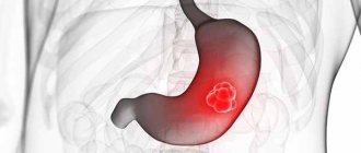
As we have already indicated, stomach cancer is a fairly common disease, and this pathology can develop in any part of the affected organ with subsequent spread to other organs (this clarification especially applies to the liver, lungs and esophagus). It should also be noted that there is a specific figure corresponding to the mortality rate of stomach cancer, which is about 800,000 people annually, which was determined on the basis of the studied indicators for 2008. In addition, it was found that this disease is more often diagnosed in men.
In approximately 80% of cases of gastric cancer, metastases develop. Let us recall that metastasis (metastasis) is understood as a process in which tumor cells begin to spread to other organs, thereby forming a pathological process in them in the form of a secondary focus. Metastasis in general occurs in tumor diseases through blood vessels (which defines hematogenous metastases), through lymphatic vessels (respectively, these are lymphogenous metastases) or through the space that the body cavities are located (which defines implantation metastases).
Returning to the characteristics of stomach cancer, we can also determine survival, which can be considered as a prognosis for stomach cancer. Thus, if the disease is detected early, 6-month survival is allowed in approximately 65% of cases, while its detection within the framework of progression to more serious ones determines it only in 5-15% of cases. It should also be noted that in approximately half of the cases of patients complaining of dyspepsia (a condition in which the normal functioning of the stomach is disrupted, digestion becomes painful and generally difficult), the subsequent discovery of much more serious pathologies occurs, including, in fact, stomach cancer.
As already noted, the prevailing age group is patients aged 40-45 years, however, the possibility of developing the disease in people from 30 to 35 years old and in younger people is also possible. In about 90% of cases, the tumor is malignant, and approximately 95% of these malignant tumors are carcinomas. Gastric carcinoma in men is diagnosed mainly between the ages of 50 and 75 years.
Stomach cancer is also characterized by the fact that its course can vary significantly depending on the specific country or region where it is diagnosed in a particular patient; accordingly, on the basis of this we can assume how significant a role climatic-geographical, nutritional, household and other types of factors play in development of the pathological process.
General structure of the stomach
Diagnosis and treatment
Tumors are detected by X-ray of the stomach. On X-ray, neoplasms look like filling defects with dark contours of regular or irregular shape. If the pylorus is not closed, the organ functions normally. The gastric mucosa is stretched, so its folds are flattened.
The ulceration appears on x-ray as an irregularly shaped lesion. Sometimes there is swelling of the mucous membrane, visible in the picture.
Tumors of soft consistency, such as lipomas, are not visible on x-rays. MRI, CT and ultrasound diagnostics are used to detect them.
The diagnosis is confirmed by fibrogastroscopy with the removal of a fragment of the formation for analysis. However, the location of the tumor does not always allow this to be done. In this case, the diagnosis is made only after surgery.
Fibrogastroscopy
There are no non-surgical treatments for tumors, so the tumor must be removed. Depending on the size and location, the tumor is excised endoscopically or using partial (wedge) resection.
After the operation, the patient is monitored so as not to miss the formation of cancer.
ONLINE REGISTRATION at the DIANA clinic
You can sign up by calling the toll-free phone number 8-800-707-15-60 or filling out the contact form. In this case, we will contact you ourselves.
If you find an error, please select a piece of text and press Ctrl+Enter
Stomach cancer: causes
It is currently not possible to identify a specific cause that provokes the development of stomach cancer. Meanwhile, certain predisposing factors for the development of the pathological process and, in fact, the disease directly related to it, can still be called relevant, among them the following:
- Helicobacter pylori bacterium . It is quite possible that the reader has an idea of what kind of bacterium this is, but we will briefly highlight the features that are inherent in it, defining what it is and what it provokes. And it provokes nothing more than gastritis and an equally common pathology - gastric ulcer. This bacterium also acts as a predisposing factor to the development of stomach cancer, and this bacterium, if present, increases this risk several times.
- Stomach polyps. They represent a growth of the mucous membrane of an organ of a benign nature; generally, gastric polyps develop against the background of existing chronic diseases that are relevant to the stomach. An example of a predisposing factor in this case is the chronic form of atrophic gastritis, in which the gastric mucosa is subject to depletion, and the production of gastric juice is significantly reduced. As in the previous case of considering a predisposing factor, a gastric polyp increases the risk of developing stomach cancer several times, and precisely in the area where it is located. This risk is supplemented by such conditions for the existing polyp as certain height indicators (if it is 2 or more centimeters), if it contains cells that cause the production of mucus, and if more than one polyp is concentrated on the gastric mucosa.
- The presence of chronic diseases of the gastric mucosa. Here, again, we can highlight the already noted atrophic gastritis, which also acts as a serious predisposing factor to the development of stomach cancer in a patient against this background.
- Heredity. Here, one can traditionally highlight the relevance of stomach diseases, and in particular stomach cancer, among the closest relatives of a particular patient - the risk of developing pathological diseases of this organ and the disease, which is the main one in our consideration, increases.
- Impact of nutritional factors. In particular, in this case, the dietary habits of patients are considered, namely, addiction to fried, spicy, canned and fatty foods. Chemically active substances damage the gastric mucosa by getting on it, which destroys its protective layer of mucus lining the surface of the epithelium, after which carcinogenic substances freely penetrate into the cells (namely, due to them, the development of cancer becomes possible), after which these cells are subject to destruction, or are reborn. In contrast, it can be noted that if the patient’s diet contains a significant amount of fruits and vegetables, vitamins and microelements, the risk of stomach cancer is significantly reduced.
- Smoking, alcohol. These impact factors are almost traditional in identifying factors that contribute to the development of a particular disease or pathological process; stomach cancer is no exception to this.
- Features of the body's hormonal activity, constitutional features. In considering this point, in particular, overweight and obesity in general are considered as background predisposing diseases, not only traditionally for the organs of the reproductive system, but also for the gastrointestinal tract, which, accordingly, determines a significant risk for the development of stomach cancer.
Peptic ulcer and stomach cancer
Peptic ulcer, indicated above as consideration in the context of the impact of Helicobacter on the development of stomach cancer, in its recurrent form, as well as duodenal ulcer - all this is considered as one of the main factors causing the development of stomach cancer. The process of degeneration of an ulcer into stomach cancer sometimes takes many years, but it is the very possibility of such degeneration that is a serious risk.
We will look at the symptoms of stomach cancer below; among them, you can also identify signs indicating that an old ulcer has degenerated into cancer. Stomach cancer develops somewhat less frequently on its own, that is, without a predisposing factor in the form of a previous ulcer. Here, cancer may resemble an ulcer in its manifestations, but the exact difference can only be made by a doctor and only as part of a diagnostic test, which is a biopsy.
Stomach cancer: classification
Based on what cells make up the tumor, the following types of forms of stomach cancer are distinguished:
- Adenocarcinoma (carcinoma). This form, as we initially noted in the general review, acts as the most common form among the types of stomach cancer. This type of stomach cancer involves the formation of a tumor based on cells that produce mucus.
- Solid gastric cancer. This form of pathology is extremely rare; dense tissue acts in this case as the basis of the tumor.
- Signet ring cell cancer of the stomach. This form of cancer is based on cells, examination of which under a microscope reveals their similarity to a ring, which, in turn, was the reason for this definition of cancer. For this gastric cancer, the characteristic features include the rapid growth of tumor formation, as well as early metastasis.
- Leiomyosarcoma. Based on this form of cancer, one can determine its peculiarity, which is the formation of a tumor formation based on the muscle cells of the organ (i.e., the stomach).
- Lymphoma. In this case, the tumor formation in stomach cancer is based on lymphatic cells concentrated within the region of this organ.
The classification of stomach cancer according to its forms does not end there; a separate part of it is based on the specific part in which the tumor developed; the following options are distinguished:
- Cardiac gastric cancer. This form of cancer develops in the upper part of the stomach organ, specifically in the place where it “joins” the esophagus.
- Body cancer of the stomach. In this form, the cancer affects the middle part of the organ.
- Cancer of the lesser curvature of the stomach. Here the cancer covers the area of the right gastric wall.
- Cancer of the pylorus (pylorus). In this variant, cancer develops from the side from which the anatomical transition of the organ to the duodenum occurs.
And finally, depending on the characteristics of the appearance of the tumor formation, a specific number of forms of stomach cancer are also distinguished, in particular, the following options include:
- Endophytic gastric cancer. This type of cancer is characterized by the fact that the tumor grows deep into the gastric wall, which, due to such features, determines its similarity to a stomach ulcer (saucer-shaped stomach cancer, ulcer-cancer).
- Exophytic gastric cancer. This type of cancer is characterized by the fact that the tumor grows into the lumen of the stomach. This type of cancer can manifest itself in a polypoid form (that is, the characteristics of its growth determine its resemblance to a stomach polyp), in a mushroom form (the growth characteristics equate it to a mushroom), and also in a nodular form (the tumor manifests itself in the form of a dense type of node protruding from the side organ walls).
Main parts of the stomach
Transabdominal ultrasound in the diagnosis of endophytic gastric cancer
Ultrasound scanner HS60
Professional diagnostic tools.
Assessment of tissue elasticity, advanced 3D/4D/5D scanning capabilities, BI-RADS classifier, options for expert cardiological studies.
Introduction
Gastric cancer still remains one of the most pressing medical problems [21-23, 35]. Unfortunately, the five-year survival rate of patients in most countries of the world ranges from 5.5-25%, despite the increasingly widespread use of the latest medical advances [3, 33, 34, 37]. Currently, the vast majority of authors believe that only timely and early diagnosis of gastric cancer can improve the results of its treatment and the subsequent prognosis for the patient’s life.
Numerous studies conducted in different countries of the world have shown that the leading role among all anatomical forms of gastric cancer is played by endophytically growing tumors (more than 65% of all detected cases), which pose the greatest difficulties for diagnosis [1, 4, 5, 13, 16, 20 ]. Even with the help of the latest diagnostic tools, it is often not possible to obtain a sufficient amount of information to clearly support the existence of an intramural neoplastic process [12,27,38].
For many years, the main methods for diagnosing gastric cancer remained X-ray and endoscopic. Only in the last few years have other radiation methods, such as X-ray computed tomography (X-ray computed tomography), ultrasound, and endosonography, begun to be used in clinical practice.
Currently, transabdominal ultrasound has firmly taken its place as the main method in the diagnosis of diseases of the abdominal organs. The capabilities of the method in assessing the condition of the stomach and duodenum have been studied to a much lesser extent [15]. Cost-effectiveness, harmlessness, and the possibility of simultaneous examination of many organs and systems of the abdominal cavity and retroperitoneal space make echography the most preferred method of primary examination of patients not only with diseases of parenchymal organs, but also of the gastrointestinal tract [8, 10, 19, 25].
Materials and methods
The purpose of this work is to study the potential of transabdominal ultrasound in the diagnosis of endophytic gastric tumors. The analyzed material includes 40 cases of endophytic gastric cancer, of which infiltrative-ulcerative forms accounted for 28 (70%) cases, diffuse forms - 12 (30%), "small" and early forms of gastric cancer - 11 (27.5%) cases. Most patients were subjected to surgical treatment; in three (in the group of patients with early gastric cancer), endoscopic treatment methods and photodynamic therapy were used. All cases were morphologically verified.
Transabdominal ultrasound of the stomach was performed as an additional research method after a preliminary comprehensive X-ray endoscopic examination. Computed tomography and endosonography were performed according to indications. The obtained ultrasound data were compared with data from other radiation and endoscopic research methods.
All ultrasounds were performed on a MEDISON SA-8800 scanner. At the first stage, the abdominal cavity was examined on an empty stomach. The purpose of the second stage was a detailed study of the walls of the stomach and its cavity, and therefore the examination was carried out with the stomach filled with liquid in standard projections (lying on the back, on the right side, on the left side, standing) with subsequent recording of the obtained data into the computer memory for subsequent repeated detailed study of them. The developed technique made it possible to visualize all parts of the stomach quite well, including the upper third of the body and the fundus of the stomach. Transabdominal ultrasound, in contrast to traditional X-ray and endoscopic research methods, made it possible to visualize not only the entire thickness of the gastric wall, but also to differentiate its layered structure, as well as anatomical structures located outside the gastric wall.
results
The main features that were analyzed during ultrasound of the stomach were elasticity, straightening of the stomach wall in all sections, thickness, its layered structure, and the presence of additional formations. For an objective and detailed study of the stomach and adequate straightening of its walls, the optimal volume was 1 liter of liquid. The criterion for satisfactory visualization of the stomach walls was considered to be clear differentiation of three layers (in some cases five), which corresponded to the mucous (hyperechoic linear structure), muscular (hypoechoic linear structure) and serous (hyperechoic linear structure) layers (Fig. 1a, b). In order to more objectively assess the thickness of the stomach wall and its layered structure, a control group of 30 apparently healthy patients was formed, in whom, along with examination of the abdominal organs, the stomach was examined using the method described above. After analyzing the results of the study in the main (with stomach tumors) and control groups, it was found that the gastric wall under normal conditions, with adequate expansion, has a thickness not exceeding 0.4 cm, with clearly differentiated layers (in most cases, three), whereas in the zone of tumor damage, the wall thickness in almost all cases exceeded 0.5 cm and was accompanied by a violation of its layer-by-layer differentiation (p
Rice. 1.
Variants of normal echograms of the stomach.
The walls are elastic, the three-layer structure of the wall is clearly visualized.
In the process of analyzing the results obtained, characteristic signs of endophytic tumor lesions of the stomach were identified, characterizing both the tumor process itself and the degree of its spread deep into and beyond the organ. This:
- wall thickening of more than 0.5 cm at the site of the lesion, accompanied by the absence of clear layer-by-layer differentiation;
- violation of the elasticity of the wall at the site of the lesion;
- unevenness, bumpiness of the contours of the affected area;
- lack of visualization of the outer layer of the wall, corresponding to the serous layer, its unevenness, vagueness (signs of tumor growth beyond the organ with the involvement of neighboring anatomical structures in the process) (Fig. 2-5).
Rice. 2.
Endophytic cancer of the posterior wall of the stomach.
Thickening of the wall up to 1 cm (arrows) over a considerable extent. The layers of the wall at the site of the lesion are not clearly differentiated.
Rice. 3.
Limited intramural tumor infiltrate (arrow) of the lower third of the body of the stomach.
Thickening of the wall up to 1.2 cm with loss of layer-by-layer structure at the site of the lesion.
Rice. 4.
Intrawall tumor infiltrate of the fundus of the stomach.
There is a thickening of the wall at the site of the lesion up to 3 cm with bumpy contours. The outer (serous) layer at the site of infiltration is not clearly visible and is uneven (signs of tumor growth beyond the stomach wall).
Rice. 5.
Circular endophytic cancer.
A)
Body of the stomach - there is a circular thickening of the wall of the body of the stomach up to 1.5 cm with loss of elasticity and layered structure at the site of the lesion.
b)
Antrum of the stomach - there is a circular thickening of the walls of the antrum of the stomach up to 2.5 cm with loss of elasticity and layered structure.
Discussion
In recent years, a fairly detailed examination technique and echographic signs of gastric cancer, signs of benign and malignant gastric ulcers have been developed [9, 11, 15]. And yet, ultrasound has received the greatest use as a method for clarifying the extent of the process, searching for metastases, and tumor growth beyond the stomach [2, 6, 17, 28, 31]. The data we obtained indicate that modern high-resolution ultrasound scanners make it possible to confidently diagnose endophytic tumors of the stomach, differentiate the layer-by-layer structure of the wall and use echography as an independent method for diagnosing tumor pathology of the stomach.
The literature also discusses the issue of wall thickness during ultrasound, both on an empty stomach and with a full stomach. Some authors believe that with ultrasound it should not exceed 1 cm [32], others consider a normal thickness of 0.4-0.6 cm [7, 24, 30]. According to L.M. Portnoy et al. (1993) it makes no sense at all to judge the thickness of the organ wall on an empty stomach. According to our data, when the walls of the stomach are adequately straightened (which is achieved by taking at least 1 liter of liquid), their thickness does not exceed 0.4 cm in all sections, and the layers are quite clearly visualized, which is one of the main criteria in distinguishing an unchanged wall from its tumor lesion .
Analysis of the results showed that in the formation of the echographic picture of a gastric neoplasm, factors such as the localization of the tumor, the form of its growth, size, and extent are of leading importance. In the endophytic form, the picture depends on the degree of wall invasion. According to some authors, the greatest difficulties arise when visualizing limited infiltration of the stomach wall, when a small heterogeneous area of invasion with uneven contours is revealed. Thus, ultrasound can be used to diagnose gastric tumors, but mainly in the late stages of the disease (T3, T4) [14, 17]. According to our data, transabdominal ultrasound can successfully detect not only advanced forms of stomach cancer, but also “small”, early forms (T1, T2), including quite accurately determining the degree of tumor invasion into the stomach wall, its prevalence, which allows Ultimately, it is enough to accurately formulate a diagnosis according to the international TNM system.
conclusions
According to our data, the effectiveness of transabdominal ultrasound of the stomach in detecting endophytic tumors of the stomach is: sensitivity - 92.5%, specificity - 89.8%, accuracy - 91.6%. However, this method serves as an additional method, and indications for it should be based on the results of previous studies (x-ray and endoscopic) [6]. For accurate diagnosis of endophytic gastric cancer, rational and comprehensive use of the entire arsenal of modern diagnostic techniques is necessary.
Literature
- Antonovich V.B. X-ray diagnosis of diseases of the esophagus, stomach, intestines.-M.: Medicine, 1987.-400 p.
- Balter S.A., Fisher M.E. and others. Ultrasound diagnosis of the prevalence of gastric cancer // Healthcare of Belarus. -1986.-N2.P.24-26.
- Berezkin D.P., Filatov V.N., Ekimov V.I. Survival of patients with gastric cancer. // Issues of oncology. -1989.-N3.-P.305-318.
- Blokhin N.N., Klimenkov A.A., Plotnikov V.I. Recurrence of stomach cancer. - M.: Medicine, 1981.P.6-7, 92-93.
- Vasilenko V.Kh., Rapoport S.I., Salman M.M. and others. Tumors of the stomach. Clinic and diagnostics. M.: Medicine, 1989.-288 p.
- Vashakmadze L.A., Shipulo N.V. and others. Ultrasound examination to determine the degree of spread of cancer of the proximal stomach // Sov.medicine.-1991.-N6.-P.63-66.
- Kugaevskaya R.A., Ignatiev Yu.T. Ultrasound examination of the stomach // Soviet medicine. 1991.-N2.-P.69-70.
- Lemeshko Z.A., Grigorieva G.A. Ultrasound research method in the diagnosis of diseases of the stomach and intestines // Sov.medicine.-1985.N3.-P.99-102.
- Lemeshko Z.A. Algorithm for ultrasound differential diagnosis of chronic ulcers and malignant tumors of the stomach. // Materials of the Fourth Russian Gastroenterological Week. Abstract. report M.: 1998.-P.27.
- Pimanov S.I., Satrapinsky V.Yu., Gordeev V.F. Ultrasound diagnosis of motor-evacuation disorders of the stomach // Soviet medicine.-1991.-N2.-P.5-8.
- Pimanov S.I. Ultrasound diagnosis of chronic ulcers and gastric cancer, taking into account their echographic types. // Materials of the Second Russian Gastroenterological Week. Abstract. report M., 1996.-P.64-65.
- Portnoy L.M., Dibirov M.P. Radiation diagnosis of endophytic gastric cancer.-M.: Medicine, 1993.-P.3-4, 9-18, 203-230
- Portnoy L.M., Nefedova V.O., Chekunova E.V. Possibilities of modern computed tomography in the diagnosis of stomach cancer // Bulletin of Radiology.-1997.-N3.-P.7-15.
- Portnoy L.M., Legostaeva T.B., Tripakhti S., Yaurova N.V. Transabdominal ultrasonography in the diagnosis of diffuse gastric cancer // Russian Journal of Gastr., Hepat., Colopr. 1998.-N3.-P.50-55.
- Prozorovsky K.V., Pruchansky V.S. Features and limits of possibilities of sonographic examination of the stomach and duodenum // Materials of the VII All-Russian Congress of Radiologists and Radiologists. Abstract. report Vladimir.-1996.-P.73.
- Samsonov V.A. Tumors and tumor-like formations of the stomach. - M.: Medicine, 1989.240 p.
- Shkondin L.A., Shkondin A.N. X-ray ultrasound parallels in tumors of the stomach and intestines // Bulletin of rentgenol.-1991.-N5.P.25-32.
- Khazanov A.I., Dzhanashiya E.A., Nekrasova N.N. Causes of death and mortality in diseases of the digestive system in the Russian Federation and European countries // Russian Journal of Gastr., Hepat., Colopr.-1996.-N1.-P.14-19.
- Alkarawi M, Bagi ME, Mohamed AE Transcutaneous ultrasound of gastric pathology // Digestion.-1998.-V.59 (suppl. 3).-P.543.
- AngelesEAAngeles A., CandanedoEGonzalez F., et al. A clinicopathologic variant of intramucosal early gastric cancer with widespread dissemination: report of three cases // J.Clin.Gastroenterol.-1998.V.27.-P.173-177.
- Cenitagoya GF, Bergh CK, KlingerERoitman J. A prospective study of gastric cancer. “Real 5-year survival rates and mortality rates in a country with high incidence // Dig.Surg.-1998.-V.15.-P.317-322.
- Eguchi T., Takahashi Y., et al. Gastric cancer in young patients // J.Am.Coll.Surg.-1999.-V.188.P.22-26.
- Faivre J., Benhamiche AM Epidemiology and etiology of malignant gastric tumors // Rev.Prat.1997.-V.47.-P.833-836.
- Goerg C., Schwerk WB Sonographic staging of gastrointestinal lymphoma // Bildgebung.-imaging.1990.-V.57.-P.21-23.
- Hollerbach S., Geissler A., et al. The accuracy of abdominal ultrasound in the assessment of bowel disorders // Scand-J-Gastroenterol.-1998.-V.33(11).P.1201-1208.
- Ikeda Y., Mori M., et al. Features of early gastric cancer detected by modern diagnostic technique // J.Clin.Gastroenterol.-1998.-V.27.-P.60-62.
- Kaneko K., Kondo H., et al. Early gastric stump cancer following distal gastrectomy // Gut.-1998.V.43.-P.342-344.
- Kim JJ, Jung HC, et al. Preoperative evaluation of the curative resectability of gastric cancer by abdominal computed tomography and ultrasonography: a prospective comparison study // Korean-JIntern-Med.-1997.-V.12(1).-P.1-6.
- Martin IG, Young S., et al. Delays in the diagnosis of oesophagogastric cancer: a consecutive case series // BMJ.-1997.-V.314.-P.467-470.
- Miyamoto Y., Nakatani M. et al. Ultrasonographic findings in gastric cancer: in vitro and in vivo studies // J.Clin.Ultrasound.-1989.-V.17.-P.309-318.
- Mortensen MB, ScheelEHincke JD, et al. Prospective evaluation of different imaging modalities in the pretherapeutic identification of patients with non-resectable upper GI tract cancer // Digestion.-1998.-V.59(suppl.3).-P.41.
- Muglia P., Gagliardi M. Apporto dell echotomografia nelle maligne neoplasia dello stomacho // Acto Chir.Mediterr.-1990.-V.6.-P.5962.
- Murakami R., Tsukuma H., Ubukata T. et al. Estimation of validity of mass screening program for gastric cancer in Osaka, Japan // Cancer.-1990.V.65.-P.1255-1260.
- Nakamura K., Ueyama T., et al. Pathology and prognosis of gastric carcinoma. Findings in 10,000 patients who underwent primary gastrectomy // Cancer.-1992.-V.70.-P.1030-1037.
- Perrot L., Champault G. What`s new in cancer of the stomach? An overview of the literature of this year // J.Chir.Paris.-1998.-V.135.-P.148-154.
- Shimizu S., Tada M., Kawai K. Early gastric cancer: its surveillance and natural course // Endoscopy.-1995.-V.27.-P.27-31.
- Tersmette AS, Guardiello FM et al. Geographical variance in the risk of gastric stump cancer: no increased risk in Japan? // Jap.J.Cancer.Res.-1991.-V.82.-P.266-272.
- Zilling Th.L., Walher B.S. et al. Delay in diagnosis of gastric cancer: a prospective study evaluating doctors and patients delay and its influence on five year survival // Anticancer.Res.-1990.-V.10.-P.411416.
Ultrasound scanner HS60
Professional diagnostic tools.
Assessment of tissue elasticity, advanced 3D/4D/5D scanning capabilities, BI-RADS classifier, options for expert cardiological studies.
Stomach cancer: stages (degrees)
Based on how intensively the tumor spreads, a number of stages of stomach cancer corresponding to the process are determined.
- Stage 0. This stage is characterized by the fact that, within the framework of its manifestation, tumor cells are limited in damage only to the inner layer of the organ, without concomitant spread into depth. With timely diagnosis of pathology during this stage of its course, very significant positive results can be achieved.
- Stage I. This stage of cancer is characterized by a 5-year survival rate in approximately 80% of cases. It is also divided into the following substages:
- 1A. This substage indicates that the tumor formation has not spread in depth to the inner layer of the organ.
- 1B. This substage determines the spread of the tumor to nearby lymph nodes or indicates its penetration into the muscle layer of the organ.
- 2A. A characteristic feature of the progression of the pathological process within this stage is the lack of spread to the inner layer of the stomach, but at the same time there are already cancer cells in the nearby lymph nodes 3-6 lymph nodes. It is also possible that the tumor has spread to the muscle layer, as well as to 1-2 lymph nodes located nearby. And finally, an option is allowed in which the tumor has grown into all layers of the gastric wall, but with the exception of spread to the lymph nodes.
- 2B. This substage can also manifest itself in several ways. Thus, an option is allowed in which the tumor is limited only to the inner gastric layer while simultaneously spreading to the lymph nodes (7 or more). It is also possible that the tumor grows into the muscle layer while simultaneously spreading to the lymph nodes (1-2). And finally, such an option is allowed within this stage, in which the tumor is outside the outer gastric layer, but with the exception of the pathological process in this scenario of spread to the lymph nodes.
- 3A. This substage of the disease indicates that the tumor has spread to the muscular gastric layer, as well as to nearby lymph nodes (7 or more). In another variant of the course of the disease, within this substage, tumor growth is allowed to all layers of the stomach with the simultaneous presence of tumor cells in nearby lymph nodes (in the amount of 1-2).
- 3B. This substage indicates that the tumor has grown to the outer gastric wall while simultaneously spreading to nearby lymph nodes (7 or more). It is also possible for a tumor to grow into tissues surrounding the stomach while there are simultaneous presence of cancer cells in nearby lymph nodes (damage to 1-2 lymph nodes).
- 3C. Within this substage, the course of the disease is based on the spread of the tumor beyond the outer gastric wall, as well as its spread to nearby lymph nodes (7 or more). In another variant of the course of the pathological process within this substage, the spread of the tumor process occurs to the tissues surrounding the gastric organ with spread to 3-6 lymph nodes.
Stomach cancer: symptoms
In fact, this disease does not have any symptoms specific to it. Meanwhile, certain manifestations can be considered as signs of stomach cancer. To begin with, we can define two main groups to which such signs (symptoms) can be attributed.
- The symptoms are not specific to the stomach. These types of manifestations include weakness, fever, weight change (loss), change in appetite (it is either reduced or completely absent). In addition, patients may develop depression, which manifests itself in loss of interest in what is happening around them, alienation and apathy. In this case, special attention is paid to changes in the patient’s typical symptoms (ulcerative or gastric), in particular, to their intensification and addition of other manifestations.
- Symptoms of a specific nature of manifestation. In this case, specific manifestations of symptoms mean such manifestations that are generally inherent in stomach diseases:
- Nausea, vomiting. These manifestations usually indicate a variety of diseases of the stomach, indicating peptic ulcers, acute gastritis, etc. With stomach cancer, these symptoms are caused by the fact that the tumor formation, having reached a significant size, begins to block the space at the exit of the stomach.
- Specific vomiting. In particular, this refers to vomiting with stagnant contents, that is, with such contents that contain food eaten 1-2 days before. This is accompanied by painful sensations for the patient and his general exhaustion.
- Liquid black stools, vomit that resembles coffee grounds. These symptoms indicate that bleeding has occurred from an ulcer or a tumor has developed in the stomach. In any case, all this requires immediate medical attention, which, in turn, will be aimed at stopping the bleeding that occurs.
- Abdominal pain. This symptom can manifest itself in different ways, because the pain can be dull, aching or pulling; it is concentrated in the epigastric region (on the left side under the edge of the ribs). As for the nature of the manifestation, the pain is mostly periodic, most often its appearance is noted after eating. If it appears constantly, then this indicates that an inflammatory process has joined the pathological process or that the tumor has begun to grow into neighboring organs.
- Difficulty passing food. This symptom can reach a stage at which even the passage of food is impossible. In this case, this manifestation can be considered either as a symptom of esophageal cancer, or as a symptom of cancer of the initial gastric region.
- Quick satiety with food, a feeling of fullness and heaviness in the stomach, general discomfort in its area.
- Increased symptoms of belching and heartburn. Patients notice such manifestations themselves; this, in fact, may be a reason to contact a specialist to clarify the diagnosis.
You should also pay attention to manifestations that indicate an advanced pathological process. These include the following options:
- palpable pain in the abdominal area;
- change in the size of the abdomen (it increases, which occurs against the background of accumulation of fluid in the abdominal cavity (defined as ascites), or this can occur as a result of an enlarged liver);
- pallor of the skin and mucous membranes (against the background of anemia), yellowness of the skin and mucous membranes; as part of later manifestations of the pathology, the skin may acquire an earthy tint; in general, in the early stages, patients do not experience any special external changes;
- an increase in the size of the lymph nodes on the left side in the area above the collarbone, as well as lymph nodes located in the axillary region on the left and lymph nodes concentrated in the navel (these changes indicate metastasis of the pathological process).
Let us repeat that the above-mentioned vomiting, which resembles “coffee grounds” in specificity, requires immediate assistance, so you need to immediately call an ambulance.
Stomach cancer: organ damage
Exophytic tumors
A huge number of epithelial tumors grow into the organ cavity or outward. And if the cavity is large enough, like the stomach, then symptoms will appear late. But when the neoplasm grows into a narrow duct and, when it reaches a small size, blocks it, characteristic symptoms begin to appear. It is even easier to notice the appearance of a tumor with exophytic growth outward. Then the localization becomes visible, which helps to recognize the presence of the disease at an early stage.
The exophytic form of tumor growth is characteristic of neoplasms of hollow organs and skin. They can be noticed during endoscopic examinations, during surgery, as well as during examination by an otolaryngologist, therapist, or gynecologist. This speeds up diagnosis and treatment and allows for a better prognosis for the patient.
Diagnosis
The main methods for diagnosing stomach cancer are the following:
- gastroscopy is a method of visualizing the gastric mucosa for current changes, which also allows for a biopsy (removal of tissue in the affected area for subsequent examination);
- fluoroscopy - this method involves the oral administration of a contrast agent, which uses barium sulfate, thereby determining the possibility of identifying the specific location of the lesion and the extent of its extent along the gastric wall;
- Ultrasound is an ultrasound research method aimed at studying the abdominal cavity and the retroperitoneal space (it is used without fail for stomach cancer, it also allows one to determine the relevance of metastasis);
- CT - computed tomography, makes it possible to diagnose stomach cancer, although it is used for a slightly different purpose, which is to assess the extent of the spread of the pathological process and to identify metastases;
- Laparoscopy is a method that allows you to determine the stage of the pathological process in stomach cancer, determine the presence of metastases in the peritoneum and liver that are not detected by CT and ultrasound.
- tumor markers - this method is highly specific, although insensitive; the sensitivity of use increases with actual metastasis.