Symptoms of pyloroduodenal stenosis
There are stages of compensation, subcompensation and decompensation of pyloroduodenal stenosis. This division is based on the characteristics of the clinical picture and data from instrumental research methods.
The compensation stage does not have pronounced clinical signs. Against the background of the usual symptoms of peptic ulcer, patients note a longer feeling of heaviness and fullness in the upper abdomen after eating; heartburn and sour belching occur more often than with uncomplicated peptic ulcer disease; Occasional vomiting of increased amounts of gastric contents occurs. Vomiting brings relief to the patient. In the subcompensation stage, patients experience an increased feeling of heaviness and fullness in the upper abdomen, and belching appears with an unpleasant smell of rotten eggs due to prolonged retention of food in the stomach. Patients are often bothered by colicky pain associated with increased gastric peristalsis, accompanied by rumbling in the abdomen. Almost every day there is profuse vomiting, which brings relief, so patients often induce it artificially. Vomit contains an admixture of undigested food. The subcompensation stage is characterized by a decrease in body weight. When examining the abdomen in thin patients, wave-like peristalsis of the stomach is visible, changing the contours of the abdominal wall. On an empty stomach, the sound of splashing in the stomach is detected. In the stage of decompensation, gastrostasis and gastric atony progress. Overdistension of the stomach leads to thinning of its wall and loss of the possibility of restoring motor-evacuation function. The patient's condition worsens significantly. Repeated vomiting is noted. The feeling of fullness in the upper abdomen becomes painful, forcing patients to artificially induce vomiting or flush the stomach through a tube. The vomit (several liters) contains foul-smelling, decaying remains of food from many days ago. Patients with decompensated stenosis are usually exhausted, dehydrated, and inactive; they are worried about thirst and decreased urination. The skin is dry, its elasticity is reduced. The tongue and mucous membranes of the oral cavity are dry. In patients who have lost weight, the contours of a distended stomach may be visible through the abdominal wall. A jerky shaking of the abdominal wall with the hand causes a splashing noise in the stomach.
In patients with pyloroduodenal stenosis, due to the exclusion of normal oral nutrition and the loss of large amounts of gastric juice through vomit, dehydration, progressive exhaustion, and disturbances in the electrolyte balance of the acid-base state are observed. Signs of water and electrolyte disorders are dizziness and fainting when the patient suddenly moves from a horizontal to a vertical position, rapid pulse, decreased blood pressure, a tendency to collapse, pallor and coldness of the skin, and decreased diuresis. Hypokalemia (potassium concentration below 3.5 mmol/l) is clinically manifested by muscle weakness. A decrease in the level of potassium ions in plasma to 1.5 mmol/l leads to paralysis of the intercostal muscles, possible paralysis of the diaphragm, respiratory and cardiac arrest. With hypokalemia, a decrease in blood pressure (mainly diastolic), disturbances in the rhythm of heart contractions, expansion of the borders of the heart, and systolic murmur at its apex are observed. Cardiac arrest may occur in systole. Hypokalemia is associated with dynamic intestinal obstruction, manifested by flatulence. As a result of dehydration of the body, renal blood flow decreases, glomerular filtration and urinary volume decrease, and azotemia appears. Due to renal failure, acidic metabolic products are not removed from the blood. Blood acidity decreases, hypokalemic alkalosis turns into acidosis. Hypokalemia gives way to hyperkalemia. As a result of disturbances in the content of electrolytes in the blood, neuromuscular excitability changes; in severe cases, gastric tetany develops: general convulsions, trismus (tonic contraction of the masticatory muscles), contraction of the hands (“obstetrician’s hand” - Trousseau’s symptom), twitching of the facial muscles when effleurage area of the facial nerve trunk (Chvostek's sign). Hypochloremic and hypokalemic alkalosis, combined with azotemia, can become incompatible with life if not properly treated.
Treatment
Patients with signs of exacerbation of peptic ulcer disease undergo a course of conservative antiulcer treatment for 2-3 weeks, as a result of which swelling and periulcerous (periulcerous) infiltrate disappear, and the ulcer can heal. This reduces the risk of surgery.
Patients with compensated stenosis can be operated on after a short (5-7 days) period of intensive antiulcer treatment. Patients with subcompensated and decompensated stenosis, who have severe disorders of water-electrolyte balance and acid-base status, require more thorough comprehensive preoperative preparation, which should include the following measures.
- Normalization of water and electrolyte disturbances (introduction of solutions of dextran, albumin, protein, balanced solutions containing K, Na and Cl ions). Potassium supplements can be administered only after urination has been restored. To maintain fluid balance, the patient should receive an isotonic glucose solution of 500 ml every 6-8 hours. The effectiveness of the treatment is judged by the general condition of the patient, blood circulation indicators (pulse, blood pressure, central venous pressure, shock index, hourly diuresis, circulating blood volume ), indicators of acid-base status, concentration of plasma electrolytes (K, Na), hemoglobin, hematocrit (the ratio of the volume of blood cells to the volume of plasma), creatinine, blood urea.
- Intravenous nutrition, which provides the body’s energy needs through the administration of solutions of glucose, amino acids, fat emulsion, etc.
- Antiulcer treatment. Preference should be given to H2 receptor blockers, which, by suppressing the secretion of gastric juice, reduce the loss of electrolytes H, K, Na, Cl.
- Systematic gastric decompression (suction of gastric contents through a tube). The restoration of patency through a stenotic (narrowed) section can be judged by the rate of evacuation of isotonic sodium chloride solution (500-700 ml) introduced into the stomach: if after 30 minutes more than half of the injected amount remains in the stomach, then patency has not yet been restored. You can also use temporary clamping of the probe after aspiration of the stomach contents: if less than 100 ml of juice accumulates in the stomach within 4 hours, then patency is partially restored. In this case, you can start feeding by mouth. These patency tests eliminate the need for repeated X-ray monitoring.
Surgical treatment is carried out to eliminate the obstruction of the stenotic section and cure peptic ulcer (see). Surgeries commonly used for peptic ulcers may be used. In case of compensated stenosis, in case of sufficient patency of the pyloroduodenal zone, selective proximal vagotomy can be performed. If during the operation it is not possible to pass a thick gastric tube through this area, then vagotomy is supplemented with pyloroplasty or duodenoplasty. In case of subcompensated stenosis, when the contractility of the stomach muscles is preserved, vagotomy with stomach drainage surgery is indicated.
Removal of half the stomach or pyloroantrumectomy with truncal vagotomy is indicated for decompensated stenosis and type II ulcers, when there is a gastric ulcer along with pyloroduodenal stenosis. Long-term results of proper surgical treatment of stenotic duodenal ulcers do not differ from the results of treatment of uncomplicated ulcers.
Diagnosis of pyloroduodenal stenosis
Differential diagnosis is carried out, as a rule, with pyloric stenosis of blastomatous origin, tuberculosis and syphilis of the stomach, and a tumor of the head of the pancreas.
Instrumental diagnostic methods:
- X-ray examination: Stage I - the stomach is of normal size or slightly dilated, peristalsis is increased. The pyloroduodenal canal is narrowed. Evacuation delay up to 6–12 hours; Stage II - the stomach is dilated, fluid is detected on an empty stomach, peristalsis is weakened. The pyloroduodenal canal is sharply narrowed. Evacuation delay for 12–24 hours; Stage III - the stomach is sharply distended, a large amount of contents is detected on an empty stomach, peristalsis is sharply weakened. Evacuation delay of more than 24 hours.
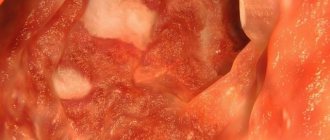
- Endoscopic examination: stage I - severe cicatricial and ulcerative deformation of the pyloroduodenal canal with a narrowing of its lumen to 1–0.5 cm; Stage II - the stomach is distended, the pyloroduodenal canal is narrowed to 1–0.3 cm due to sharp cicatricial deformation. Peristaltic activity of the stomach is reduced; Stage III - a huge stomach. Atrophy of the mucous membrane. Marked narrowing of the pyloroduodenal canal.
Laboratory diagnostics: signs of progressive dehydration, hypoproteinemia, hypokalemia, severe alkalosis.
Material and methods
The work was carried out in a systematic review design and represents the so-called secondary scientific research. Unlike a literature review, a systematic review, thanks to the methods of Evidence Based Medicine and Critical Appraisal Tools, allows us to identify and summarize the results of the most reliable primary studies in a particular branch of medical science.
The inclusion criteria for the systematic analysis were randomized clinical trials (RCTs) studying the surgical treatment of ulcerative pyloroduodenal stenosis in patients over 18 years of age.
Methodologically, the work was carried out taking into account the recommendations of the Prism group [2] and the mechanisms of critical analysis from the Oxford Center for Evidence-Based Medicine, used to assess the likelihood of systematic errors in selected primary scientific works [3]. To determine the degree of reliability of the information obtained, the Oxford gradation of evidence, edited by J. Howick, was used [4]. According to this hierarchy of evidence, reliable information can be obtained from studies that correspond to the first level of evidence, which include RCTs. To determine whether primary scientific studies meet the stated highest level of evidence, they were scored based on the possible risks of major systematic errors, as recommended by Cochrane experts [5].
Electronic search of evidence was carried out in Russian and English. From Russian databases, the elibrary electronic library was used; from English-language ones, the Cochrane library and the PubMed database of medical and biological publications were used. Search keywords: peptic ulcer disease, pyloric stenosis, gastric outlet obstruction, vagotomy, antrectomy, randomized controlled trials, meta-analyses ( metaanalysis).
The primary search for evidence is limited to 2014–2018. However, due to the fact that currently there is practically no research on the topic of surgical treatment of ulcerative pyloroduodenal stenosis, in order to give the systematic review the necessary power, we were forced to expand the recommended time frame to the full possible depth in these databases. In addition, references to literature sources from all scientific papers selected for this systematic review were carefully reviewed.
Treatment of pyloroduodenal stenosis
- Conservative antiulcer treatment for 2–3 weeks may precede surgery.
- Surgical treatment - in case of compensated stenosis with sufficient patency of the pyloroduodenal zone, selective proximal vagotomy with pyloroplasty or anastomosis between the stomach and small intestine (stomach drainage operation) can be performed. In other cases, resection is performed in various ways.
Essential drugs
There are contraindications. Specialist consultation is required.
- Omeprazole (proton pump inhibitor and antiulcer agent). Dosage regimen: orally with a small amount of water at a dose of 20 mg 2 times a day for 2-3 weeks.
- Clarithromycin (broad spectrum antibacterial agent). Dosage regimen: orally at a dose of 500 mg 2 times a day for 2-3 weeks.
- Amoxicillin (bactericidal antibacterial agent). Dosage regimen: orally at a dose of 1000 mg 2 times a day for 2-3 weeks.
Treatment of the disease
Surgery is the surest way to treat pyloroduodenal stenosis. Before the operation, the patient undergoes preparation for it, which consists of conservative treatment:
- Establishing water and electrolyte balance using solutions of “Protein”, “Dextran”, “Albumin”. The success of the procedure is assessed by a biochemical blood test.
- Introducing glucose, fat emulsion and amino acids parenterally to replenish the body's energy costs.
- Therapy aimed at eliminating the ulcer.
- Aspiration of gastric contents using a probe.
After the preparatory measures have been taken, the person is sent for surgery, which depends on the stage of stenosis of the pyloroduodenal zone. Stenosis is distinguished:
To make the organ passable again, duodenoplasty may be necessary.
- Compensated. If patency is complete, selective proximal vagotomy is performed. If the probe cannot be inserted, duodenoplasty or pyloroplasty is performed in the process.
- Subcompensated. A vagotomy with drainage operation is performed.
- Decompensated. Part of the stomach is removed.
Incidence (per 100,000 people)
| Men | Women | |||||||||||||
| Age, years | 0-1 | 1-3 | 3-14 | 14-25 | 25-40 | 40-60 | 60 + | 0-1 | 1-3 | 3-14 | 14-25 | 25-40 | 40-60 | 60 + |
| Number of sick people | 0 | 0 | 28 | 43.6 | 43.6 | 28 | 20 | 0 | 0 | 16 | 18 | 18 | 13 | 8 |
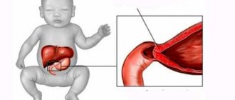
PYLORODUODENAL SCAR STENOSIS
Pyloroduodenal cicatricial stenosis is a complication of duodenal ulcer or ulcer of the pyloric or prepyloric stomach, in which there is a disruption in the movement of gastric contents from the stomach to the duodenum due to cicatricial narrowing of the lumen of the organ. It is observed in 5–10% of patients with peptic ulcer.
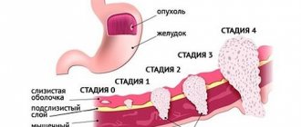
Classification. Depending on the severity of the narrowing of the pylorus and the associated difficulty in the passage of stomach contents into the duodenum, 3 stages of pyloroduodenal stenosis are distinguished: 1) compensation stage, 2) subcompensation stage, 3) decompensation stage.
The stage of stenosis is determined by the rate of emptying of the stomach into the duodenum.
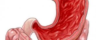
Etiology and pathogenesis. Pyloroduodenal stenosis develops as a result of scarring of the ulcer, inflammatory infiltration or proliferation of fibrous connective tissue of the pylorus or duodenum in the area of the callous ulcer. Alternating phases of exacerbation and remission of ulcers in the pyloroduodenal zone lead to scarring, deformation and narrowing of the lumen of the outlet of the stomach. As a result, difficulty arises in emptying the stomach into the duodenum, which is initially compensated by increased peristalsis of the stomach, hypertrophy of its muscle layer, and then leads to significant stretching and increase in the volume of the stomach, in which food and liquid begin to stagnate for a long time. A significant reduction in the volume of food, liquid and gastric juice penetrating from the stomach into the duodenum and their ever-increasing loss through vomiting lead to exhaustion of the patient, loss of body weight, dehydration, loss of sodium, potassium, hydrogen, and chlorine ions.
The patient's complaints depend on the stage of the disease.
1. Compensation stage
. The general condition does not suffer noticeably outside of exacerbation of peptic ulcer disease. During exacerbation, the pain is intense and prolonged, there is a feeling of heaviness and fullness in the epigastrium after eating, sour belching and, occasionally, vomiting with food.
2. Subcompensation stage
. Patients experience rotten belching, rumbling in the abdomen, and at times cramping pain, and an increased feeling of heaviness and fullness in the epigastrium. In the evening - profuse vomiting mixed with undigested food taken several hours before. Vomiting brings relief. Patients gradually experience weakness, fatigue, and weight loss.
3. Stage of decompensation
. Patients complain of dull arching pain in the epigastrium, constant, rotten belching, frequent vomiting, often with food eaten the day before. To alleviate the condition, patients artificially induce vomiting. The patients are exhausted. After repeated vomiting, general weakness, lethargy, headache, paresthesia and pain in the muscles of the arms and legs appear. Tonic spasms of various muscle groups, disturbances of consciousness, and a decrease in daily diuresis may be observed.
Anamnesis. Most patients have an ulcer history of varying duration (usually long) with multiple alternations of exacerbations and remissions of the disease.
Examination of the patient.
In the compensation stage
physical examination usually does not reveal pathological changes.
In the stage of subcompensation
Pallor and dryness of the skin and weight loss may be noted. The pulse is moderately increased, blood pressure is normal or slightly reduced. The tongue is dry and covered with a white coating. The abdomen may be somewhat swollen in the epigastrium; sometimes in thin patients, wave-like peristaltic movements of the stomach can be seen here. Palpation reveals moderate pain in the epigastrium and right hypochondrium. When the abdominal wall is shaken above the stomach, a “splashing noise” is detected, and with percussion, a tympanic sound is detected. During auscultation, increased peristalsis is periodically heard.
In the stage of decompensation
the condition of patients is moderate or severe.
The skin is dry, pale, skin turgor is reduced, it easily folds and slowly straightens when released. The patients are exhausted and their body weight is reduced. Convulsions, contraction of the hands in the “obstetrician’s hand” type ( Trousseau’s symptom ), twitching of the facial muscles when the zygomatic bone is tapped anterior to the external auditory canal ( Chvostek’s symptom ) may be detected.
The pulse is increased, blood pressure is decreased. The tongue and oral mucosa are dry. The abdomen is sharply swollen in the epigastrium, sometimes convulsive contractions of the stomach can be seen here. On palpation, pain is noted in the epigastrium and right hypochondrium. Significantly enlarged contours of the stomach are palpated. Percussion produces a tympanic sound. Auscultation reveals rare, sluggish peristaltic sounds. Diagnostics.
Laboratory research:
A general blood test in patients in the stage of subcompensation or decompensation reveals blood thickening (increased hemoglobin, red blood cells, increased hematocrit).
Determination of BCC reveals a deficiency in circulating blood volume.
A biochemical blood test reveals a decrease in the level of sodium, potassium, calcium, chlorine, and protein ions, and also reveals an increase in the content of urea and creatinine in the blood, and a violation of the acid-base state towards alkalosis.
A general urine test reveals oliguria, increased specific gravity of urine, microhematuria, and hypochloruria.
Instrumental studies:
1. Fluoroscopy and radiography of the stomach and duodenum with determination of the rate of evacuation of barium sulfate suspension from the stomach into the duodenum is necessary to determine the stage of pyloroduodenal cicatricial stenosis. In the compensation stage
a moderate narrowing of the pyloroduodenal canal is determined, the stomach is of normal size or slightly dilated, peristalsis is enhanced, the evacuation of the contrast agent is slightly slowed down.
After 12 hours there is no contrast agent in the stomach. In the stage of subcompensation
in patients, the stomach is significantly expanded, contains fluid on an empty stomach, its peristalsis is weakened, and the pyloroduodenal canal is significantly narrowed.
There is a pronounced slowdown in the evacuation of barium suspension from the stomach. After 12 hours, the contrast agent still remains in the stomach cavity, but after 24 hours it is no longer in the stomach. In the stage of decompensation
, the stomach is sharply dilated and on an empty stomach contains a large amount of liquid.
Contrast material accumulates in the lower part of the stomach in the form of a cup ( "cup sign" ).
The greater curvature of the stomach is located low, sometimes at the level of the symphysis pubis. The pyloric canal is sharply narrowed or is not filled with barium sulfate. Peristalsis of the stomach is very weakened. The evacuation of the contrast mass from the stomach slows down sharply - after 24 hours, a suspension of barium sulfate remains in the stomach, which can linger here for up to several days. 2. Fibrogastroduodenoscopy (FEGDS). In the compensation stage
– pronounced scar-ulcerative deformation of the pyloroduodenal canal with a narrowing of its lumen to 1–1.5 cm.
In the subcompensation stage,
the stomach is distended, contains fluid, the pyloroduodenal canal is narrowed to 0.3–1 cm due to a sharp cicatricial deformation.
In the stage of decompensation
, the stomach is sharply dilated, contains a lot of fluid, atrophy of the mucous membrane and erosive gastritis are noted. A pronounced narrowing of the pyloroduodenal canal up to 0.1 - 0.3 cm is detected.
Treatment. It is impossible to cure cicatricial pyloroduodenal stenosis with conservative methods, and the progression of stenosis worsens the patient’s condition and poses a threat to life. Therefore, ulcerative cicatricial pyloroduodenal stenosis is an absolute indication for surgical treatment. However, it should be remembered that symptoms of impaired pyloric patency can be caused not only by the cicatricial process, but also by the inflammatory edematous process that accompanies an exacerbation of peptic ulcer disease. Therefore, the true stage of cicatricial pyloroduodenal stenosis can be judged after a course of conservative antiulcer therapy, if the patient’s condition allows it.
Conservative therapy can play the role of preoperative preparation and consists of the following components: 1) daily gastric lavage for 3-5 days; 2) correction of the water and electrolyte composition of the blood through intravenous infusions of saline solutions; 3) intravenous administration of protein drugs, glucose, vitamins, parenteral nutrition.
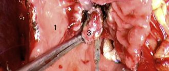
Surgical treatment consists of resection of a significant part of the distended stomach (2/3 or 3/4 or more) with suturing of the duodenal stump tightly and the formation of a gastrojejunostomy (Billroth-2 modification). Gastroduodenoanastomosis (according to Billroth-1) in this case is undesirable due to cicatricial deformation of the duodenal bulb.