Causes of pancreatic cancer
The exact causes of pancreatic cancer are still unknown. Only a few factors have been identified that increase the likelihood of developing the disease:
- smoking
- diabetes mellitus for a long time
- chronic pancreatitis (inflammation of the pancreas)
- stomach ulcer
- existing pancreatic cancer in blood relatives
- genetic syndromes (BRCA2 gene mutation, Lynch syndrome, familial atypical multiple melanoma)
- obesity.
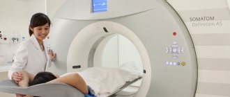
In what cases is ultrasound diagnostics needed?
An ultrasound is prescribed if the patient has signs characteristic of organ damage. The triad of symptoms of pancreatic cancer are:
- jaundice,
- epigastric pain,
- sudden weight loss.
Symptoms indicate tumor progression, accompanied by cancer intoxication.
Also indications for the study are:
- trauma in the projection of the organ,
- upset stool, flatulence,
- diabetes,
- abdominal discomfort associated with eating,
- changes in laboratory parameters in blood and urine,
- suspicion of a malignant process in the pancreas,
- metastases in the peritoneum from tumors of another location,
- management of chronic pancreatitis.
A type of ultrasound is Dopplerography. During the examination, using a special sensor, the sonologist evaluates the nature of the blood flow of the organ. Tortuous vessels indicate a malignant process.
Types of pancreatic cancer
There are two main types:
- Adenocarcinoma (exocrine cancer) accounts for the majority of all pancreatic malignancies.
- Endocrine cancer grows from neuroendocrine (hormone-producing) cells and is also called islet cell cancer or neuroendocrine tumor.
In ICD-10, pancreatic cancer has code C25, with additional junior-level codes set depending on the location in different departments (for example, C25.0 - cancer of the head of the pancreas, C25.1 - cancer of the body of the pancreas); There is a separate code for pancreatic cancer that goes beyond the boundaries of one localization.
Where does pancreatic cancer metastasize?
This malignant tumor is characterized by rapid spread to nearby areas. The pancreas is surrounded by several important structures of the abdominal cavity, which makes the spread of cancer cells rapid. The tumor, even in the early stages, often grows into the liver, gall bladder, intestines, and peritoneum.
Examination algorithm using ultrasound
Based on ultrasound data, a general examination algorithm has been created for accurate diagnosis of pancreatic cancer:
- transcutaneous examination in B-mode in real time - used to detect tumors and is a screening method with which the examination of the patient begins,
- color Doppler scanning or ultrasound in B-mode using CO2 as contrast - for differential diagnosis of malignant formation and inflammation of the pancreas,
- color Doppler scanning in the color Doppler mapping (CDC) or EDC (power Doppler mapping) mode - establishes the relationship of the tumor with nearby vessels.
Transcutaneous B-mode imaging
Transcutaneous ultrasound in B-mode is the most common method for studying parenchymal organs. Provides a two-dimensional, two-color image of the organ being studied on the screen. Allows you to see:
- circuit,
- localization,
- tumor stage.
It is one of the screening methods for pancreatic pathology.
Real-time B-mode ultrasound for pancreatic cancer is based on detecting direct and indirect signs of the disease. Direct signs are the identification of a single focus or cavity with a clear boundary of heterogeneous density between the tumor and normal tissue. Changes in the parenchyma of the pancreas are the main sign of an existing neoplasm; they are observed with ultrasound in 80% of cases. Based on the intensity of signal reflection, a tumor (its echogenicity) can be:
- hypoechoic,
- hyperechoic,
- isoechoic,
- mixed.
Indirect signs are:
- expansion of the common bile duct and pancreatic duct (more than 3 mm),
- a significant increase in the size of the pancreas in the area of the neoplasm.
Dilatation of the bile ducts in the liver is possible in the presence of a tumor in the head of the pancreas or lymph nodes in the area of the hepatoduodenal ligament.
Depending on the size of the formation, the size of the gland may be unchanged or locally or diffusely increased.
Color Doppler scanning
To diagnose pancreatic cancer, a method with the Doppler effect, named after the Austrian physicist who discovered it, is used. In this case, a color image of the reflected ultrasonic signal is obtained. Unlike conventional ultrasound, this method is based on the reflection of ultrasound by moving objects, for example, blood cells in the vascular bed - red blood cells. Due to the changing frequency of the signal reflected from them, vascular pathologies are identified, the differences between benign neoplasms and malignant tumors are determined, and the following is assessed:
- speed,
- direction of flow,
- blockage of blood vessels,
- collaterals.
This procedure has no contraindications, no side effects are observed, the patient is not exposed to radiation - it is absolutely safe. With the help of color flow, a color image of the blood flow is displayed, any circulatory disorders in the organ are visible.
Color Doppler scanning using Color Doppler or EDC modes
An additional method for the differential diagnosis of a malignant process and inflammatory changes in the pancreatic parenchyma is the use of color Doppler examination in the Color Doppler and EDC modes.
When using the methods, it is determined:
- presence and patency of blood vessels,
- the nature and speed of blood circulation in them.
If pancreatic cancer is suspected in a patient during duplex scanning, the following is noted:
- absence or sharp decrease in blood flow through the vessels inside the tumor,
- the presence of collateral vascular networks with arterial blood flow.
EDC is essentially a more advanced color Doppler mapping. Its difference from Color Doppler, in which the color-coded frequency shift of the Doppler signal is visualized on the screen, lies in the display of numerous signals from moving objects (red blood cells) - this method maps the energy characteristics of the signals.
Symptoms and signs of pancreatic cancer
Symptoms depend on the location of the cancer cell and the degree of involvement of the gland tissue.
In the early stages, the cancer does not manifest itself in any way, but then the patient may notice:
- jaundice
- darkening of urine and lightening of stool
- pain in the upper or middle third of the abdomen and back
- loss of appetite
- weight loss for no apparent reason
- general weakness, fatigue
- tendency to thrombosis.
If several of these symptoms appear, it is better to consult a doctor immediately.
In case of pancreatic cancer with metastases to the liver, a feeling of fullness in the stomach after eating (even if a small amount of food was eaten), itching of the skin, and pain in the right hypochondrium are also noted.
Ultrasound signs
The picture of a cancerous tumor on an ultrasound of the pancreas looks like a formation of regular or irregular shape with uneven, unclear contours. The echogenicity of such formations is reduced, in rare cases significantly increased. The structure can also be different, both homogeneous and heterogeneous. If the tumor tissue is of increased echo density, then there may be areas with increased echo structure and vice versa.
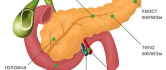
The ability to detect pancreatic tumors depends on their location and size. Tumors of the head and body are determined in the early stages, since they are accessible to research; tumors more than one and a half centimeters in diameter can be detected. Tail tumors are much more difficult to detect due to their anatomical location and poor accessibility to an ultrasound machine. It is easier for a specialist to identify a tumor with a diameter of 3 cm located in the head than a tumor with a diameter of 5 cm, which is localized in the tail.
Localization of the tumor in the tail of the gland
It is difficult to differentiate a tumor from pseudotumor pancreatitis, so it is necessary to carefully examine the pancreatic duct. With pseudotumorous pancreatitis (as with other types of pancreatitis), the main pancreatic duct is dilated and uneven in diameter along its entire length. With cancer, the duct is widened only below the formation.
Ultrasound allows you to assess whether surgery on the tumor is possible. To do this, the sonologist evaluates how the cancerous formation is located in relation to large vessels: the inferior vena cava and portal vein, aorta, superior mesenteric artery. If the tumor is in close contact with one or more vessels over a large area, then its resection is impossible or presents great difficulty.
If a malignant process in the pancreas is suspected, an ultrasound will check whether there are metastases in the liver and lymph nodes. Metastases look like round formations of low echo density. The most accurate sign of metastasis is the absence of an echo-negative rim around it. Metastatic lesions of the lymph nodes look like hypoechoic round formations with smooth contours; they are located next to the aorta or pancreas.
Ultrasound is not informative only for one type of pancreatic tumor. These are so-called hormonally active tumors (insulinomas). They do not reach large sizes and are located in the body or tail of the gland, so they are difficult to visualize. Patients suffering from this type of cancer, as a rule, have undergone multiple operations; they have an active adhesive process in the abdominal cavity, which complicates conditions for diagnosis. The picture that the doctor sees on ultrasound is not unique to this tumor; to make a diagnosis, it is necessary to take into account clinical data and the results of other studies.
Diagnosis of pancreatic cancer
Instrumental examinations may include:
- ultrasound examination of the abdominal organs
- computed tomography (CT)
- magnetic resonance imaging (MRI)
- positron emission tomography (PET). This is a more accurate and modern method that allows you to detect small lesions.
Does an ultrasound of the pancreas show signs of cancer? Since the pancreas is covered by the small intestine, liver with gall bladder, and spleen, the organ is quite difficult to visualize and signs of a tumor are not always visible. Also, small formations are not visible on ultrasound. Therefore, if a malignant tumor is suspected, the doctor may prescribe:
- Endoluminal endoscopic sonography. With this method, an ultrasound probe is inserted into the intestine to obtain more detailed images of the gland.
- Endoscopic retrograde cholangiopancreatography (ERCP), in which a special dye is injected into the ducts of the gallbladder and pancreas, and then X-rays are taken, which can reveal the tumor.
- Laparoscopy is the introduction of an optical system into the abdominal cavity to examine internal organs.
- Peritoneal fluid cytology, where a sample of abdominal effusion is examined.
For a final diagnosis, a biopsy of the pancreas is often performed, followed by examination of the resulting fragment under a microscope.
The presence of the tumor marker CA 19-9 in the blood is currently not considered a reliable diagnostic sign, but a change in its level may indicate the effectiveness of the treatment. A biochemical blood test can reveal an increase or decrease in bilirubin levels.
What will the study show in the case of cancer?
No specific preparation is required for ultrasound diagnostics. To better view the organ, the doctor asks you to inflate your stomach and hold your breath for a while.
If patients experience bloating, they are advised to follow a slag-free diet for 3 days before the ultrasound and drink adsorbent drugs. This is activated carbon, taken 2 pills 3 times a day. You should not have dinner before the ultrasound. The last meal takes place 7 hours before the examination.
When performing diagnostics, an ultrasound scan of the pancreas shows:
- with a single tumor localization, there is one cancer focus;
- in case of progression, a tumor of a dense structure is visible.
Basically, research is carried out not only on the glands, but also on the entire digestive system - the abdominal cavity, pelvis, soft tissues of the organ.
A type of ultrasound is Dopplerography, during which a sonologist evaluates the characteristics of blood flow using a special sensor. If tortuous vessels are detected on ultrasound, this indicates cancer.
In addition to cancer, ultrasound reveals a number of diseases.
- Chronic pancreatitis, pancreatic necrosis.
- Acute form of pancreatitis.
- Cysts.
- Abscesses.
- Lipomastosis.
acute pancreatitis
Instrumental techniques include CT and magnetic resonance scanning. Multispectral tomography can detect metastases of pancreatic cancer. Laparoscopy is a surgical method of examination that can detect cancer in the peritoneum. During the procedure, a cancer biopsy is performed for subsequent histological analysis.
Stages of pancreatic cancer
The stages of the disease are determined to select the optimal treatment tactics.
- Stage 1. The cancer is limited to the pancreas and can be completely removed by surgery. The size of the tumor does not exceed 2 cm.
- Stage 2. The tumor affects nearby organs and lymph nodes, but can still be completely removed by surgery.
- Stage 3. The tumor grows into the large blood vessels around it and affects the lymph nodes, and its removal by surgery is not possible in all cases.
- Stage 4. There are metastases to the abdominal organs, peritoneum. It is impossible to remove the tumor surgically.
Pancreatic cancer treatment
The main goal of treatment is the complete removal of all cancer cells in the gland and surrounding tissues. If this is not possible, then it is necessary to prevent the growth of the tumor, its spread and thereby reduce the symptoms. The choice of method depends on the stage of the disease and the patient's condition.
Surgical treatment (possible options)
- Whipple's operation: the head of the pancreas, a fragment of the stomach, the gallbladder, bile ducts and a section of the small intestine are removed. The remaining portion of the pancreas is sufficient to produce insulin and digestive enzymes.
- Total pancreatectomy with resection of the entire gland, parts of the stomach, small intestine, common bile duct, spleen, gallbladder and regional lymph nodes.
- Distal resection, when the body and tail of the pancreas and the spleen are removed.
Chemotherapy
- Regional therapy, with the introduction of medicine directly into the gland.
- Systemic therapy by injecting drugs that kill cancer cells into a muscle or vein. Drugs used: gemcitabine, paclitaxel, 5-fluorouracil, irinotecan, oxaliplatin, capecitabine, cisplatin, docetaxel.
Radiation therapy
Two types of radiation therapy are used:
- Internal therapy, when a radioactive substance is delivered into the tumor tissue using catheters or needles.
- Treatment using external radiation generated by special installations.
Targeted therapy
Special substances attack only cancer cells without damaging healthy ones. Tyrosine kinase inhibitors (erlotinib, masitinib, trametinib) are used to block signals that cause tumor growth.
Biological therapy (immunotherapy)
Synthesized substances, for example, nivolumab, ipilimumab, pembrolizumab, demcizumab, stimulate the body’s own immune system to fight cancer cells. Most of these drugs are only undergoing clinical trials for the treatment of pancreatic cancer.
Pain due to pancreatic cancer is treated according to a three-step scheme, i.e. first they use non-narcotic painkillers, and then switch to narcotic ones, including potent ones (morphine).
Possible symptoms
Due to the peculiarities of the placement of the pancreas, which is in contact with all digestive organs and has a direct working relationship with them, the development of pronounced symptoms of the disease indicates a painful process that has already gone quite far.
The signs of a pancreatic tumor are somewhat different, it all depends on the location of the organ damage. Regardless of the location of common cancer formations, the following symptoms appear:
- pain syndrome;
- depression;
- yellowing of the skin;
- body weight is lost.
Painful discomfort sometimes does not have a clear localization, but is often detected in the epigastric region, under the left and right ribs, radiating to the back.
The area where the symptom of pain is formed depends on the area of the gland damaged by the formation:
- in blockage of the common bile duct or Wirsung canal;
- peritoneal phenomena due to exacerbation of the concomitant inflammatory process that develops in nearby tissues;
- in compression of nerve trunks.
As pancreatic tumors form, dyspeptic disorders increase.
- There is no desire to eat.
- It makes me feel nauseous.
- Belching of air appears due to bloating.
- Vomiting begins.
- Abnormal stool – diarrhea, constipation.
The main early symptom of pancreatic cancer is rapid weight loss. The victim loses a lot of weight as a result of changes in the digestive process and cancer poisoning, which is determined by the decomposition products of the formation. Atypical, rapidly dividing cells of the formation lose their activity without producing enzymes. Since the Wirsung canal or common bile duct is compressed by the expanding formation, the duodenal canal ends in entry.
- Digestive juice with enzymes and their breakdown of carbohydrates, fats, proteins.
- Bile, in the presence of which fats are broken down.
At the last stage of pancreatic disease, tumor manifestations progress and new ones appear. With cancer of the body and tail, vascular thrombosis is observed, which develops more often than cancerous damage to the head.
As cancer develops, symptoms change and may develop:
- diabetes;
- decreased glucose levels;
- pancreatic stool;
- yellowing of the skin and mucous membranes.
If the patient has not previously experienced diabetes, the disease affects the organ when the cancer covers the tail part of the gland. This is combined with the large distribution of the islets of Langerhans in this zone with beta cells that produce glucose, and alpha cells that synthesize the insulin antagonist - glucagon.
Due to inadequate insulin production, the formation of glycogen from glucose is disrupted, which subsequently leads to the development of diabetes mellitus.
Diabetes, a consequence of cancer, is characterized by a number of symptoms.
- Headache.
- Inability to concentrate.
- Increased fatigue.
- Increased amount of urine excreted.
- Dry mouth.
- The occurrence of hallucinations.
- Loss of mind.
- Inappropriate behavior.
- Presence of seizures.
- Development of coma.
If you have diabetes, you need to pay attention to your health and be monitored by a doctor.
Pancreatic stool is characterized by the following manifestations:
- gray color of stool;
- greasy shine;
- stinking smell;
- emptying up to several times a day.
Jaundice forms as the cancer spreads. The patient's skin, sclera, and mucous membranes turn yellow, excrement becomes discolored, urine darkens, and dilation of the bile ducts is also observed. The increase in hysteria is formed from the development of pathology. The patient complains of excruciating itching, which intensifies at night, and severe pain. These symptoms may develop in later stages when tumors grow into the hilum of the liver.
In pregnant women and children, symptomatic signs require treatment, which will be constantly monitored by a doctor. Cancer therapy requires the development of an individual regimen.
When carrying a child, treatment is selected depending on the week of pregnancy and the stage of the tumor. If cancer is detected in the thirty-first week, symptomatic therapy is carried out to eliminate pain, the main treatment is carried out after the birth of the child. Before 12 weeks of cancer detection, the woman is offered to terminate the pregnancy.
In case of cancer development, the gland needs diagnostics to confirm the diagnosis. Ultrasound examinations are prescribed. As an additional diagnosis, tests are taken for pancreatic cancer, blood and urine parameters are examined, and X-ray and other diagnostic methods are prescribed.
Rehabilitation after surgical treatment
After surgery, it is necessary to adequately anesthetize the patient and improve the functioning of the gastrointestinal tract, since any operation on the abdominal organs causes a temporary disruption of intestinal function. This means that you need to follow a diet (hence, there are restrictions on food intake). In the early postoperative period, they are first allowed to drink a little, then gradually switch to semi-liquid food.
Gradually, the patient begins to move more and exercise for at least 30 minutes a day.
As a rule, after tumor removal, a 6-month course of chemotherapy is given. Good contact with the doctor and strict adherence to the treatment plan are essential for successful recovery after surgery. It is very important to receive psychological support in a timely manner.
Complications
- Jaundice occurs when a tumor blocks the bile ducts and the bilirubin pigment found in bile is absorbed into the blood in large quantities. This causes the skin and whites of the eyes to turn yellow. Urine becomes dark, and feces, on the contrary, become light. There is usually no abdominal pain with jaundice. To eliminate it, stenting is performed, i.e. placing a plastic or metal tube into the duct through which bile is discharged into the intestines. Sometimes, instead of a stent, a new duct is formed during surgery to drain bile into the intestines.
- Pain in pancreatic cancer occurs due to damage to nerve endings by the tumor. Painkillers are used to relieve pain; sometimes radiation therapy can help; in severe cases, a block of the celiac plexus nerves is performed.
- Small bowel obstruction occurs when a tumor grows into or puts pressure on the outside of the intestine (and therefore the movement of food through it is disrupted or completely stopped). In these cases, a stent is inserted into the small intestine to prevent it from being blocked, or an operation is performed in which a bypass for the food mass is created, connecting the stomach with the section of the intestine located below the tumor.
- Weight loss is associated with insufficient production of pancreatic juice, impaired absorption of nutrients, as well as nausea and vomiting. In this case, split meals in small portions and taking enzymes can help.
- Recurrence of pancreatic cancer.
How does the disease manifest itself?
The first signs of cancer are characterized by pain, which indicates that the tumor has grown into the nerve endings. The intensity of pain varies. This will be discomfort or an acute attack. The localization of the syndrome depends on the part of the organ affected, the head, body or tail of the gland.
Episodic pain occurs between the shoulder blades, in the navel and lower back. The severity of pain in the pancreas can vary, it all depends on the position of the body.
The patient also develops a dislike for heavy meals, and increases sensitivity to alcohol, caffeine and pills.
Pathology at an early stage is accompanied by the following signs:
- hematological;
- cutaneous;
- nervous;
- pancreatic.
For intestinal indications, symptoms of pancreatic cancer vary in course. Cancer is diagnosed by clarifying the signs that are associated with the abdominal area.
- The stool has a fatty coating.
- Pale stool.
- Weight drops sharply.
- I suffer from heartburn all the time.
- Unreasonable bloating.
- Frequent diarrhea.
- Pain in the abdominal area.
heartburn
And also with pancreatic cancer there is chronic fatigue and fatigue. Tests of the patient will help identify tumor formation and the presence of metastases.
When the excretory system is disrupted, dark urine is observed. A common symptom is the appearance of pain in the pancreas area. At the same time, many patients also experience weight loss, which results in an incorrect prognosis. Loss of body weight has no connection with this symptom, since it occurs due to changes in the activity of the pancreas.
When a neoplasm develops in the pancreas, the patient may experience changes of a nervous nature.
The nervous system is dependent on the presence of toxins and waste in the blood. The pancreas takes part in the process of filtering harmful elements. The occurrence of a formation in its area leads to disruption of this work, compression of the bile ducts is observed, and the contents of bile enter the tissue. As a result, inhibition of nerve cells is recorded, which is characterized as follows:
- the patient's anxiety increases;
- suffers from insomnia or the patient gets tired quickly;
- the body is inhibited, the manifestation of reflexes is also slow.
Skin manifestations are common. It is possible to change the shade of the skin if the neck of the pancreas is damaged or with other cancer formations.
The main manifestation on the skin is jaundice. When the functioning of an organ changes, bile does not flow normally, which leads to intoxication of the body and jaundice of the epidermis. The patient's skin is itchy in any part of the body. There is no rash with itching, but it grows as the pathology develops and may not stand out until the end, as a sign that accompanies cancer.
When the disease is advanced, when the formations of the pancreas spread to nearby organs, metastases develop, and other signs are observed.
- Liver function is impaired.
- Foods are poorly digested.
- There is an accumulation of fluid in the peritoneum.
accumulation of fluid in the abdomen
When the formation envelops the blood vessels and leads to their rupture, internal blood loss is recorded. This complication becomes a factor in the development of anemia. If the cancer spreads to the lungs, the patient experiences a painful cough attack that cannot be relieved with anti-cough medications, shortness of breath, and spitting up blood.
Bone metastases lead to pain in the spine, legs and arms. At the last stage of the disease, the patient completely loses his ability to work, pain worries him all the time, and it is difficult to overcome cancer poisoning.
With the development of diabetes mellitus, the following is recorded:
- dry mouth;
- thirst;
- urine is excreted in large volumes;
- night trips to the toilet.
During pregnancy, the clinical development of pancreatitis depends on the stage of changes in the pancreas. When acute swelling of an organ occurs, the course is often mild. If pancreatic necrosis is hemorrhagic, the situation of patients is extremely difficult. Pancreatitis in pregnant women often manifests itself in a painless form, characterized by shock and signs of damage to the central nervous system.
Patient survival prognosis
The chances of recovery depend on:
- location of the tumor and the possibility of its complete removal
- degree of germination of atypical cells into nearby organs
- presence of distant metastases
- general health.
Exocrine cancer is poorly treated, with a five-year survival rate of 6%.
For patients with a small tumor (less than 2 cm) that does not extend beyond the gland capsule and does not have metastases to the lymph nodes, after surgical treatment, the five-year survival rate ranges from 18% to 24%.
Life expectancy for stage 4 pancreatic cancer depends on the tumor's resistance to chemotherapy and radiation. In this case, treatment is carried out to improve the quality of life, which may include:
- anesthesia
- surgical or radiation biliary decompression
- normalization of gastric motility
- psychological help.
Diet for pancreatic cancer
It is quite difficult for patients to maintain weight, since appetite is reduced from the disease and from drugs for its treatment, and the lack of digestive enzymes impairs the digestion of food. In addition, cancer cells produce substances that destroy muscle and fat tissue, which can lead to wasting. The diet includes:
- Refusal of fried, fatty foods. As a source of fats, you can use vegetable oil (olive, canola, peanut), nuts and avocados;
- Small meals up to 6-8 times a day;
- Consume at least 2 servings of fruits or vegetables per day.
It is recommended to drink at least 6 glasses of liquid per day.
Treatment with folk remedies for pancreatic cancer
There is no scientific data on the effectiveness of alternative methods for pancreatic cancer.
The fact that some anticancer drugs are synthesized from plants (for example, docetaxel is obtained from Yew berry (Táxus baccáta)) does not indicate the possibility of treatment with herbs. The content of herbal components in medicines is standardized, which makes it possible to calculate the correct dosage and minimize side effects.
The information is for reference purposes only and is not intended for self-diagnosis and treatment. There are contraindications. Specialist consultation is required.