Structure (anatomy) of the organ
Anatomically, the duodenum is divided into 4 sections: upper, descending, lower and ascending.
The first upper part or pars superior is from 5 to 6 cm long. It is directed obliquely, from front to back, from left to right, then forms an arched bend (superior curvature), and passes into the descending part.
The next part is the descending part or pars descendens, located to the right of the spine at the level of the lumbar region, at the point of transition to the lower part it forms the lower curvature. The length of this part is from 7 to 12 cm. On the medial wall of this part of the intestine there is a longitudinal fold of the mucous membrane. This fold has a papilla on its surface with ducts opening on it.
The horizontal part of the duodenum, pars inferior, 6 to 8 cm long, is directed from right to left, transverse to the spine, then bends upward, where it becomes the ascending part.
The ascending part or pars ascendens, relatively short (4-5 cm), this part forms a curvature called the duodenum-jejunum. This anatomical formation is located to the left of the spinal column at the level of the lumbar region. Sometimes this part of the intestine is not expressed. The initial section of the upper part is called the duodenal bulb.
The duodenum has a variable shape: the most common is a horseshoe shape, less often ring-shaped and angular.
The location of the duodenum is also variable, depending on weight, age and many other factors. In overweight people and young people, the intestine lies slightly higher than in thin and elderly people.
In relation to the spine, the sections of the intestine are also not always located in the same way. The following options for the location of parts of the intestine are more common: the upper part is located at the level of the first lumbar vertebra, the descending part is located to the right of the spine at the level of the second and third vertebrae. The lower part of the duodenum corresponds to the space from the third to the fifth lumbar vertebrae. Less often it descends to the pelvis.
The duodenum is covered by peritoneum unevenly, over a small area. The upper part of the intestine is in contact with the pancreas, common bile duct, portal vein, and gastroduodenal artery. In this place the intestine is devoid of cover. Therefore, this section is located mesoperitoneally. The ascending intestine is approximately similarly covered with peritoneum.
The descending and lower sections of the intestine are located retroperitoneally; they have peritoneal cover only in the front.
From the walls of the duodenum to the organs of the retroperitoneal space there are connective tissue fibers that fix it. The main function of fixing the intestine is performed by the peritoneum, which covers it in front. The second role is played by the root of the mesentery of the transverse colon. The pancreas has a certain role in fixation.
The least fixed part of the intestine is the upper one, therefore it is the most mobile. She can easily move in different directions.
What systems surround the duodenum?
In front and above the upper part of the intestine are the following organs: the quadrate lobe of the liver, the neck and the body of the gallbladder. The hepatoduodenal ligament is located between the upper part of the intestine and the left lobe of the liver (lower surface). At the base of this ligament lies the common bile duct. The upper part of the duodenum, not covered by peritoneum, is in contact with the portal vein, common bile duct, and arteries (gastroduodenal and pancreatic-duodenal).
The posterior surface of the descending part of the intestine is in contact with the pelvis of the right kidney, ureter, and renal vessels. The hilum of the kidney is slightly covered by the duodenum. The descending part of the duodenum is in contact with: the right curvature of the colon, the ascending colon, and the head of the pancreas.
The common bile duct is often located in the parenchyma of the head of the pancreas, then it connects with the pancreatic duct and flows into the duodenum. On the surface of the intestinal mucosa there is a small longitudinal fold (up to 1 cm long), on the surface of which there is a large duodenal papilla, on which the pancreatic duct opens. Sometimes there is an accessory pancreatic duct that opens higher than the main one.
Between the descending part of the duodenum and the head of the pancreas, a groove is formed in which the common bile duct lies. The superior mesenteric vein and artery are adjacent to the lower part of the duodenum. These vessels are located at the root of the mesentery. Further along this section the transverse colon and loops of the small intestine are adjacent.
Sometimes the duodenum can be compressed by the superior mesenteric arteries, leading to obstruction. In the clinic, such cases are called arterio-mesenteric obstruction, which often occurs with prolapse of the small intestine and some other diseases of the abdominal organs. Retroperitoneal tissue and loops of the small intestine surround the ascending part of the duodenum. The lower part of the intestine crosses the aorta at different levels: above or below the bifurcation.
The intestinal walls are constructed as follows:
- serous membrane;
- muscle layer;
- mucous membrane.
Between the mucosa and the muscle layer is the submucosa. The mucous membrane is covered with many villi. The villi are lined with prismatic bordered epithelium, which significantly increases the absorption capacity of this part of the gastrointestinal tract. Between these epithelial cells are goblet enterocytes that produce glycoproteins and glycosaminoglycans. The hormones enteroglucagon, gastrin and secretin are produced by other cells - Paneth or intestinal enterocytes. The duodenal bulb has longitudinal folds, the rest of the small intestine has circular folds. Due to the common bile and pancreatic ducts, a longitudinal fold of the mucous membrane is formed.
The mucosa contains lymphatic follicles, lymphocytes and plasma cells. The submucosa contains duodenal or Brunner's glands, the ducts of which open on the surface of the intestinal crypts.
The muscular layer of the intestine consists of non-striated muscle fibers arranged in two layers. The outer layer is longitudinal, the inner layer is circular. Serosa covers the duodenum partially, for a short distance. The remaining parts of the intestine are covered with adventitia, which is loose connective tissue. Numerous vessels and nerves pass through it.
Which environment predominates in the lumen of the duodenum depends on the stage of digestion. Before digestion begins, the environment is slightly alkaline (pH from 7.2 to 8.0). When the acidic contents of the stomach enter the intestine, the reaction changes towards acidic, but is then quickly neutralized and restored to its previous levels. The acidic environment is neutralized by pancreatic juice and other digestive juices.
Duodenum
The duodenum, duodenum (see Fig. 504, 534, 535, 536), begins under the liver at the level of the body of the XII thoracic or I lumbar vertebra, to the right of the spinal column. Starting from the pylorus of the stomach, the intestine goes from left to right and backward, then turns down and descends in front of the right kidney to level II or the upper edge of the III lumbar vertebra; then it turns to the left, is located at first almost horizontally, crossing the inferior vena cava in front, and then goes obliquely upward in front of the abdominal aorta and, finally, at the level of the body of the I or II lumbar vertebra, to the left of it, passes into the jejunum. Thus, the duodenum forms, as it were, a horseshoe or an incomplete ring, covering the head and partly the body of the pancreas from above, to the right and below.
The initial section of the intestine is the upper part, pars superior, which is initially slightly expanded and forms an ampulla, ampulla; the second section is the descending part, pars descendens, then the horizontal (lower) part, pars horizontalis (inferior), which passes into the last section - the ascending part, pars ascendens. When the upper part passes into the descending part, the upper bend of the duodenum, flexura duodeni superior, is noticeable, and when the descending part passes into the horizontal, the lower bend of the duodenum, flexura duodeni inferior. Finally, during the transition of the duodenum to the jejunum, the steepest duodenal-jejunal bend, flexura duodenojejunalis, is formed. The muscle that suspends the duodenum, m., approaches the posterior surface of the bend. suspensorius duodeni, which is a muscle-connective tissue cord attached to the left leg of the diaphragm. The length of the duodenum is 27-30 cm, the diameter of the widest descending part is 4.7 cm. A slight narrowing of the lumen of the duodenum is noted at the level of the middle length of the descending part, in the place where it is crossed by the right colon artery, and at the border between the horizontal and ascending parts where the intestine is crossed from top to bottom by the superior mesenteric vessels.
The wall of the duodenum consists of three membranes: mucous, muscular and serous. Only the beginning of the upper part (for 2.5-5 cm) is covered with peritoneum on three sides; the descending and lower parts are located retroperitoneally and are covered with adventitia.
The muscular layer, tunica muscularis, of the duodenum has a thickness of 0.3-0.5 mm, greater than the thickness of the rest of the small intestine. It consists of two layers of smooth muscle: the outer - longitudinal layer, stratum longitudinale, and the inner - circular layer, stratum circulare.
The mucous membrane, tunica mucosa, consists of an epithelial layer with an underlying connective tissue plate, a muscular plate of the mucous membrane, lamina muscularis mucosae, and a layer of submucosal loose fiber that separates the mucous membrane from the muscularis. In the upper part of the duodenum, the mucous membrane forms longitudinal folds, in the descending and horizontal (lower) parts - circular folds, plicae circulares. Circular folds are permanent and occupy 1/2 or 2/3 of the circumference of the intestine. In the lower half of the descending part of the duodenum (less often in the upper half) on the medial section of the posterior wall there is a longitudinal fold of the duodenum, plica longitudinalis duodeni, up to 11 mm long, distally it ends with a tubercle - the major papilla of the duodenum, papilla duodeni major, at the top of which is located the mouth of the common bile duct and pancreatic duct (see Fig. 535). Slightly above it, at the apex of the minor duodenal papilla, papilla duodeni minor, there is the mouth of the accessory pancreatic duct, which occurs in some cases.
Features of digestion and organ functions
From the stomach, food in a liquid or semi-liquid state enters the duodenum. The pyloric part of the stomach performs peristaltic contractions and pushes the bolus of food towards the sphincter. The latter's receptors send a signal along the vagus nerve, and the sphincter opens. The opposite happens when the food bolus irritates the organ's receptors. The muscle sphincter remains closed until the chyme moves further through the intestines. The main functions of the duodenum: participation in the process of digestion and absorption of nutrients. The next stage of digestion begins in this section of the intestine. The common bile and pancreatic ducts open on the surface of the nipple of Vater. The process of digestion of food is carried out by pancreatic juice, intestinal juice and bile. All these components have a pronounced alkaline reaction, so the acidic environment of food coming from the stomach is quickly neutralized. Pancreatic juice contains organic and inorganic substances. The first include proteolytic, lipolytic, aminolytic enzymes. Proteins are broken down by trypsin, chymotrypsin, elastase and carboxypetidase. Inhibitors of proteolytic enzymes protect the pancreas from self-digestion. Lactase, maltase and amylase break down carbohydrates into monosaccharides. Phospholipase and lipase break down fats into simpler components (glycerol and fatty acids). Liver cells secrete bile into the intestinal lumen. Externally, it is a liquid with an alkaline reaction, golden yellow. Dry residue contains up to 2.5%. Main components: pigments, bile acids, cholesterol. From 0.5 to 1.2 liters are produced per day. This process is continuous, however, bile enters the intestines only during meals. The rest of the time, bile is in the gallbladder. Bile is necessary to activate pancreatic enzymes. In particular, lipases. Emulsification of neutral fats is carried out by fatty acids. In addition, bile enhances the peristalsis of the intestinal walls, increases the production of pancreatic juice, and acts as a bacteriostatic substance, preventing the development of putrefactive bacteria.
How it hurts: symptoms
Diseases of this part of the digestive tract can be of inflammatory and non-inflammatory nature.
The most common inflammatory disease is duodenitis. The following bacterial and fungal infections are less common: tuberculosis, actinomycosis. Typically these infections are acquired from other organs. Duodenitis can simulate the symptoms of a peptic ulcer.
The second most common diseases are erosive and ulcerative diseases. Often these diseases are combined with liver pathology, intestinal neoplasms, diseases of the respiratory and cardiovascular systems, and kidneys. Therefore, all patients who have been found to have erosions of the stomach or duodenum are subject to careful examination.
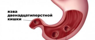
Approximately 10% of the world's population suffers from peptic ulcer disease. Peptic ulcer disease is mainly provoked by stressful situations, bacterial infection, smoking, the use of certain drugs and poor diet. In approximately half of patients, this disease is hereditary. The disease worsens in the spring. Often the duodenal bulb has scar changes after the healing of the ulcer.
Diseases of a tumor nature are relatively rare. These can be both malignant and benign neoplasms. The risk of developing the disease increases with age. Among malignant tumors, the first place belongs to sarcoma. The predominant localization is the descending part of the organ. The main factors in the development of the disease are diffuse polyposis, adenomas, hereditary intestinal diseases and Crohn's disease. Benign tumors can be multiple or solitary. Often, when large in size, they can contribute to the development of complications of the disease: intestinal obstruction and bleeding. Specific examination methods will help confirm or refute the presence of the disease.
Main symptoms of diseases.
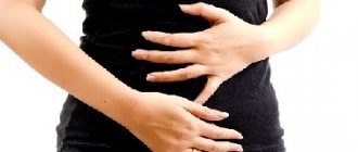
- All diseases of this organ are accompanied by pain. With ulcers and erosions, the patient is bothered by “hunger pains” and frequent night attacks. The peak of pain occurs after eating (after 2 hours). The patient will note that the pain subsides after eating. With an ulcer of the intestinal bulb, the pain subsides after vomiting. Localization of pain: right hypochondrium, epigastrium, radiates to the back and right arm. With peptic ulcer disease, pain is the main clinical concern.
- Bleeding. This symptom occurs in approximately 20% of patients. It manifests itself as melena (black stool), bloody vomit, and vomit the color of coffee grounds. The blood test shows low hemoglobin, the patient is weak and prone to developing collapsing conditions.
- Dyspeptic disorders. The patient is concerned about the following symptoms: heartburn, loose stools, or, conversely, constipation.
- Increased appetite. More often, this symptom is due to the patient’s desire to drown out “hunger pains.”
- Intestinal diseases are accompanied by irritability, poor health, and general symptoms.
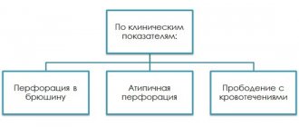
The diseases may have the following complications: bleeding, organ perforation, cicatricial deformities, narrowing of the lumen, precancerous conditions, malignancy of benign tumors, reactive pancreatitis, liver and gallbladder diseases.
How a common disease manifests itself: duodenitis (video)
In general, the diseases have a favorable prognosis, with the exception of some complicated situations. Patients are able to work, but the type of activity should be changed if it is associated with stressful situations, poor diet, or heavy physical exertion.
Prevention of diseases of this part of the intestine is not difficult. The main thing for the patient is to follow a diet, exclude certain foods from the menu, and give up bad habits. Diseases of the duodenum of inflammatory and infectious origin have a relapsing course.
Patients with diseases of the duodenum are under constant medical supervision and undergo an anti-relapse course of treatment in the spring and autumn. In the absence of a proper regimen and full treatment, all symptoms of the disease recur. A preventive course of treatment should be taken every 3-5 years, even if there are no obvious symptoms of the disease.
A case of ultrasound diagnosis of duodenal maltoma
Ultrasound machine HS40
Top seller in high class.
21.5″ high-definition monitor, advanced cardio package (Strain+, Stress Echo), expert capabilities for 3D ultrasound in obstetrics and gynecology practice (STIC, Crystal Vue, 5D Follicle), high-density sensors.
Introduction
Maltoma (B-cell MALT /mucous-associated lymphoid tissue/ lymphoma of low malignancy) is a tumor originating from lymphoid tissue associated with the mucous membrane, having a special pathomorphology and course, which is caused by chromosomal mutations that disrupt the structure of genes that regulate proliferation and apoptosis. Maltomas are non-Hodgkin's lymphomas, which are relatively rare. Meanwhile, in recent years, perhaps due to the improvement in the quality of instrumental diagnostics, a noticeable increase in the incidence of non-Hodgkin's lymphomas has been noted. Thus, in the USA, the incidence of all types of non-Hodgkin lymphoma increased from 1973 to 1989 by 60%, and if in 1950 it was 5.9 per 100,000 population, then in 1989 it was already 13.7 [1]. Such an increase in incidence has not been observed for any other cancer.
Non-Hodgkin's lymphomas predominantly affect people over fifty years of age, although age limits range from 20 to 80 years, and women predominate among patients. Isolated cases of the disease have also been reported in children.
However, with a wide variety of clinical variants of non-Hodgkin lymphomas, a special place is occupied by extranodal manifestations of the disease, which account for from 24 to 40.7% [1, 2, 3].
The organs of the gastrointestinal tract are the most common extranodal localization of non-Hodgkin lymphomas: the incidence of damage reaches 20% of all clinical forms of the disease and 30-45% of its extranodal manifestations. The pathological process most often involves the stomach (from 40 to 75%), for example, in comparison with epithelial tumors, gastric lymphomas account for 1 to 5% of all malignant neoplasms [1, 2, 4]. Much less frequently, non-Hodgkin lymphomas are localized in the esophagus (1%), duodenum (5%), small and large intestines.
Macroscopically, half of the lymphomas of the gastrointestinal tract are polypoid formations, somewhat less often - ulcerative, and a very small part is infiltrative ulceration. Lymphomas are often discovered as incidental findings when studying biopsies of patients suffering from erosive gastritis and other benign diseases of the stomach [5].
In recent years, increased interest in the study of primary gastric maltomas is due to the clinical uniqueness and features in therapeutic approaches.
In the early 90s of the last century, a causal relationship was established between the microbe Helicobacter pylori and the development of primary gastric maltoma: infection is diagnosed in more than 90% of cases [1]. In 1993, it was established that Helicobacter pylori is an antigenic stimulus that triggers a cascade of complex B- and T-cell immunological reactions, the outcome of which is Helicobacter-induced B-cell lymphoproliferation in the gastric mucosa, transforming into B-cell marginal zone lymphoma of the MALT type [7 , 8]. It is generally accepted that maltoma is an extreme manifestation of Helicobacter pylori infection, going beyond the natural infectious process.
Clinical manifestations of primary gastric maltoma are scant. The main symptom is pain (94%) in the epigastric region of varying intensity, less often weight loss (33%) of the body and nausea (12%) [4]. Most patients (99%) have a long-term “gastroenterological history” (chronic gastritis, peptic ulcer). Gastroduodenoscopy with multiple biopsies from different parts of the stomach and subsequent cyto- and histological examination, immunophenotyping are the main diagnostic methods [4].
We have not come across any descriptions of cases of successful ultrasound diagnosis of non-Hodgkin lymphomas of the gastrointestinal tract and, in particular, duodenal maltoma in the literature. That is why in this message we want to share our experience.
Description of observation
The patient, born in 1941, suffering from coronary heart disease, exertional angina, FC 2, paroxysmal atrial fibrillation, was referred by the local physician for an annual dispensary ultrasound examination of the abdominal organs. He did not have any complaints characteristic of diseases of the digestive system.
Upon inspection:
satisfactory condition. Normal build. The skin and visible mucous membranes are clean and of normal color. Breathing is vesicular, no wheezing. Heart sounds are muffled. Pulse 80 beats per minute, rhythmic, satisfactory filling. Blood pressure - 150/90 mm Hg. Art. The tongue is moist and covered with a white coating. The abdomen is soft and painless on palpation in all parts. Pasternatsky's symptom is negative on both sides. Urination is not impaired.
During ultrasound examination:
the liver is enlarged (the anteroposterior size of the left lobe is 8.0 cm, the right one is 16.2 cm), the tissue has increased echogenicity, diffusely heterogeneous. The portal vein, intra- and extrahepatic bile ducts are not dilated. The gallbladder is of medium size, the wall is unevenly thickened to 0.4 cm, compacted, the contents are homogeneous. The pancreas is not enlarged, the structure is homogeneous, and echogenicity is increased. In the right hypochondrium next to the gallbladder, in the projection of the duodenal bulb, a round formation measuring 7.8 x 5.2 x 5.8 cm with a central hyperechoic zone occupying about 1/3 of the diameter and a hypoechoic peripheral part is determined (Fig. 1 ). The spleen is of normal size and structure, the splenic vein is not dilated. No pathology was detected on the part of the kidneys. Conclusion: diffuse changes in the liver and pancreas. Ultrasound picture of a space-occupying lesion in the duodenum. To clarify the diagnosis, esophagogastrodeodenoscopy (EGD) and fluoroscopy of the stomach are recommended.
Rice. 1.
Sonographic picture of maltoma.
X-ray
performed on the same day showed that the act of swallowing was not impaired. The esophagus and cardia are freely passable. The stomach is usually located, empty on an empty stomach, and normotonic. Folds of the mucous membrane can be traced in all sections, of medium caliber, somewhat convoluted. Stomach motility is preserved. Peristalsis is uniform. The evacuation was slow and occurred after a prolonged spasm. The duodenal bulb is turned posteriorly, contains mucus, along its posterior and lateral walls there are pillow-shaped thickened folds of the mucosa, which pass into the descending part of the duodenum (Fig. 2 a, b). Conclusion: X-ray picture of gastritis, duodenitis with mucosal hyperplasia? Mass formation of the duodenum?
Rice. 2.
X-ray of the bulb and descending duodenum.
A)
Formation along the back wall of the bulb with clear, even contours.
b)
With compression, defects are noted along the upper and lower walls of the intestine.
During endoscopic examination
carried out a day later, it was established: the esophagus is not changed, the cardia closes completely. The gastric mucosa is pink, slightly swollen, the folds are longitudinal, elastic, straightened. Peristalsis is sluggish. The pylorus and duodenal bulb are deformed, the lesser curvature of the latter shows compression, tuberosity of the mucous membrane with a slight coating of fibrin; on biopsy, bleeding is increased, the tissue is rigid, dense, and cannot be displaced. In the postbulbar sections the mucous membrane is swollen. Conclusion: severe deformation of the duodenal bulb; duodenal tumor; germination from the outside cannot be ruled out.
, magnetic resonance imaging was performed
organs of the abdominal cavity and retroperitoneal space: on a series of MRI scans, the liver is slightly enlarged (the lower edge of the right lobe reaches the lower pole of the right kidney), with a homogeneous structure. The liver vessels and bile ducts are not dilated. The duodenum is presented in the form of a volumetric formation (located between the head of the pancreas and the liver) along a length of up to 70 mm with clear tuberous contours, without visible growth of the head of the pancreas, with unevenly thickened walls (wall thickness varies from 23 mm to 40-45 mm) and a narrowed deformed lumen. The pancreas is not enlarged, with clear contours, without visible pathological changes, homogeneous structure. The spleen is not enlarged. There were no signs of enlarged lymph nodes. Conclusion: MRI shows a mass formation in the duodenum (leiomyoma?).
In the biopsy
The following picture was observed in the material: in the first preparation there was a microfragment of the duodenal mucosa with densely diffuse infiltration of the stroma, small-sized lymphoid elements such as mature lymphocytes and prolymphocytes, among which there were single cells of medium size. Stroma with foci of sclerosis and hyalinosis. The mucous membrane in the surrounding areas has moderately severe active inflammation. In the second microfragment of the duodenal mucosa, there was polypoid hyperplasia and signs of severe active chronic inflammation. Conclusion: histological picture of diffuse lymphoma.
Immunomorphological study
allowed us to draw a conclusion about the presence of B-cell lymphoma with damage to the duodenal mucosa. In the smears studied, the cellular composition was consistent with small cell lymphoma, most likely MALT lymphoma.
Conclusion
This observation proves that echography allows, during a screening examination of the upper parts of the digestive tract, to reliably identify its space-occupying formations, determining the further examination plan.
In contrast to endoscopic, which is the “gold standard” for diagnosing diseases of the gastrointestinal tract, radiation diagnostic methods, in addition to identifying the primary focus, make it possible to visualize surrounding tissues, determine the organ affiliation of the tumor, and clarify the extent of the process. At the same time, priority when conducting screening examinations should be given to the harmless, non-burdensome, and no contraindications ultrasound method.
Literature
- Aruin L.I., Kapuller L.L., Isakov V.A. Morphological diagnosis of diseases of the stomach and intestines. M., 1998. pp. 232-245.
- Moskalenko O. A., Osmanov D. Sh., Probatova N.A. et al. Primary MALT lymphomas of the stomach in elderly patients // Modern Oncology, 2000. N 4. P. 146-151.
- Sutcliffe SBGospodarowicz MK // Haematological Oncology Cambrige, Cambrige University Press, 1992. Pp. 189-222.
- Poddubnaya I.V. Clinical hematology // Sub. ed. M.A. Volkova. M.: “Medicine”, 2001. P. 336-375.
- Taal BG, Boot H, van-Heerde P et al. Primary non-Hodgkin lymphoma of the stomach: endoscopic pattern and prognosis in low versus high grade malignancy in relation to the MALT concept // GUT, 1996. N 39. Pp. 556-561.
- Harris NL, Jaffe EF, Diebold]. et al. // Annals of Oncology. 1999. N 10. Pp. 1419-1432.
- Isaacson PG //Annals Oncology. 1995. N 6. Pp. 319-320.
- Morgner A., Alpen B., Thiede C. et al. //GUT. 2002. N 3. Pp. 204-205.
Ultrasound machine HS40
Top seller in high class.
21.5″ high-definition monitor, advanced cardio package (Strain+, Stress Echo), expert capabilities for 3D ultrasound in obstetrics and gynecology practice (STIC, Crystal Vue, 5D Follicle), high-density sensors.