Stomach examination
If a person suspects he has gastritis, his visit to a gastroenterologist should be normal. In practice, we have to deal with the opposite phenomenon: patients shy away from the stomach examination room, because once, having undergone gastroscopy, the pleasant sensations do not remain for a long time. Unfortunately, swallowing the probe remains the most accurate procedure for accurately diagnosing a patient. Without gastroscopy, it is not possible to identify the nature and degree of development of the disease, and also to see how hyperemic the inner wall of the digestive organ is. Gastroscopy allows you to establish the etiology of the disease, which helps prescribe the correct treatment regimen for the pathology.
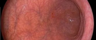
Before seeing pathology, it is necessary to examine the mucous membranes of different people several times in order to identify the pattern of histomorphology of a healthy digestive organ. The stomach of a healthy person reflects the light of the endoscope from the inside, and the secreted mucus gives shine to the transparent main gastric cells with a transparent light-refracting medium. Examination of an empty stomach reveals a folded surface with a height of convolutions of no more than 1 cm. Inflating the stomach with air straightens the folds and the inner surface of the mucous membrane becomes smooth, showing the smallest shades of color and integrity of the integument. You should know that the pyloric region of the digestive organ is somewhat paler than the rest of the organ. The pylorus area is distinguished by more massive folds, which is considered normal. A yellow tint to the inner surface of the stomach in some patients is not considered a pathology.
What to do if the gastric mucosa is hyperemic?
Normally, the gastric mucosa has a light pink color, which becomes brighter closer to the pyloric region.
In some patients they have a yellowish tint, which is not a pathology. During examination, the epithelium reflects the light of the endoscope, so it appears shiny. Numerous folds of the mucosa have a thickness of 6-10 mm.
Their size gradually increases closer to the antrum. When air is introduced into the stomach cavity, the folds of the mucous membrane are smoothed out, and this allows you to examine the entire surface.
If the diagnostician notes that the gastric mucosa is hyperemic, what does this mean? External signs of hyperemia are redness and swelling of the folds of the stomach. The color change is due to blood flow.
The mucous and submucosal layers of the wall have a branched capillary network, with numerous anastomoses between them. Therefore, an increase in blood inflow and a decrease in blood outflow causes the filling of capillaries, which shine through the epithelial layer, changing the color of the mucous membrane.
The chronic form of gastritis often does not have significant manifestations and manifests itself only in a slight decrease in the overall quality of life of the patient. Mild forms of the disease are characterized by stool disorders, and severe forms are characterized by anemia, frequent passage of gas, and bad breath.
Depending on the state of the acidic environment of the stomach, the symptoms of the disease differ slightly. So, with increased acidity, the following signs are noted:
- pain in the middle of the hypochondrium that goes away after eating
- diarrhea
- heartburn after eating acidic foods
- frequent belching
Reflux esophagitis
Gastritis
An accurately identified form of gastritis is the key to successful treatment, which is comprehensive. Neglect of the pathology and failure to comply with doctor’s instructions complicates the treatment of gastritis. For this reason, the outcome of the disease depends only on the patient’s desire to quickly eliminate the stomach problem. A twice-a-year examination by a gastroenterologist will relieve sudden onset pathologies.
Diseases due to gastric hyperemia
Hyperemic gastric lining occurs in several varieties. The type of hyperemia determines the diagnosis of the disease.
With superficial gastritis, hyperemia reaches a moderate degree. The inflammatory process can cover a separate area or become widespread. During the acute course of the disease, the endoscope reveals white foam, the folds of the organ look thicker than normal. When gas is injected, it is not possible to completely achieve a smooth inner wall.
Atrophic gastritis is characterized by focal thinning of the membrane. The vascular pattern in this place is clearly visible, the areas of the mucosa around the atrophic zone look paler.
If the hyperemic gastric mucosa is accompanied by the release of purulent masses, such gastritis has a fibrous form. The disease rarely has independent factors of genesis; in most cases, scarlet fever or measles have consequences in the form of hyperemia of the mucous membrane, followed by vomiting with blood. This is how areas of dead mucous membrane with pus are rejected and is accompanied by intense pain.
Recommendations for the treatment and prevention of gastric hyperemia
An accurately identified form of gastritis is the key to successful treatment, which is comprehensive. Neglect of the pathology and failure to comply with doctor’s instructions complicates the treatment of gastritis. For this reason, the outcome of the disease depends only on the patient’s desire to quickly eliminate the stomach problem. A twice-a-year examination by a gastroenterologist will relieve sudden onset pathologies.
In various pathological conditions of the stomach, redness and swelling of its walls appear. This condition is fraught with the development of serious complications.
Hyperemia of the gastric mucosa is often diagnosed during endoscopic examination of the digestive organs. This phenomenon usually requires medical attention.
Prevention of hyperemia of the gastric mucosa
The key to favorable progress and complete cure of diseases, and therefore of symptoms, is an accurate diagnosis with determination of the root cause of its occurrence. Treatment becomes more complicated if the doctor’s recommendations are not followed. Therefore, it is important to undergo a physical examination and gastroscopy with a gastroenetrologist twice a year, which will avoid sudden problems with parts of the stomach.
In addition, it is important to eat right, get rid of bad habits, avoid stress, and not abuse gastro-soluble medications.
What is hyperemia of the gastric mucosa
In medicine, the term “hyperemia” means redness and swelling, in particular of the mucous membranes and skin. This phenomenon occurs as a result of the vessels in the affected area overflowing with blood.
If during gastroscopy it is discovered that the gastric mucosa is swollen and hyperemic, then this condition indicates that the inflammatory process of the organ wall has begun. Hyperemia can be localized diffusely or focally.
This pathology is a symptom of many stomach diseases. Normally, when the mucous membrane has a pink tint, it reflects the glare of the endoscope, and its thickness ranges from five to eight millimeters.
When wrinkles expand under the influence of air, they quickly smooth out. It is considered normal when the epithelium in the antrum is pale pink.
Why does my stomach hurt: nuances
Mechanical or even chemical irritants in the form of eating spicy foods, pepper, mustard or hydrochloric acid, alcohol do not cause pain. When the walls of the stomach stretch and the stomach itself becomes full of food, then it begins to hurt. Symptoms appear in the form of colic or dull aching pain. If the membrane is inflamed and hyperemic, then pain appears from any mechanical irritant.
The shell does not have a protective layer, and exposure to gastric juice rich in hydrochloric acid leads to pain and heaviness in the stomach. With angina pectoris, hernia, or gastric ulcer, the skin segments act on the vagus nerve, which also leads to pain. The cause of pain in the pancreas may be intercostal neuralgia or affected abdominal wall muscles.
Main causes
Hyperemia of the mucous membrane occurs due to the following diseases:
In addition, the following factors can provoke this condition:
- mechanical damage to an organ with a sharp object;
- improper and irrational nutrition;
- measles infection, scarlet fever;
- entry into the body of the bacterium Helicobacter pylori;
- renal failure;
- depression for a long time;
- stressful situations.
In some cases, the mucous layer may turn red due to an inflammatory process in the walls of the organ.
Symptoms of the disease, dangerous signs
Hyperemic gastric mucosa may be accompanied by the following symptoms:
- pain in the epigastric zone;
- heartburn;
- nausea;
- vomit;
- difficulty urinating;
- drowsiness;
- swelling of the limbs, face;
- tachycardia;
- weight gain or loss;
- lack of coordination.
If these signs occur, it is important to contact an experienced specialist who will refute or confirm the diagnosis.
The form of gastritis is determined by the nature and localization of hyperemia:
- Moderately hyperemic mucosa with edema, accompanied by a foam-like white coating on the surface, in which the affected areas are distinguished, indicate a mild inflammatory process.
- If the redness is local, the mucous folds are thin and pale, with pronounced blood vessels, then this phenomenon indicates atrophic gastritis.
- With foci of hyperemia, there may be a phlegmonous form, which occurs when the organ is damaged by something sharp.
- Severe focal redness, in which a purulent process is observed, raises suspicion of the fibrous form. A dangerous sign in this case is vomiting with blood.
- When hyperemia is diffuse, a superficial form of gastritis is possible.
If the patient has a bulbitis, then edema with hyperemia of the surface of the stomach wall and a thickened layer of the antral epithelium are diagnosed.
Classification of mucosal hyperemia
There are passive hyperemia, which is characterized by excessive blood flow, and active (when the flow of blood from the wall of the organ is impaired). The passive type of hyperemic mucosa is a violation of the venous circulation in the organ. The active form is arterial hyperemia.
In the first case, the organ continues to be damaged as a result of oxygen deficiency. Staying active promotes recovery.
In addition, hyperemia can be focal or diffuse depending on the location.
How to treat
Arterial hyperemia is not treated in 85% of cases. In such a situation, intensive blood supply to tissues begins as a consequence of independent regeneration of damage. The cells of the stomach receive the necessary amount of oxygen and nutritional components, due to which metabolic processes are accelerated. As a result, cells begin to divide rapidly, and healthy tissues replace damaged structures. In the arterial form of the pathology, doctors only prescribe proper nutrition and help balance the diet.
In the remaining 25% of cases, active hyperemia, as well as venous blood filling, indicate the presence of gastritis. In case of inflammation of the mucous membrane, complex treatment is carried out. The patient must follow a strict diet, take antibiotics to suppress the growth of Helicobacter pylori and other medications to accelerate tissue regeneration. You are allowed to drink herbal infusions and eat honey.
Important information: How to treat hemophilia in children and what are the clinical symptoms of blood incoagulation in newborns
Diagnostic methods
A gastroenterologist will help diagnose the problem. He first examines the patient and collects anamnesis.
After a medical examination, a gastroscopy is performed. It is performed using a special device - an endoscope. It is equipped with viewing optics and a camera.
This diagnosis is an unpleasant and painful procedure, but it allows you to accurately determine the condition of the organ, identify the causes of hyperemia, thanks to which the doctor prescribes the appropriate treatment tactics. In addition, using this method, a biopsy is performed, i.e., tissue is taken for examination.
Diagnostics
Having looked at the statistics, we can conclude that almost 90% of people need consultation with a gastroenterologist. To make a correct diagnosis, a specialist prescribes an examination, which is divided into laboratory and instrumental diagnostics.
Laboratory methods include: studies of gastric juice, blood, urine and feces. With their help, you can determine the secretory function, bacterial composition of the gastrointestinal tract, enzyme activity and other important functions. But without instrumental methods, the analysis results are uninformative.
Laboratory methods include: studies of gastric juice, blood, urine and feces. With their help, you can determine the secretory function, bacterial composition of the gastrointestinal tract, enzyme activity and other important functions. But without instrumental methods, the analysis results are uninformative.
The gastric mucosa is focally hyperemic, there is a coating with whitish foamy mucus on the surfaces of the organ in “mucus lakes”, the folds are compacted and are not completely smoothed out with the help of air.
To detect the disease - even if there are almost no problems with the stomach - make an appointment with a gastroenterologist. Gastroscopy is an excellent diagnostic option. Diagnostics involves a procedure carried out by a probe, camera and inspection optics. Using this method, you can assess the condition of organs, take a tissue biopsy, find out the diagnosis and prescribe therapy.
A gastroenterologist will help diagnose the problem. He first examines the patient and collects anamnesis.
After a medical examination, a gastroscopy is performed. It is performed using a special device - an endoscope. It is equipped with viewing optics and a camera.
This diagnosis is an unpleasant and painful procedure, but it allows you to accurately determine the condition of the organ, identify the causes of hyperemia, thanks to which the doctor prescribes the appropriate treatment tactics. In addition, using this method, a biopsy is performed, i.e., tissue is taken for examination.
For preventive purposes, as well as at the slightest suspicion of gastrointestinal diseases, you should seek help from a gastroenterologist. The most accurate and reliable research method is gastroscopy. This procedure involves attaching a camera and optics to the probe, as well as the use of other auxiliary instruments that are inserted deep into the esophagus and stomach.
Gastroscopy allows you to accurately determine the condition of the internal organs of the gastrointestinal tract, identify pathological changes and, if necessary, take a tissue sample for further study.
A doctor can easily notice pathological changes in the epithelium, since in a healthy state they are characterized by transparent mucus, as well as a shiny pink surface. Diagnosis is made on an empty stomach, which allows you to notice the slightest changes not only in the epithelium itself, but also in its thickness.
Gastric hyperemia is characterized by redness of the mucous membrane and indicates the onset of the development of many gastrointestinal diseases. Early diagnosis allows you to determine the cause of its development, as well as eliminate it as quickly and effectively as possible.
Treatment methods
Treatment of hyperemia of the gastric mucosa depends on the nature and severity of the disease. Basically, treatment is carried out with an integrated approach. Therapy may include the use of drugs from the following groups:
- Antibacterial agents. Antibiotics are prescribed in case of bacterial infection, for example, Helicobacter pylori.
- Antacids. The most commonly prescribed are Rennie, Maalox, Almagel, Gastal, Phosphalugel, Gelusil, Taltsid.
- Histamine receptor blockers (for example, Ranitidine).
- Drugs that stimulate gastric secretion. These include plantain juice or Plantaglucid.
- Proton pump inhibitors. Omeprazole, Zolser, Ultop or Bioprazole are widely used in the treatment of gastritis and ulcers.
- Enzymes. Drugs such as Mezim, Festal or Mexaza improve digestive processes.
In some cases, nitrofuran derivatives and bismuth subcitrate (De-nol) are prescribed. The use of vitamin B12 is also necessary.
Only a qualified doctor can prescribe these medications, taking into account the diagnosis, the severity of the disease, as well as the individual characteristics of the body.
In addition, physiotherapeutic procedures contribute to recovery. It is important to stop drinking alcohol and smoking during treatment.
An important component of the treatment of stomach diseases is dietary nutrition. In frequent cases, the Pevzner diet is recommended. The choice of foods for the diet is also based on whether gastric secretion is increased or decreased.
In addition, alternative medicine is an auxiliary therapy.
Possible complications and prognosis
After the underlying disease of the stomach is cured, a symptom such as redness of the mucous membrane goes away on its own.
However, if this problem is ignored, the following complications may develop:
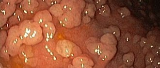
- polyposis;
- stomach bleeding;
- malignant tumor;
- Iron-deficiency anemia;
- Ménétrier's disease;
- chronic pancreatitis;
- cholecystitis.
In addition, any form of gastritis can lead to peptic ulcers, which can even be fatal if severe.
If you have stomach problems, the condition of your nails, skin and hair worsens.
To avoid the development of undesirable consequences, it is important to promptly diagnose diseases that are accompanied by gastric hyperemia and begin timely treatment. Therefore, if there are any signs of diseases of the digestive organs, you must consult a gastroenterologist.
Prevention measures
To prevent the development of hyperemia of the gastric wall, you need to adhere to the basic rules of prevention. First of all, it is important that the diet is balanced and rational. Therefore, it is necessary to include healthy foods in your diet and avoid junk food.
In addition, preventive measures include:
- Full sleep.
- Compliance with hygiene rules.
- Doing physical exercise daily.
- Annual preventive examinations.
- Compliance with medical recommendations.
- Avoiding stressful situations.
- Alternating physical activity with rest.
Following these recommendations will reduce the risk of developing hyperemia several times.
Hyperemia of the walls of the stomach affects the surface of the organ mucosa. It is a sign of various organ diseases that can cause serious complications. Therefore, it is important to consult a doctor in time to determine the pathology and undergo appropriate treatment. Therapy depends on the underlying diagnosis and its severity.
Have you been struggling with GASTRITIS and ULCERS for many years without success?
“You will be amazed at how easy it is to cure gastritis and ulcers just by taking it every day.
An additional symptom of many diseases of the digestive system is swelling and redness of the mucous membrane. The fact that the gastric mucosa is hyperemic is clearly visualized during the patient's gastroduodenoscopy. Such an examination is prescribed by a doctor if an ulcer, gastritis, pancreatitis is suspected, which are accompanied by other symptoms (pain in the epigastric region, nausea, belching, flatulence).
Types and characteristic symptoms
Chronic hyper- or normacid gastritis . Although usually mass cell death does not occur with this type of inflammation, when the process spreads deeper in places, focal atrophy of the mucosa in the antrum of the stomach is observed. Inflammation also often affects the duodenum, causing duodenitis. In this case, the following may be observed:
- constipation;
- heartburn;
- belching with sour air;
- occasional nausea.
Pain with this form of gastritis, especially in combination with duodenitis, often resembles that of a peptic ulcer - at night, on an empty stomach, 1.5-2 hours after eating (however, unlike an ulcer, they are less intense and less likely to be associated with seasonality, subside when following a diet and occur when it is inaccurate).
Symptoms revealed using laboratory and instrumental methods:
- Contrast radiography - impaired motility, thickening of the folds of the inner lining of the stomach, signs of hypersecretion of gastric juice on an empty stomach.
- FGDS - redness, swelling of the mucous membrane, the presence of mucus or bile in the stomach.
- Histology - a picture of superficial gastritis and focal atrophy in the antrum of the stomach, unchanged mucosa or superficial gastritis in the fundus.
- Palpation of the abdomen - moderate or slight pain in the epigastric region and slightly to the right.
- pH-metry - normal or increased secretion of HCl.
What is the problem
If the results of gastroscopy indicate that the gastric mucosa is focally hyperemic, this indicates the development of the initial stage of the inflammatory process in the gastric walls. This is not a separate disease, but an additional symptom of the main pathological processes developing in the epigastric region.
It is very important to consult a doctor in time, do not ignore pain in the upper stomach, nausea, and heartburn. Focal hyperemia of the gastric mucosa accompanies most diseases localized in this area, but it is detected only during diagnosis.
Normally, the gastric mucosa is pink, smooth, and reflects the shine of endoscopic equipment. The thickness of the folds should not be less than 5 mm, more than 8 mm. When expanded with air, the folds should be completely straightened.
Diseases accompanied by symptoms
Hyperemia can be a signal of many dysfunctions in the organs of the digestive system. Based on how pronounced a given symptom is and where it is localized, conclusions can be drawn about the type of disease. Often, in the presence of such a clinical sign, a diagnosis of gastritis, gastroduodenia, or peptic ulcer is made. Diseases not related to the gastrointestinal tract are very rarely detected.
Gastritis of various forms provokes the appearance of such symptoms:
- A mild inflammatory process is accompanied by mild hyperemia, the lesion is focal. The surface of the mucous membrane is swollen, covered with a white coating, the folds are thickened, and when stretched with air, they do not smooth out.
- If an atrophic process occurs, this is accompanied by severe thinning and pallor of the mucosa, and a red vascular pattern is clearly visible. The lesion is local in nature.
- The fibrous form of gastritis is accompanied by severe hyperemia, which is focal in nature with a purulent process. The main provoking factor of this pathological process is the impact of infectious agents during the development of scarlet fever and measles. Additionally, symptoms such as vomiting with blood appear. This indicates that the festering film has begun to come off.
- If the phlegmous form of the disease develops, lesions are visualized, which are caused by trauma to the stomach with a sharp object (for example, a fish bone).
- In the presence of bulbitis, swelling with redness is noted, the folds are thickened in the antrum. The mucous membrane is swollen, its surface is red. The main causes of this disease are considered to be an incorrect diet and the influence of the bacterium Helicobacter pylori.
- In the presence of dysfunction in the kidneys, most patients experience swelling or hyperemia of the mucous membrane, expressed in different intensities (depending on the degree of development of the pathological process).
- Hyperemia can be caused by such provoking factors as long-term depression, chronic stress, and regular emotional overstimulation. Under the influence of such unfavorable psychological factors, the vascular walls of the stomach become overfilled with blood fluid.
Causes and symptoms
The following factors can provoke the development of a pathological process:
- mechanical damage to the stomach due to gastritis or ulcers;
- unbalanced diet;
- severe infectious lesions;
- overgrowth of Helicobacter pylori;
- renal failure;
- chronic stress;
- depressive state.
In clinical practice, 2 types of disease are classified, each of which is characterized by an individual symptomatic picture and causes:
- Arterial hyperemia or active form of pathology. The stomach is intensively supplied with oxygen and nutrients, so in most cases there is a positive dynamics of treatment and rapid recovery. Diffuse foci of hyperemia disappear within 1-2 weeks. The arterial form of pathology is possible with increased functional activity of the myocardium or due to low pressure in the vessels. In some cases, the cause of blood filling is a violation of innervation: dilated vessels, degeneration of nerve endings in the smooth muscles of the bloodstream.
- Venous-passive redness. The organ stops receiving the elements it needs, so the process of tissue regeneration stops. The thickness of the stomach walls increases, body temperature decreases. The mucous membrane atrophies due to oxygen starvation of cells. The passive form of the pathology is characteristic of increased pressure in the venous vessels and mechanical damage.
Important information: How to treat marble disease (deadly marble disease) in children and symptoms of osteopetrosis
If a person develops venous congestion, he requires urgent treatment. The presence of a passive form of pathology can be suspected based on the following symptoms:
- the appearance of excess body weight;
- swelling of the face and legs;
- tachycardia;
- high blood pressure;
- drowsiness, chronic fatigue;
- violation of coordination in space.
In 9 out of 10 patients, hyperemia develops due to gastritis, intestinal inflammation or peptic ulcer of the stomach and duodenum. In rare cases, the disease can progress regardless of the state of the digestive system. The following signs are characteristic of different forms of hyperemia:
- As the tissue dies, the surface of the mucosa becomes thinner and begins to turn pale. In such a situation, when the vessels are full, no redness is observed, the vascular network is clearly visible.
- With increased acidity, foci of redness of various locations appear on the mucous membrane of the organ. A coating of foamy white mucus is visible on the walls of the stomach; the folds of smooth muscle become dense and practically do not smooth out when exposed to air.
- Against the background of fibrous gastritis, severe focal hyperemia is observed, characterized by the presence of pus. In such a situation, measles or scarlet fever can provoke the development of a pathological process. The patient often feels sick, and blood clots are found in the vomit.
- When the duodenal bulb becomes inflamed, the folds of the stomach become dense and begin to thicken. Congestion of the vessels leads to the appearance of severe swelling and bright red spots on the surface of the mucous membrane. This condition can be provoked by Helicobacter infection and an unbalanced diet.
- With phlegmonous gastritis, the cause of hyperemia is mechanical trauma to the mucous membrane with sharp objects: plastic, glass, fish bones.
- The superficial form of gastritis provokes the development of hyperemia on the body and in the antrum of the organ.
Important information: What is venous thromboembolism: symptoms and treatment
With renal failure, severe swelling is observed.
Diagnostic measures
If any suspicious symptoms appear (for example, nausea, bloating, heartburn, belching, pain), it is very important to visit a gastroenterologist in a timely manner. One of the main methods for studying the condition of the mucosa is a procedure called esophagogastroduodenoscopy. It is performed using an endoscope - a special probe with a camera at one end to view the internal state.
Using this method, you can accurately assess the condition of the body and the internal walls of the stomach, take a piece of tissue for analysis (biopsy), visualize the presence of a pathological process, and prescribe the correct course of therapy.
A competent specialist will easily determine the presence of pathology in hyperemic epithelium, since healthy tissue looks shiny and produces mucus normally. During the examination, when inflated with air, the folds are straightened, the surface is smooth, the epithelial cover is intact and clean, without wounds or ulcerations. The color is pink, there may be a yellowish coating.
As the disease develops, the symptoms worsen, redness of the epithelial layer appears, the folds increase, do not straighten when inflated, and swelling is observed.