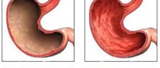
According to statistics, up to 15% of all bleeding from the digestive tract is due to Mallory-Weiss syndrome. It was described in 1932 by pathologist Kenneth Mallory and physician Soma Weiss from Boston, after whom the syndrome was named. It is a rupture (crack) in the area of the gastroesophageal junction, the lower third of the esophagus or the cardiac part of the stomach, which occurs during retching or repeated vomiting. Isolated esophageal localization of the rupture is 8%, in the cardiac part of the stomach - 44%, and in almost 50% the mucosal crack is located in the esophageal-gastric junction.
Men aged 30–65 years who are prone to alcohol abuse are more likely to suffer. It is rare in children.
Factors leading to mucosal rupture
The reasons leading to an increase in pressure in the lumen of the digestive tract and, subsequently, to the occurrence of a crack are the following:
- repeated vomiting and retching;
- continuous long-term hiccups;
- paroxysmal severe cough;
- blunt trauma to the upper abdomen;
- cardiopulmonary resuscitation measures;
- diaphragmatic hernia (found in more than 50% of patients);
- medical procedures (endoscopy).
In some cases, the cause cannot be determined.
Trauma to the oral mucosa
Acute mechanical injury. Occurs as a result of biting or injury with instruments and is uncommon. It manifests itself in the form of a hematoma (intratissue hemorrhages without violating the integrity of the epithelium), erosion or ulcers. With interstitial hemorrhage, pain is noted at the site of injury, which quickly (after 1 - 3 days) disappears, but if the integrity of the epithelium is damaged, a painful erosion or ulcer with infiltration is formed. Once infected for the second time, the wound turns into a long-term non-healing ulcer.
Chronic mechanical injury. Happens frequently. The traumatic factor can be sharp edges of teeth, bridges and removable dentures, tartar, spicy and hot food, etc. The clinical picture and course of the process largely depend on the location of the damage (presence or absence of the submucosal layer), the age of the patient, secondary infection, the strength of the irritant factor a. Damage to the mucous membrane caused by chronic irritants is more common in older people. This is facilitated by a decrease in the turgor of the mucous membrane, a decrease in the height of the bite due to abrasion of the hard tissues of the teeth, loss of teeth, and their displacement. In older people, the regeneration process is slowed down, which causes slow healing of the damaged mucous membrane.
Changes in the mucous membrane during chronic mechanical trauma may not bother the patient for a long time, causing only a feeling of awkwardness, discomfort, minor pain, and swelling. When examining the mucous membrane, catarrhal inflammation (edema, hyperemia), a violation of its integrity (erosion, aphthae, ulcers), proliferative changes (hypertrophy of the gingival papillae, gingival margin), hypertrophy of the tongue papillae such as papillomatosis, and increased keratinization (leukoplakia) are revealed. These symptoms can occur in combination with each other. It should be remembered that simultaneously with mechanical trauma, the mucous membrane is exposed to the microflora of the oral cavity, which is often reflected in the clinical picture and course of the process.
Most often, with chronic trauma, catarrhal inflammation occurs: hyperemia, edema with infiltration, proliferation. The severity of these changes depends on the strength and duration of the stimulus. In this case, the process may be accompanied by exudation (at first the secretion is serous and then purulent).
The course of catarrhal inflammation can be acute or chronic. Acute inflammation lasts up to 7-10 days and disappears quickly when the irritant is eliminated. In the absence of treatment, chronic focal or generalized inflammation is observed. A traumatic ulcer that occurs as a result of local damage is also called decubital.
Of the factors that can cause irritation and damage to the oral mucosa, dentures should be highlighted. A removable denture transmits chewing pressure to the mucous membrane, delays self-cleaning of the oral cavity, which leads to a disruption of the established balance between different types of microorganisms, and changes the analyzer function of the mucous membrane receptors. In the occurrence of inflammation of the mucous membrane under the prosthesis, in addition to the traumatic factor, the sensitization factor should also be taken into account.
Inflammation of the mucous membrane of the prosthetic bed can be focal - in the form of pinpoint hyperemia or large hyperemic spots and diffuse, often occupying the entire surface of the prosthetic bed. Against the background of an inflamed and edematous mucous membrane, pinpoint hemorrhages, erosions, as well as foci of hyperplasia of the mucous membrane in the form of granularity or lobulation may occur. Changes in the mucous membrane can also be observed when using fixed dentures.
Habitual biting of the mucous membrane. Habitual biting of the mucous membrane of the cheeks and lips is a common occurrence among neuropathists, mainly young people (high school students and students). It may not be noticed by patients or be conscious. The habit of constantly biting the mucous membrane leads to chronic damage localized along the line of closure of the teeth and adjacent areas accessible to biting. The mucous membrane swells and acquires a whitish, macerated, flaky surface in the form of diffuse spots or poorly defined large areas. The epithelium is unevenly desquamated, has a fringed appearance due to multiple small flaps, and is easily removed by scraping.
During the normal course of the process there is no pain. In severe cases, painful erosions may occur. Pathohistologically, the phenomena of parakeratosis are detected.
Injuries to the oral cavity during sexual activity . Damage to the frenulum of the tongue and soft palate is a common lesion of the oral mucosa associated with sexual activity. Ulceration of the frenulum of the tongue is possible as a result of injury to it on the lower incisors during orogenital sex. Often in such cases, an ulcer with a grayish-white fibrinous exudate is detected, surrounded by a thin belt of hyperemia. The diagnosis is easy to establish based on the history.
Abstaining from sexual activity promotes healing. Chronic irritation can cause secondary bacterial infection, the development of leukoplakia or traumatic fibroma, and HPV infection. During fellatio, damage to the soft tissues of the oral cavity is possible in the form of erythema and submucosal hemorrhages on the soft palate. Isolated bright red petechiae are usually noted initially. They gradually merge into a large spot that extends across the midline of the palate. Hemorrhages are painless, do not fade on diascopy and resemble petechiae in infectious mononucleosis, but unlike it, they are not accompanied by fever and lymphadenopathy. Petechiae gradually turn pale and disappear within one week.
The most common causes of rupture of the gastric mucosa
The mechanism of occurrence of Mallory-Weiss syndrome depends on those factors that lead to a crack in the mucosa and further bleeding from it.
- Vomiting is the first and most common cause of the formation of a crack in the gastric mucosa. It occurs when the patient has: alcoholism, pregnancy, bulimia (sharp attacks of hunger, during which a large amount of food is eaten at once and vomiting is artificially induced). When vomiting, the pressure in the cardiac part of the stomach increases sharply, its wall is stretched and the mucous membrane ruptures.
- The second most common cause of mucosal rupture is the presence of a diaphragmatic hernia: during endoscopy with strong urge to vomit, the gastric mucosa prolapses into the esophagus and is pinched in the cardiac sphincter. At this point, a crack may form in the stomach.
- In contrast, iatrogenic injury (caused by endoscopy) is extremely rare but has been reported in the literature.
Forecast
If the injured person seeks medical help in a timely manner (within 3 hours after injury), then the prognosis for him will be quite favorable. With minor damage, tears in the mucous membrane can heal on their own within 3 days, provided that you take medications prescribed by doctors and follow their recommendations. If the patient goes to a hospital 5 hours after the incident (no later than 65 hours), then serious complications may already begin.
According to statistics, with complete rupture of the stomach, the mortality rate reaches 85%. Perforation of the walls of the organ, leading to the development of peritonitis, ends in death in 40% of cases. The mortality rate among patients whose injuries did not cause damage to the organ walls reaches 3%.
In the case where severe vomiting led to rupture of the mucous membrane, the patient should continue to take precautions:
- do not overeat;
- stop or minimize the consumption of alcoholic beverages;
- engage in timely treatment of gastrointestinal diseases whose symptoms include vomiting.
When damage to the abdomen occurs without compromising the integrity of the skin, gastric rupture is possible. The main reason is mechanical impact. The walls of the organ and ligaments may be damaged. Symptoms depend on the severity of the injury. Such an injury is dangerous, so patients are urgently hospitalized. The surgical method of eliminating the problem is more often used. Medicines are intended to relieve symptoms and prevent infection with inflammation. Diet allows the digestive system to adapt to the post-injury state.
Clinical picture and complications of massive bleeding
Clinically, the syndrome is manifested by the presence of fresh blood in the vomit, which is observed in 30–50% of patients.
Less common are melena (black stool as a result of bleeding), abdominal pain, weakness, lethargy, dizziness, and loss of consciousness.
Mallory-Weiss syndrome is dangerous due to its severe complications, which include:
- hypovolemic shock;
- decreased blood supply to the myocardium up to the development of myocardial infarction;
- perforation of the mucosa at the site of rupture;
- ischemia and tissue infarction that occur after endovascular embolization of vessels damaged by rupture of the mucous membrane for therapeutic purposes.
Symptoms
After an injury, a person is in a state of shock for some time. At this time, he cannot give an objective assessment of his feelings. As a result, the clinical picture appears much later.
Patients should pay attention to the following symptoms:
- sharp pain appears;
- The abdominal muscles become very tense;
- due to the accumulation of blood in the abdominal cavity, a dull and short sound appears when tapped;
- the abdominal area swells and becomes hard as a rock;
- due to blood loss, spots appear before the eyes;
- a person may be in prostration;
- the skin acquires a pale grayish tint;
- Coordination of movements is impaired, the lower limbs become weak and give way.
How is the diagnosis made?
A fissure in the digestive tract can be suspected taking into account the following diagnostic criteria:
- Clinical manifestations are the presence of scarlet blood in the vomit, mainly in patients under the influence of alcohol. With massive bleeding, signs of hemorrhagic shock are observed: severe pallor, tachycardia, profuse sweating, low blood pressure, orthostatic collapse (loss of consciousness when changing body position).
- Data from the main diagnostic and at the same time therapeutic method - endoscopic.
- General clinical blood test - makes it possible to assess the presence of anemia and its severity.
All diagnostic techniques are necessary to establish:
- severity of bleeding;
- localization of mucosal rupture;
- early detection of complications.
Diagnostic measures
According to statistics, the majority of patients who were taken to a hospital with such symptoms undergo immediate surgical treatment. As a rule, during surgery, surgeons identify a mucosal rupture, its location and size.
If the patient is delivered to a hospital in satisfactory condition, surgeons will carry out the following diagnostic measures:
- Tests (blood, stool, urine) are prescribed.
- An x-ray is taken with contrast liquid. This procedure is prescribed to patients only after the exacerbation has been relieved and the pain syndrome has been relieved.
- A procedure called laparocentesis is performed.
- An endoscopy is performed, during which surgeons carry out not only diagnostics, but also therapeutic manipulations.
- Laparoscopy is performed. This surgery is performed under general anesthesia. The patient undergoes several punctures in the abdominal cavity. Through these holes, the surgeon inserts a special instrument, on the tip of which a microcamera is fixed. Thanks to laparoscopy, it is possible not only to visually, through a monitor, assess the severity of the lesion, but also to carry out the necessary surgical manipulations.
- The “gentle catheter” method is used, etc.
Treatment methods
Treatment for a crack in the gastric mucosa includes several stages:
- measures to eliminate bleeding;
- restoration of hemodynamic disorders;
- normalization of the patient's condition;
- measures to prevent the development of complications.
If the condition is not extremely severe, conservative therapy is used (currently 90% of all methods; mortality with this treatment is 3%):
- infusions of colloid and crystalloid solutions to restore lost fluid (5% glucose, Ringer's solution, Lactasol), sometimes - transfusion of blood products;
- hemostatic therapy (aminocaproic acid, Dicynon);
- if necessary, a Blackmore probe, which stops bleeding by mechanical compression;
- drugs that block the formation of hydrochloric acid; in mild cases, it is possible to take drugs orally (PPIs are used - proton pump inhibitors: Pantoprazole, Omeprazole, Esomeprazole);
- antiemetics (metoclopramide / Cerucal / 10 mg in tablet form or by injection 3 times a day);
- antacids, bismuth preparations and coating agents (Venter, De-nol, Almagel, Gaviscon, etc.).
If drug treatment is ineffective, they switch to endoscopic methods or perform an emergency operation to close the defect.
How to recognize the problem?
Main symptoms
Acute stabbing pain radiating to the lower back may be the result of a complete rupture. Diagnosis becomes difficult when, along with the damaged stomach, there are lesions of other tissues and organs.
Against the background of severe abdominal pain, a person experiences weakness.
Shock is the main symptom when the stomach ruptures. Later, a deceptive period of “imaginary well-being” begins, at the end of which the patient feels acute unbearable pain - acute abdomen syndrome. A short dull sound when tapping the injured area is explained by the presence of fluid in the peritoneum and the risk of developing peritonitis - inflammation of the abdominal cavity. And:
- presence of gases;
- weakness;
- temperature increase;
- nausea, vomiting blood.
Symptoms of bleeding that occurs during rupture:
- lowering blood pressure;
- decreased heart rate;
- weakness.
Return to contents
Diagnostic methods
The exact extent of the mucosal rupture is determined only during surgery. The following methods allow you to make the right decision in making a diagnosis:
The extent of organ damage can be assessed during laparoscopic surgery.
- Laparoscopy. An invasive technique that involves inserting a device with a microcamera into the peritoneum through small incisions and simultaneous treatment - repairing the rupture.
- Laparocentesis. It involves a puncture in the anterior abdominal wall to detect or exclude the presence of blood, bile, exudate, gas in the abdominal cavity. Often performed before laparoscopy.
- A variation of abdominal puncture with the yaw catheter technique for visualization of blood clots.
- Contrast fluoroscopy, which is used after relief of inflammation and pain to evaluate the final result of the operation.
- Blood, urine, and stool tests necessary to monitor the patient’s condition.
Return to contents